
postural drainage/handwritten notes of postural drainage techniques
postural drainage positions techniques chest physiotherapy
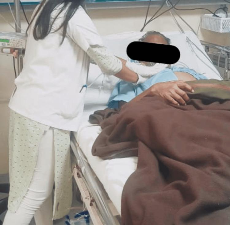
This clinical photograph illustrates a healthcare professional, likely a physical therapist, performing chest physiotherapy on a patient in an inpatient setting. The clinician is seen standing at the bedside, applying manual chest vibration techniques to the patient's anterior thoracic region. The patient is positioned supine in a hospital bed, equipped with side rails and integrated medical devices. Visible equipment includes a bedside monitor or infusion pump on a vertical pole, along with various wires and tubing consistent with continuous clinical monitoring or oxygen therapy. The educational focus of the image is on respiratory rehabilitation interventions, specifically the manual clearance of secretions (airway clearance therapy) in patients with conditions like pneumonia or chronic obstructive pulmonary disease. The image demonstrates the practical application of chest PT, emphasizing the tactile and manual nature of vibration and percussion to facilitate mucus drainage and improve pulmonary function.
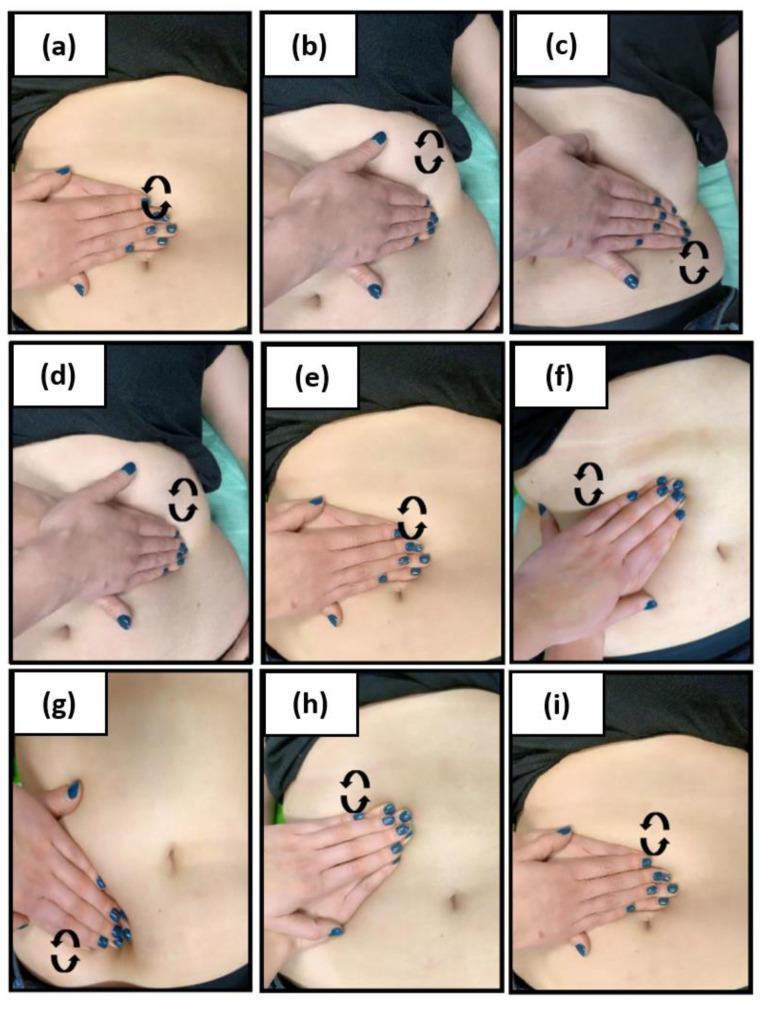
A series of nine clinical photographs (a-i) illustrating manual lymphatic drainage (MLD) techniques for the abdominal region, specifically showing deep drainage protocols. The images demonstrate precise hand placements and orientations relative to key anatomical landmarks on a patient in the supine position. Key positions include central placement over the umbilicus (naval), positioning parallel to the left and right costal arches, and alignment with the left and right inguinal ligaments. Each sub-image includes black circular arrow icons indicating the intended direction of circular manual pressure and rhythmic manipulation required for the effleurage technique. This visual guide demonstrates the sequential steps used in physical therapy to facilitate lymphatic flow and reduce lymphostasis in patients with conditions such as obesity or lymphedema. The series serves as an educational resource for medical and physiotherapy professionals to visualize the correct hand ergonomics and directional strokes necessary for effective abdominal lymphatic drainage.
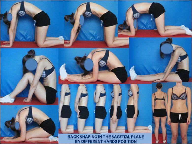
This composite educational patient photograph illustrates the 'back shaping' and symmetrical positioning techniques of the DoboMed method for scoliosis physiotherapy. The image demonstrates variations in sagittal plane spinal alignment across multiple postures, emphasizing active thoracic kyphotization and lumbar lordotization. The top row shows a patient in various quadruped (kneeling) positions; different hand and arm placements (flat hands, elbows on ground) are used to modulate thoracic convexity and vertebral mobilization. The middle sections depict sitting and long-sitting postures focusing on trunk elongation and sagittal correction. The bottom row showcases a sequence of kneeling and standing postures where subtle shifts in hand position and shoulder alignment are used to correct spinal deformity and improve postural awareness. The goal illustrated is 3D auto-correction to stabilize the primary curve and promote balanced trunk alignment. This resource is intended for physical therapists and medical students studying Conservative Treatment of Scoliosis (CTS) and Physiotherapeutic Specific Scoliosis Exercises (PSSE).
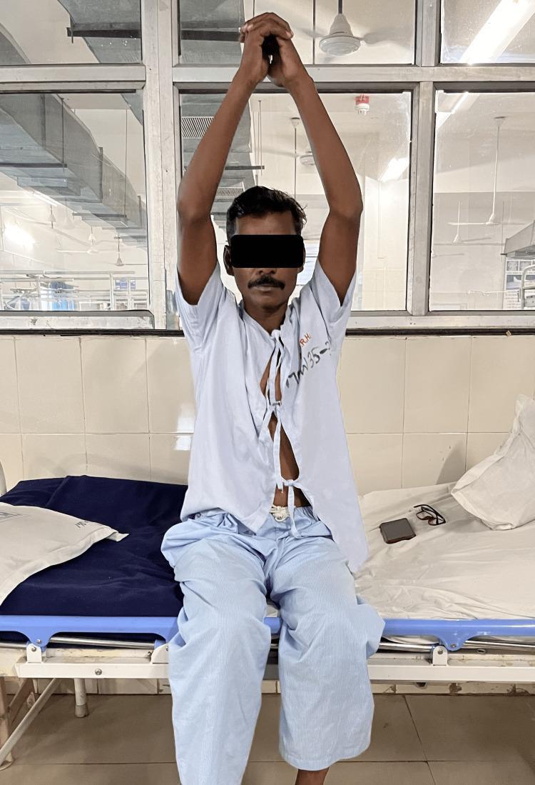
A clinical photograph of an adult male patient demonstrating thoracic expansion exercises as part of a post-operative respiratory physiotherapy regimen. The patient is sitting upright on a hospital bed with his arms extended vertically and hands clasped above his head, a position designed to maximize rib cage elevation and lung volume expansion. He is wearing an open hospital gown, and the setting includes typical clinical markers such as a blue medical mattress and tiled hospital walls. This visual illustrates active chest physical therapy techniques aimed at improving ventilation, reducing the work of breathing, and increasing exercise tolerance following procedures like intercostal drainage (ICD) tube removal. The image serves as a clinical guide for healthcare students and professionals in pulmonary rehabilitation and physiotherapy management.
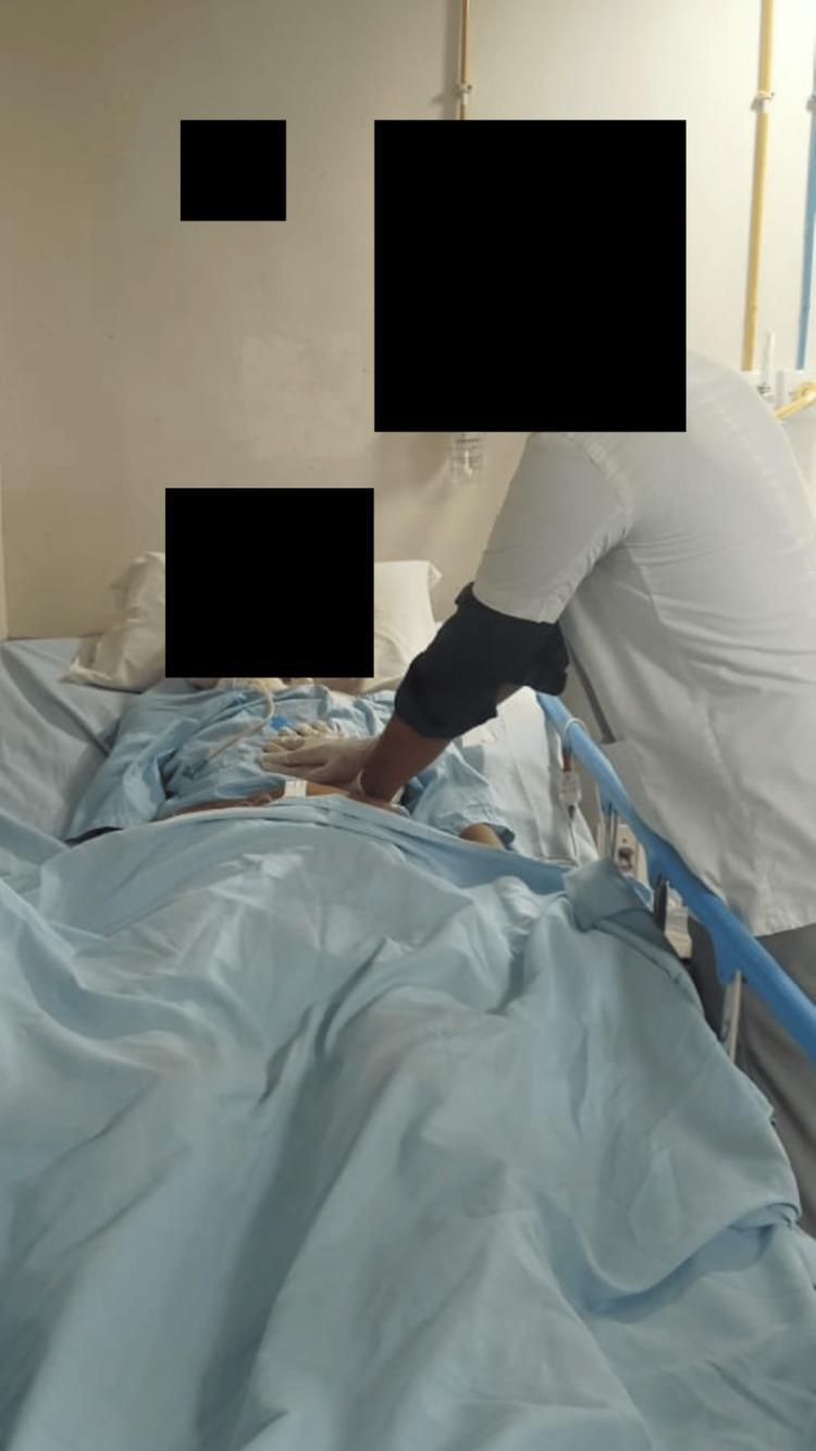
This clinical photograph illustrates a healthcare professional performing chest physiotherapy on a patient in a hospital setting. The patient is positioned supine in a hospital bed, with the clinician standing at the bedside applying a manual chest vibration technique. The clinician's hands are placed specifically over the patient's left lower lung zone, a maneuver intended to facilitate the mobilization and clearance of pulmonary secretions. The patient appears to be receiving supportive care, evidenced by the presence of medical tubing (likely an endotracheal or nasogastric tube) and hospital attire. The clinician is shown wearing protective gloves while performing the intervention. This image serves as an educational example of active pulmonary rehabilitation and airway clearance therapy within a clinical or post-operative context, highlighting manual techniques used in respiratory physiotherapy.
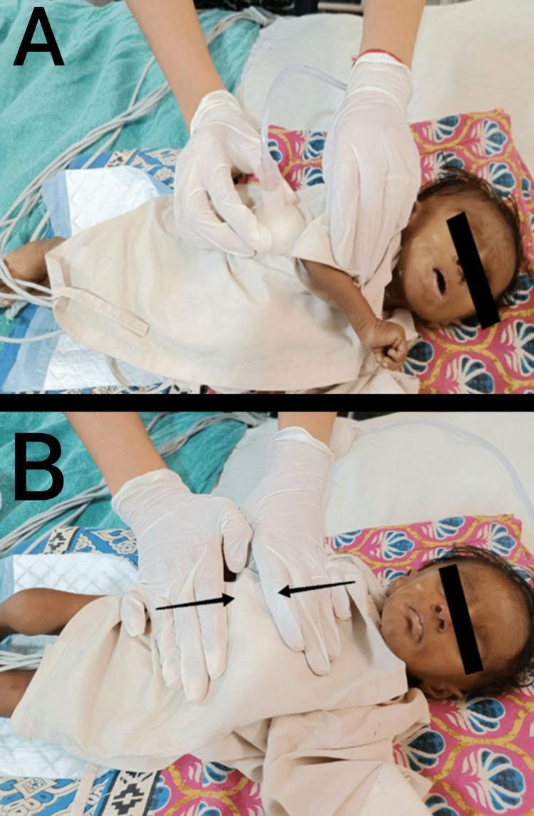
This clinical photograph consists of two panels (A and B) demonstrating postoperative pediatric chest physiotherapy techniques on a three-month-old infant in a supine position. Panel A illustrates mechanical chest vibration, where a healthcare worker in sterile gloves uses a handheld percussor cup with an attached suction/vibration tube against the infant's thoracic wall to facilitate airway clearance. Panel B depicts a manual respiratory therapy technique known as prolonged slow expiration (PSE). In this panel, the practitioner's gloved hands are positioned on the infant's thorax and abdomen, with superimposed black arrows indicating the direction of manual pressure applied to assist with expiratory flow and secretion mobilization. The infant is monitored via chest leads visible in the background and is supported on patterned bedding. These procedures are typical in neonatal or pediatric postoperative care following cardiothoracic surgery to prevent atelectasis and manage pulmonary secretions.
postural drainage lung segments positions diagram
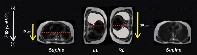
This sequence of cross-sectional thoracic CT images illustrates the concept of a postural lung recruitment maneuver in a pediatric patient. The visualization shows four anatomical states: initial Supine, Left Lateral (LL), Right Lateral (RL), and final Supine. In the supine position, the thorax is elliptical with a horizontal orientation; a red dotted line demarcates a lower lung zone predisposed to atelectasis. Transitioning to lateral positions (LL and RL) reorients the thoracic dimensions, increasing the gravity-dependent vertical distance. Yellow arrows with values (15 cm in supine vs. 25 cm in lateral) represent the trans-pulmonary pressure (Ptp) gradient. In LL, the non-dependent right lung is shown as an aerated gray schematic, indicating recruitment. In RL, the previously dependent left lung is now non-dependent and recruited. The final supine image demonstrates bilateral lung aeration, illustrating how sequential positioning can eliminate atelectasis and maintain an 'open lung' state by utilizing gravity-dependent changes in thoracic geometry and pleural pressure gradients.
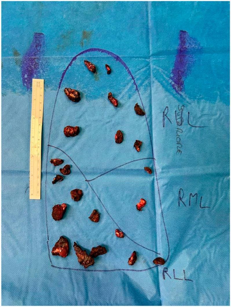
This clinical photograph illustrates a macroscopic intraoperative specimen mapping of multiple lung metastases. Set against a blue surgical drape, a diagrammatic representation of the right lung is hand-drawn, labeled with anatomical segments: RUL (Right Upper Lobe), RML (Right Middle Lobe), and RLL (Right Lower Lobe). Arranged within these marked zones are 25 dark, reddish-brown, irregular-shaped metastatic nodules, ranging from approximately 0.5 cm to 2.0 cm in size, exhibiting a firm, fleshy texture. A vertical metric ruler is positioned on the left side of the diagram for scale. This systematic arrangement is used in oncological thoracic surgery to assist pathologists in anatomical localization and histopathological assessment of multifocal metastatic disease. The image highlights the complexity of precision resections and the clinical importance of manual palpation and accurate anatomical orientation during pulmonary metastasectomy.
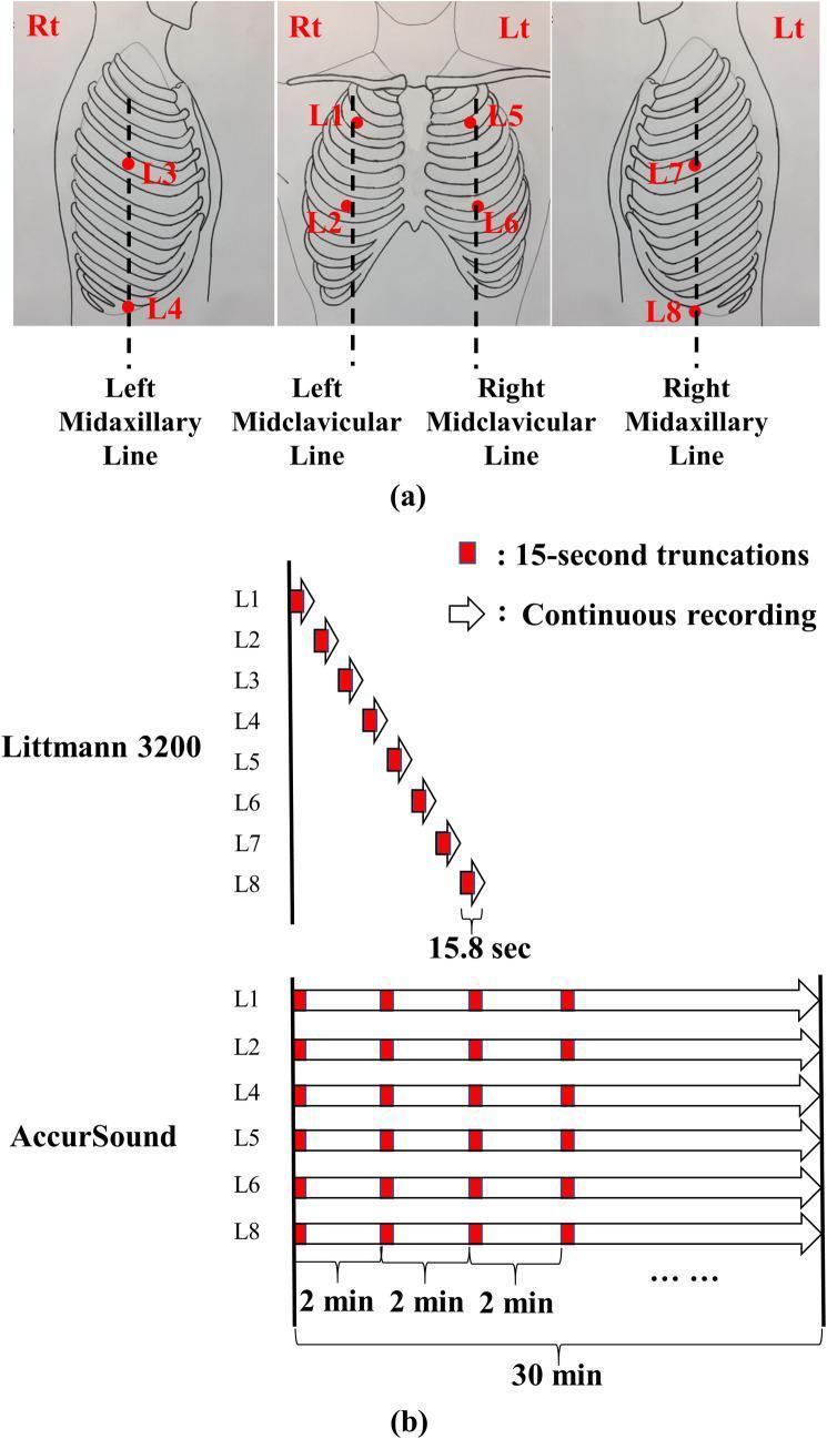
This educational graphic illustrates standardized auscultation locations and lung sound recording protocols. Panel (a) presents anatomical diagrams of the thorax showing eight specific auscultation points (L1–L8). Right-side locations include the second intercostal space (ICS) on the midclavicular line (L1), the fifth ICS on the midclavicular line (L2), the fourth ICS on the midaxillary line (L3), and the tenth ICS on the midaxillary line (L4). Left-side locations (L5–L8) mirror these positions. Panel (b) compares two digital recording methodologies. The Littmann 3200 protocol demonstrates a sequential approach, capturing 15.8-second recordings from L1 through L8 one by one. In contrast, the AccurSound (HF-Type-1) protocol depicts a simultaneous multi-channel approach, recording from six locations (L1, L2, L4, L5, L6, L8) concurrently for a continuous 30-minute duration. The diagram highlights how these recordings are truncated into 15-second segments for analysis. This visual is designed to explain standardized pulmonary physical examination techniques and data acquisition for digital stethoscopes.
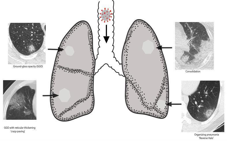
An educational anatomical diagram and comparison chart illustrating the radiological manifestations of COVID-19 lung injury. The central element is a diagram of the respiratory system, showing a SARS-CoV-2 viral particle entering the trachea, leading to bilateral lung involvement. Surrounding the diagram are four labeled chest CT scan segments demonstrating specific patterns: 1) Ground-glass opacity (GGO), appearing as hazy, increased lung attenuation that does not obscure underlying vessels; 2) Consolidation, showing dense, homogenous opacification with obscuration of bronchovascular structures; 3) GGO with reticular thickening, also known as the 'crazy-paving' pattern, characterized by thickened interlobular septa superimposed on a GGO background; and 4) Organizing pneumonia with a 'reverse halo' (atoll) sign, featuring a central area of ground-glass opacity surrounded by a ring of denser consolidation. The diagram highlights the typical peripheral and multi-lobar distribution of these findings, common in viral pneumonia cases requiring diagnostic imaging classification.
postural drainage techniques positions lung segments handwritten notes physiotherapy
https://hopkinscf.org/wp-content/uploads/2019/06/postural_dr…
Postural Drainage - Complete Notes
Definition
- Source: Fishman's Pulmonary Diseases and Disorders, Bronchial Hygiene section
- Source: AARC Clinical Practice Guideline - Postural Drainage Therapy
Components of Postural Drainage Therapy (PDT)
- Postural drainage - gravity-assisted positioning
- Percussion (cupping/clapping/tapotement)
- Vibration
- Turning - rotation around the longitudinal axis
- Cough / airway clearance techniques - essential for mobilizing loosened secretions
Techniques in Detail
1. Percussion
- Also called cupping, clapping, or tapotement
- Hand position: Cup the hand as if holding water, palm facing down - the cupped shape traps a cushion of air
- Mechanism: Rhythmically strike the thorax over the lung segment being drained to apply intermittent kinetic energy to the chest wall and lung, dislodging mucus
- Duration: 3-5 minutes per segment
- Can be performed manually or with a mechanical percussor
2. Vibration
- Fine tremorous pressure applied manually over the draining segment
- Applied during expiration only - press in the direction the ribs and soft tissue move on exhalation
- Duration: ~15 seconds (or 5 exhalations) per segment after percussion
- Used to further propel secretions toward central airways
3. Coughing / Huffing
- Follows percussion + vibration
- Patient coughs or "huffs" vigorously after each positional segment to expectorate mobilized secretions
- Huff cough (FET): Forced expiration technique - open glottis, contract abdominal muscles, produce a "huff" rather than a forced cough (less airway collapse)
Postural Drainage Positions - All Lung Segments


Summary Table of Positions
| Position | Lung Segment | Patient Position |
|---|---|---|
| #1 | Apical segments - Upper lobes | Sitting upright or leaning back ~30°; percuss above clavicles |
| #2 | Posterior segments - Upper lobes | Sitting, leaning forward 30° over pillow; percuss upper back |
| #3 | Anterior segments - Upper lobes | Supine (flat on back); percuss below clavicles on chest |
| #4 | Lingula (left upper lobe) | Left side down, foot of bed elevated 12 inches; percuss left nipple area |
| #5 | Right middle lobe | Right side down, foot of bed elevated 12 inches; percuss right nipple area |
| #6 | Superior segments - Lower lobes | Prone (face down), flat; percuss middle of back below shoulder blades |
| #7 | Anterior basal segments - Lower lobes | Supine, foot elevated 18 inches; percuss lower ribs |
| #8 & 9 | Posterior basal segments - Lower lobes | Prone, foot elevated 18 inches; percuss lower back |
| #10 | Lateral basal segments - Lower lobes | Side lying, foot elevated 18 inches; percuss lateral lower ribs |
Key rule: Foot of bed is elevated to create the head-down (Trendelenburg) tilt for lower lobe drainage. For upper lobes, no tilt or reverse tilt is used.
Duration
- Each position should be held for 3 to 15 minutes (AARC guideline)
- Standard treatment sessions are typically 20-40 minutes total
- In cystic fibrosis: usually performed twice daily
Indications
- Cystic fibrosis (CF) - most common use; Fishman's Pulmonary Diseases notes percussion + postural drainage is the most widely prescribed method
- Bronchiectasis
- Lung abscess - most acute abscesses resolve with antibiotics + postural drainage (Bailey and Love's Surgery)
- COPD with excessive secretion production
- Pre/post-operative pulmonary preparation
- Pneumonia with retained secretions
- Aspiration of foreign material (mercury, etc.)
Contraindications
| Absolute | Relative |
|---|---|
| Intracranial pressure >20 mmHg | Active hemoptysis |
| Head/neck injury - unstable | Pulmonary edema |
| Active hemorrhage with hemodynamic instability | Large pleural effusion |
| Recent spinal surgery/injury | Rib fractures, flail chest |
| Empyema | Recent esophageal surgery |
| Bronchopleural fistula | Uncontrolled hypertension |
Important Modifications
- Infants/young children with CF: Head-down (Trendelenburg) positioning is excluded because it increases gastroesophageal reflux risk and worsens cough exacerbations. Modified positions without head-down tilt are used instead. (Fishman's Pulmonary Diseases)
- Always modify based on patient tolerance and clinical condition
Alternative Airway Clearance Devices (when PD not feasible)
- HFCWO vest - high-frequency chest wall oscillation
- Flutter device - oscillating resistance during forced expiration
- Acapella / Aerobika - combines PEP + oscillating resistance
- PEP mask - positive expiratory pressure
- Autogenic drainage - self-administered breathing technique
- Intrapulmonary percussive ventilation (IPV)
Clinical Pearls
- Perform PD before meals or 1.5-2 hours after to avoid aspiration/vomiting
- Combine with bronchodilator nebulization beforehand to open airways
- Auscultate lung fields before and after to confirm effectiveness
- The treated segment is always placed uppermost (not dependent) so gravity pulls secretions down toward the large airways
- Adequate systemic hydration reduces sputum viscosity and makes drainage easier (Barash Clinical Anesthesia)