
Cervical conditions
Reading File
"Cervical conditions" could refer to several different areas - I want to make sure I give you exactly what's most useful.
Asking for Preferences
Which type of cervical conditions are you asking about?
Cervical spine conditions include a broad range - from disc disease to myelopathy, fractures, and congenital anomalies. Let me pull comprehensive coverage from the medical library.
Searching sources...
Finding Sources
Finding Sources
Searching PubMed
"cervical spine"[MeSH Terms] AND "disorders"[MeSH Terms]
Good sources found. Let me now read the key sections from Miller's Orthopaedics, Campbell's, and the radiology textbook to get comprehensive content.
Reading File
Reading File
Finding Sources
Reading File
Finding Sources
Finding Sources
Reading File
Reading File
Searching Images
cervical spine disc herniation myelopathy radiculopathy diagram
Reading File
Finding Sources
I now have comprehensive content. Let me fetch the MRI image of disc-related myelopathy from the textbook, which will be very illustrative.
Loading Image
Loading Image
Excellent - I now have comprehensive content and images to produce a thorough answer.
Cervical Spine Conditions
The cervical spine (C1-C7) is one of the most clinically important regions of the musculoskeletal system. Conditions here range from degenerative disc disease to traumatic injuries and inflammatory arthritis. Below is a systematic overview.
1. Cervical Degenerative Disc Disease (Cervical Spondylosis)
This is the most common cervical condition, peaking between ages 40-50 years. Men are more affected than women. C5-6 is the most frequently involved level, followed by C6-7. Risk factors include frequent heavy lifting, cigarette smoking, and excessive driving.
Pathoanatomy
Degeneration follows the Kirkaldy-Willis degenerative spinal cascade. In the cervical spine, the intervertebral disc interacts with four additional articulations:
- Two uncovertebral joints of Luschka (hypertrophic changes here are the primary feature, distinguishing cervical from lumbar degeneration)
- Two facet joints (capsules have sensory receptors contributing to pain and proprioception)
Progressive disc collapse leads to loss of normal lordosis, anterior cord compression across a kyphotic spine, and osteophyte formation (disc-osteophytic spurs). Biochemical mediators - matrix metalloproteinases, nitric oxide, prostaglandin E2, and interleukin-6 - have been identified in herniated cervical disc material and are implicated in pain production.
The diagram below shows the cervical nerve root anatomy and its relationship to the disc and uncinate process:
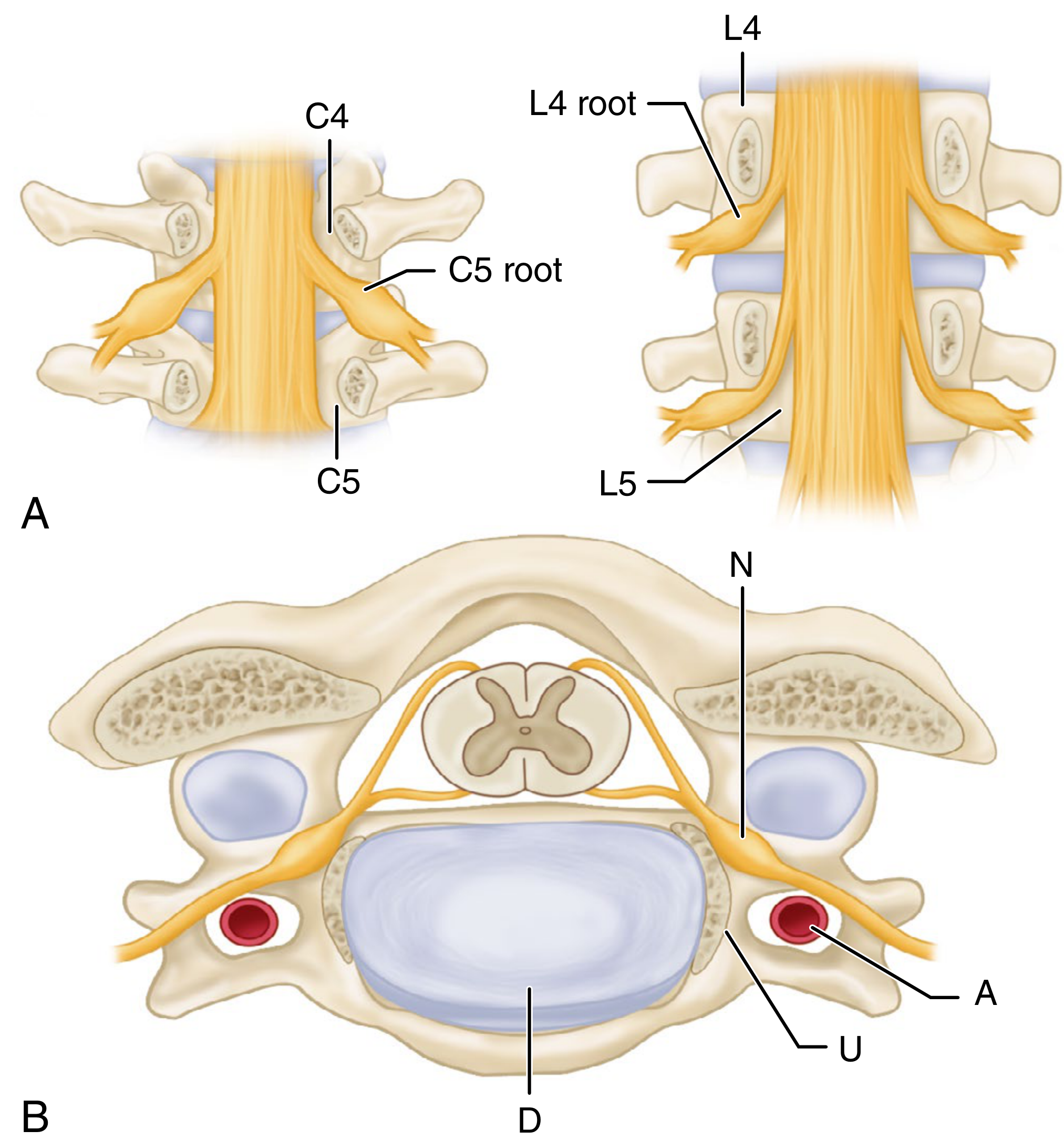
Note: The C5 root exits above C5 (at C4-C5 foramen). So a disc herniation at C5-6 compresses the C6 root, and herniation at C7-T1 affects C8.
Spondylosis produces four distinct clinical entities:
2. Discogenic Neck Pain (Axial Pain)
- Pure axial neck pain without neurological compromise
- Examination typically benign - normal motor, sensory, and reflex findings
- Cervical ROM may be reduced but without mechanical instability
- MRI: decreased T2 signal ("dark disc"), with or without annular tear or high-intensity zone (HIZ)
- Treatment: NSAIDs, symptomatic care, patient education about self-limiting natural history
- Surgical options are limited and should be avoided for pure discogenic pain
3. Cervical Radiculopathy
Definition: Nerve root compromise (motor, sensory, or both) from disc herniation, disc-osteophytic complex, facet arthropathy, thickened ligamentum flavum, or uncovertebral osteophytes.
Symptoms:
- Neck pain that evolves into arm/shoulder pain, paresthesias, and numbness
- Pain radiates in a dermatomal distribution to the upper extremity
- Motor weakness is uncommon; when present, follows the corresponding myotome
- Reflexes are normal or hyporeflexic (lower motor neuron pattern)
Key clinical tests:
- Spurling test - rotation + lateral bend + vertical head compression reproduces radicular symptoms
- Shoulder abduction sign - relief of radicular pain with shoulder abduction (hand placed on top of head)
Root levels and disc levels (cervical roots exit above their vertebra):
| Disc Level | Nerve Root Affected | Motor Deficit | Sensory | Reflex |
|---|---|---|---|---|
| C4-5 | C5 | Deltoid, biceps weakness | Lateral arm | Biceps |
| C5-6 | C6 | Wrist extensors, biceps | Lateral forearm, thumb | Brachioradialis |
| C6-7 | C7 | Triceps, wrist flexors | Middle finger | Triceps |
| C7-T1 | C8 | Finger flexors, intrinsics | Ring/little finger | None |
Treatment:
- Non-operative: NSAIDs, cervical epidural steroid injections, isometric exercises, traction
- Surgical: Anterior cervical discectomy and fusion (ACDF) is the gold standard; posterior foraminotomy is the classic choice for lateral disc fragments
4. Cervical Myelopathy
Myelopathy represents spinal cord compression - the most serious clinical entity of cervical spondylosis.
The sagittal MRI below shows disc-related myelopathy at C3-4, with high T2 signal within the cord (arrowhead) representing cord ischemia/myelomalacia:
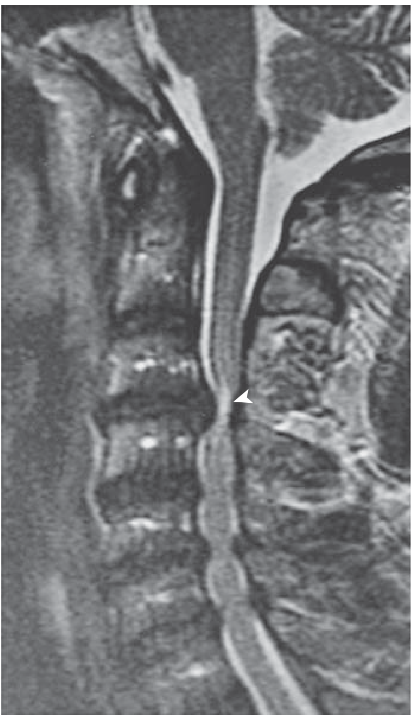
Pathogenesis: The normal A-P diameter of the cervical cord is ≤10 mm. CSM rarely develops if the congenital canal diameter exceeds 16 mm. In congenitally narrow canals, disc protrusion, osteophytes, ligamentum flavum hypertrophy, ossification of the posterior longitudinal ligament (OPLL), and vertebral body subluxation combine to compress the cord.
Clinical features (upper motor neuron pattern):
- Leg spasticity, upper extremity weakness or clumsiness
- Myelopathy hand and finger escape sign (small finger spontaneously abducts due to weak intrinsic muscles)
- Hyperreflexia, Hoffmann sign, clonus, Babinski sign
- Inverted radial reflex (finger flexion when brachioradialis reflex is elicited)
- Lhermitte sign - lightning-like sensations down the back with neck flexion
- Sphincter dysfunction (late feature, usually preceded by motor/sensory deficits)
- Neck pain is often NOT a prominent symptom
Natural history: Variable - some patients have stable deficits for years; others show gradual or stepwise deterioration. Surgical decompression is frequently indicated given the progressive natural history. Results are better when symptoms are milder, duration < 12 months, and age < 70 years.
Surgical options:
- Anterior: ACDF, anterior cervical corpectomy and fusion (ACCF) - suitable for kyphotic or lordotic alignment
- Posterior: Laminectomy ± fusion, laminoplasty - contraindicated in fixed cervical kyphosis (cannot indirectly decompress cord)
- Combined (circumferential): for multilevel corpectomy with strut reconstruction
- C5 nerve root palsy is the most common postoperative complication
5. Myeloradiculopathy
A combination of spinal cord and nerve root involvement, producing both upper and lower motor neuron signs at different levels simultaneously.
6. Disc Herniation Types
| Type | Description | Consequence |
|---|---|---|
| "Soft" disc herniation | HNP without bony osteophytes; usually posterolateral | Acute radiculopathy; rarely dysphagia (anterior) |
| "Hard" disc herniation | HNP with disc-osteophytic spur | Similar to soft; cord or root compression |
Large central herniations or spondylotic bars in a congenitally narrow canal can cause myelopathy.
7. Rheumatoid Spondylitis of the Cervical Spine
RA preferentially affects the occipitoatlantoaxial (O-A-A) joint - less common now due to widespread use of DMARDs.
- Synovitis destroys the transverse ligament and/or dens, causing atlantoaxial instability
- Most common finding: anterior C1-C2 subluxation
- Diagnosis: Flexion/extension radiographs - assess AADI (atlanto-dens interval) and PADI (posterior atlanto-dens interval)
- AADI > 9-10 mm = surgical indication
- PADI < 14 mm = suggests cord compression, usually requires surgery
Surgical options: C1-C2 transarticular screws (Magerl technique), or C1 lateral mass - C2 pedicle/pars fixation (Harms construct) - the current preferred method.
8. Summary Table: Cervical Conditions at a Glance
| Condition | Key Feature | Investigation | Treatment |
|---|---|---|---|
| Axial neck pain | No neuro deficit | MRI (dark disc) | NSAIDs, physio |
| Radiculopathy | Dermatomal arm pain, hyporeflexia | MRI, EMG | Epidural injections, ACDF |
| Myelopathy | UMN signs, Hoffmann, Lhermitte | MRI (T2 cord signal) | Surgical decompression |
| RA instability | C1-2 subluxation | Flex/ext XR, MRI | C1-C2 fusion (Harms) |
| Disc herniation (soft) | Acute radiculopathy | MRI | Conservative → ACDF |
| Disc herniation (hard) | Spondylotic complex | CT + MRI | ACDF or corpectomy |
Sources:
- Miller's Review of Orthopaedics 9th Edition, Chapter 8 (Cervical Spine)
- Campbell's Operative Orthopaedics 15th Ed 2026, Chapter 45 (Cervical Disc Disease)
- Bradley and Daroff's Neurology in Clinical Practice, Chapter 104 (Cervical Spondylotic Myelopathy)
- Firestein & Kelley's Textbook of Rheumatology (Cervical Spine in RA)
This is a shared conversation. Sign in to Orris to start your own chat.