
Nasal cavity with diagram
Reading File
Searching sources...
This is a visual anatomy topic. Let me search the medical library for nasal cavity content and diagrams simultaneously.
Finding Sources
Searching the Web
nasal cavity anatomy diagram labeled structures
Good results. Now let me read the comprehensive nasal cavity content from the textbooks and fetch the diagrams simultaneously.
Reading File
Reading File
Loading Image
Now let me get the blood supply and more detailed anatomy content from the Color Atlas and Cummings textbook, plus a more detailed diagram.
Reading File
Reading File
Loading Image
I have rich content from the textbooks. Let me now fetch the textbook anatomy diagram and read the blood supply / nerve supply sections.
Loading Image
Reading File
Excellent! I have very rich content now. Let me fetch the detailed nasal cavity anatomy diagram from the Color Atlas textbook.
Loading Image
I now have excellent diagrams and comprehensive textbook content to deliver a full, well-illustrated answer on the nasal cavity.
Nasal Cavity - Anatomy
Overview
The nasal cavity is the most superior part of the respiratory tract. It extends from the external nares (nostrils) anteriorly to the posterior choanae, where it becomes continuous with the nasopharynx. The nasal septum divides it into right and left halves. Each half has four walls: floor, roof, lateral wall, and medial wall (septum).
Diagrams
Sagittal section - nose and nasal cavities:
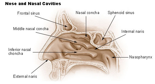
From the SEER Training Atlas - sagittal section showing the three nasal conchae, frontal and sphenoidal sinuses, and nasopharynx.
Lateral nasal wall - bony structure, mucosal appearance, and neurovascular supply (Color Atlas of Human Anatomy):
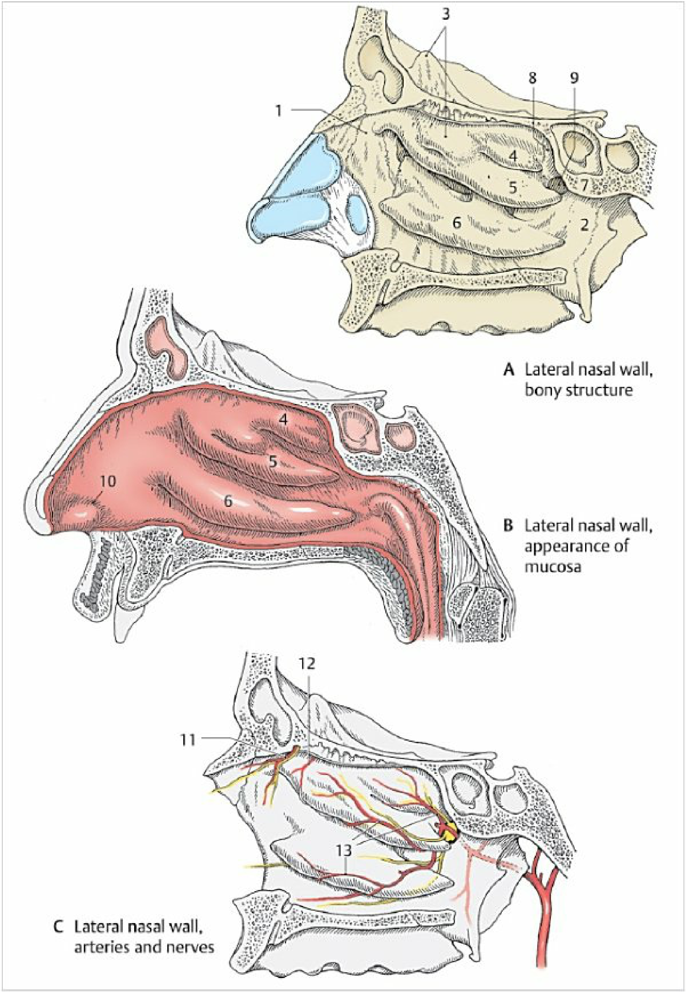
Fig. 3.3 - Color Atlas of Human Anatomy, Vol. 2. (A) Bony lateral wall with conchae labeled 4 (superior), 5 (middle), 6 (inferior), with maxilla (1), sphenoethmoidal recess (8), sphenopalatine notch (9). (B) Mucosal appearance with vestibule (10) and conchae. (C) Arterial (red) and nerve (yellow) supply.
Paranasal sinuses (anterior, lateral, and transverse views):
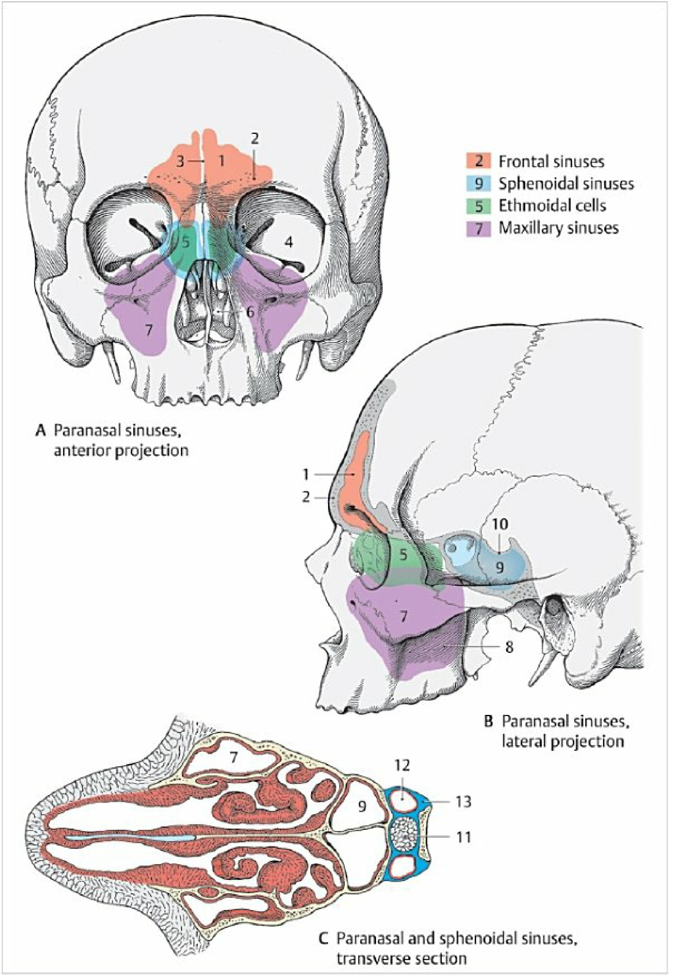
Fig. 3.5 - Color Atlas of Human Anatomy, Vol. 2. Frontal sinuses (orange), ethmoidal cells (green), sphenoidal sinuses (blue), maxillary sinuses (purple).
Boundaries
| Wall | Components |
|---|---|
| Floor | Palatine process of maxilla (anterior 3/4) + horizontal plate of palatine bone (posterior 1/4) |
| Roof | Nasal bones + nasal part of frontal bone (anterior), cribriform plate of ethmoid (central/superior), body of sphenoid (posterior) |
| Lateral wall | Maxilla (anterior), perpendicular plate of palatine bone (posterior), ethmoid (superior) |
| Medial wall (septum) | Perpendicular plate of ethmoid (upper bony), vomer (lower bony), septal cartilage (anterior) |
Lateral Wall - Conchae and Meatuses
Three curved bony shelves project from the lateral wall:
- Superior nasal concha - part of the ethmoid; smallest
- Middle nasal concha - part of the ethmoid
- Inferior nasal concha - a separate independent bone
Each concha overhangs a meatus (passage) of the same name. The meatuses are the key drainage routes:
| Meatus | What drains into it |
|---|---|
| Superior nasal meatus | Posterior ethmoidal cells (1-2 openings) |
| Sphenoethmoidal recess (above superior concha) | Sphenoidal sinus |
| Middle nasal meatus | Frontal sinus, maxillary sinus, anterior and middle ethmoidal cells; contains the hiatus semilunaris (curved crevice) bounded by the uncinate process and ethmoidal bulla |
| Inferior nasal meatus | Opening of the nasolacrimal duct |
The function of the conchae is to increase surface area so that inspired air is warmed, humidified, and filtered more effectively.
Nasal Septum (Medial Wall)
The septum has three parts:
- Bony part (posterior/inferior): Perpendicular plate of ethmoid (upper) + vomer (lower)
- Cartilaginous part: Septal (quadrangular) cartilage
- Membranous part: Soft tissue at the anterior tip
The free posterior margin of the vomer forms the medial boundary of each choana.
Mucosal Regions (3 types of epithelium)
| Region | Epithelium | Location |
|---|---|---|
| Nasal vestibule | Stratified keratinized squamous epithelium (skin) | Within the nostrils, bounded posteriorly by the limen nasi |
| Respiratory region | Pseudostratified ciliated columnar epithelium with mixed nasal glands | Remainder of nasal cavity (majority) |
| Olfactory region | Olfactory neuroepithelium | Superior septum, superior turbinate, and upper aspect of middle turbinate |
Blood Supply
The nasal cavity has a dual arterial supply:
External carotid artery system (via maxillary artery):
- Sphenopalatine artery - main supply to the posterior and inferior lateral nasal wall; also supplies the septum
Internal carotid artery system (via ophthalmic artery):
- Anterior ethmoidal artery - anterior and superior lateral wall + superior septum
- Posterior ethmoidal artery - posterior superior lateral wall
Kiesselbach's area (Little's area): A 1.5-mm zone of rich capillary anastomosis at the anteroinferior part of the nasal septum (junction of vestibule and nasal cavity). This is the most common site of epistaxis (nosebleeds). It receives contributions from the sphenopalatine, greater palatine, superior labial, and anterior ethmoidal arteries.
Nerve Supply
Sensory innervation (branches of the trigeminal nerve, CN V):
- Anterior/superior nasal mucosa: Ophthalmic division (CN V1) - anterior and posterior ethmoidal nerves
- Posterior/inferior nasal mucosa: Maxillary division (CN V2) - via the sphenopalatine ganglion
Autonomic innervation:
- Parasympathetic (secretomotor - regulates nasal secretions): Preganglionic fibers travel via the greater superficial petrosal nerve → Vidian nerve → synapse in sphenopalatine ganglion → postganglionic fibers to nasal mucosa
- Sympathetic (vasoconstriction - regulates turbinate congestion): Postganglionic fibers from the deep petrosal nerve → Vidian nerve → pass through sphenopalatine ganglion (without synapsing) → nasal mucosa
Olfactory (CN I):
- Olfactory nerve fibers from the olfactory neuroepithelium pass through the cribriform plate of the ethmoid to synapse in the olfactory bulb.
Clinical Notes
- Epistaxis (nosebleeds): Most commonly occur at Kiesselbach's area on the anteroinferior septum. Posterior bleeds arise from the sphenopalatine artery and are more severe.
- Sinusitis: Infections spread from nasal mucosa into the paranasal sinuses through their drainage openings. Obstruction of the osteomeatal complex (middle meatus area) is the most common anatomical bottleneck causing recurrent sinusitis.
- Septal deviation: A deviated nasal septum is a common anatomical variant that can obstruct airflow, contribute to sinusitis, and affect the nasal cycle.
- Concha bullosa: Pneumatisation (air cell formation) within the middle turbinate - a common variant that can obstruct the middle meatus.
- Sphenoidal sinus access: Because the roof of the nasal cavity slopes downward anteroposteriorly, surgical dissection progressing posteriorly (during endoscopic sinus surgery) approaches the skull base, which must be recognized to avoid intracranial injury.
- Pituitary surgery: The sphenoidal sinus, lying immediately posterior to the nasal cavity, provides the endoscopic transsphenoidal route for pituitary gland surgery.
Sources: Color Atlas of Human Anatomy, Vol. 2 (Internal Organs), p. 174-177; Scott-Brown's Otorhinolaryngology Head & Neck Surgery, Vol. 1; Cummings Otolaryngology Head and Neck Surgery.
This is a shared conversation. Sign in to Orris to start your own chat.