
Myocardial in farction
Reading File
Now let me search the medical library for myocardial infarction content in parallel.
Finding Sources
Searching PubMed
"myocardial infarction"[MeSH Terms] AND management
Good results. Let me now read the most comprehensive sources in parallel - Braunwald's (the gold standard cardiology text), Robbins Pathology, and Ganong's physiology.
Reading File
Reading File
Reading File
Excellent. Now let me read the Robbins Pathology section (which has morphological changes) and Goldman-Cecil (clinical management), plus search for specific management details.
Reading File
Reading File
Finding Sources
Now let me read the Harrison's treatment section and the Goldman-Cecil STEMI reperfusion section, plus get the morphological changes table from Robbins.
Reading File
Reading File
Reading File
Now let me get the morphological changes over time (the key table) and complications sections.
Reading File
Finding Sources
I now have comprehensive information from multiple authoritative textbooks. Let me compile a thorough response.
Loading Image
I now have comprehensive information from multiple authoritative sources. Let me compose the complete answer.
Myocardial Infarction (MI)
Definition
MI - commonly called a "heart attack" - is necrosis of cardiac muscle resulting from prolonged ischemia. The 2018 joint task force (ACC/AHA/ESC) defines it as "the presence of acute myocardial injury detected by abnormal cardiac biomarkers in the setting of evidence of acute myocardial ischemia."
Roughly 800,000 individuals in the United States experience an MI each year (nearly one MI every 40 seconds), causing almost 400,000 deaths annually. - Robbins, Cotran & Kumar Pathologic Basis of Disease, p. 511
Epidemiology & Risk Factors
- ~10% of MIs occur before age 40; 45% occur before age 65
- Male sex increases relative risk through middle age
- Women are relatively protected during reproductive years; this protection is lost after menopause (declining estrogen - rise in inflammatory markers, cholesterol, BP)
- IHD is the most common cause of death in older adult women
- Key risk factors: atherosclerosis, hypertension, diabetes, dyslipidemia, smoking, family history, obesity
Pathogenesis
Coronary Arterial Occlusion
The typical sequence (Robbins & Kumar Basic Pathology, p. 468):
- An atheromatous plaque is eroded or suddenly disrupted by endothelial injury, intraplaque hemorrhage, or mechanical forces - exposing subendothelial collagen and necrotic plaque contents to blood
- Platelets adhere, aggregate, and are activated - releasing thromboxane A2, ADP, and serotonin - causing further platelet aggregation and vasospasm
- Coagulation is activated by tissue factor - adding to the growing thrombus
- Within minutes, the thrombus can completely occlude the coronary artery lumen
Angiography within 4 hours of MI onset shows coronary thrombosis in almost 90% of cases. By 12-24 hours, only 60% show thrombosis even without intervention (spontaneous lysis occurs in some).
Uncommon Causes (10% of MIs)
- Vasospasm (with or without atherosclerosis) - e.g., cocaine or ephedrine
- Embolism (from AF mural thrombus, valve vegetations, prosthetic material)
- Small vessel disease (vasculitis, amyloid, sickle cell)
- Shock / severe hypertrophy with demand ischemia
Myocardial Response to Ischemia
Timeline of Events (Robbins, Cotran & Kumar, Table 12.4)
| Event | Time |
|---|---|
| ATP depletion begins | Seconds |
| Loss of contractility | < 2 minutes |
| ATP reduced to 50% | 10 minutes |
| ATP reduced to 10% | 40 minutes |
| Irreversible cell injury | 20-40 minutes |
| Microvascular injury | > 1 hour |
The first biochemical consequence is cessation of aerobic metabolism within seconds, causing inadequate ATP production and accumulation of lactic acid. Contractility ceases within ~1 minute. Only severe ischemia (blood flow ≤10% of normal) lasting 20-30 minutes or more leads to irreversible necrosis. Progressive loss of viability is complete by 6-12 hours.
Necrosis Progression
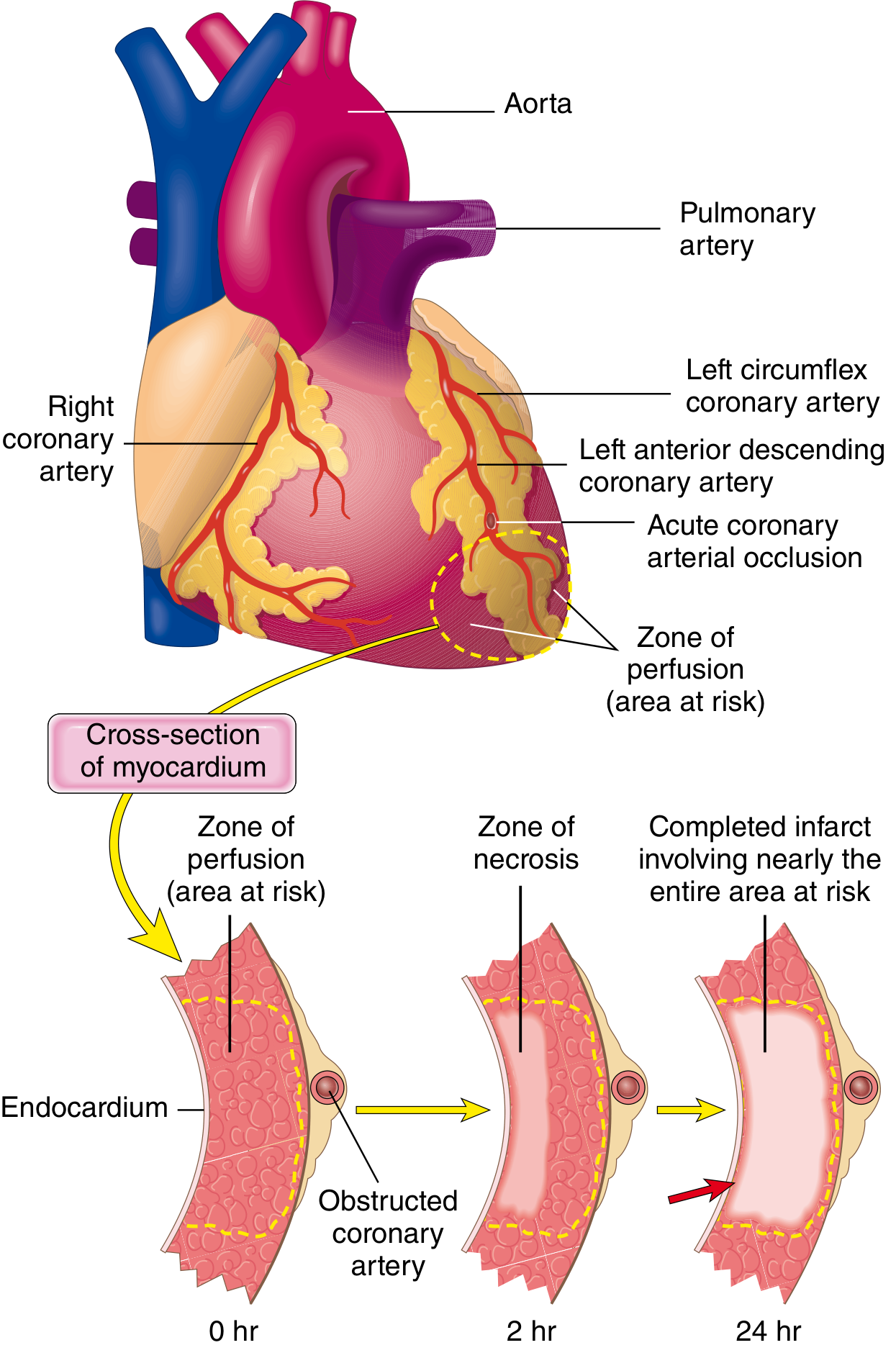
Irreversible injury first occurs in the subendocardial zone - the area most susceptible because it is:
- Last to receive blood from epicardial vessels
- Exposed to the highest intramural pressures (impeding inflow)
With prolonged ischemia, a wavefront of necrosis moves centripetally toward the epicardium (subendocardial → transmural).
Coronary Artery Distribution & Infarct Location
| Coronary Artery | Frequency | Area Infarcted |
|---|---|---|
| LAD | 40-50% | Anterior LV wall, anterior 2/3 of ventricular septum, apex |
| RCA | 30-40% | Inferior/posterior LV wall, posterior 1/3 of septum, inferior RV wall |
| LCX | 15-20% | Lateral LV wall (except apex) |
- In right-dominant circulation (~80% of individuals), the RCA perfuses the posterior septum
- RCA occlusions may extend into the RV wall in 15-30% of cases
- Isolated RV infarction is rare (1-3%)
Morphological Changes Over Time
(Robbins, Cotran & Kumar, Table 12.5)
| Time | Gross Appearance | Microscopic Findings |
|---|---|---|
| 0-4 hours | None visible | None (electron microscopy may show changes) |
| 4-12 hours | Occasional dark mottling | Early coagulative necrosis; edema; wavy fibers |
| 12-24 hours | Dark mottling | Coagulative necrosis; pyknosis of nuclei; marginal contraction band necrosis; early PMN infiltrate |
| 1-3 days | Mottling with yellow-tan softening | Coagulative necrosis; loss of nuclei; heavy PMN infiltrate |
| 3-7 days | Hyperemic border; central yellow-tan softening | Macrophages begin to appear; beginning disintegration of dead myofibers |
| 1-3 weeks | Gray-white scar forming at margins, depressed | Granulation tissue (macrophages, fibroblasts, new vessels) |
| > 2 months | White-gray scar | Dense collagenous scar (complete) |
The mechanism of cell death is predominantly ischemic coagulative necrosis. In reperfused infarcts, contraction band necrosis (hypercontracted sarcomeres) is prominent.
Cardiac Biomarkers
Disruption of sarcolemmal integrity allows intracellular proteins to leak into the circulation:
| Biomarker | Rise | Peak | Normalization |
|---|---|---|---|
| Troponin I / T (high-sensitivity) | 2-4 h | 24-48 h | 5-14 days |
| CK-MB | 3-12 h | 24 h | 48-72 h |
| Myoglobin | 1-4 h | 6-7 h | 24 h (less specific) |
High-sensitivity troponin is the preferred biomarker - most sensitive and specific for myocyte necrosis.
Classification
By ECG Pattern
- STEMI (ST-Elevation MI): complete coronary occlusion; transmural ischemia; ST elevation in leads overlying infarct
- NSTEMI (Non-ST-Elevation MI): partial occlusion or spontaneous reperfusion; subendocardial ischemia; no ST elevation (may show ST depression, T-wave changes, or normal ECG)
By Depth
- Transmural: full-thickness necrosis from endocardium to epicardium (usually corresponds to STEMI)
- Subendocardial: limited to inner 1/3-1/2 of wall (usually corresponds to NSTEMI)
ECG Changes
Three major abnormalities in acute MI (Ganong's Review of Medical Physiology, p. 534):
| Defect in Infarcted Cells | Current Flow | ECG Change in Overlying Leads |
|---|---|---|
| Rapid repolarization (K+ channel opening) | Out of infarct | ST segment elevation |
| Decreased resting membrane potential | Into infarct | TQ segment depression (recorded as ST elevation) |
| Delayed depolarization | Out of infarct | ST segment elevation |
Evolution of ECG Changes
- Acute (minutes-hours): Hyperacute T-waves → ST elevation ("tombstone" pattern)
- Evolving (hours-days): ST normalizes, T-wave inversion develops
- Old/healed: Pathological Q waves (dead tissue electrically silent; area negative during systole) - persist indefinitely
- Leads on the opposite side of the heart show reciprocal ST depression
Clinical Features
Symptoms:
- Severe, crushing, pressure-like chest pain - often radiating to left arm, jaw, shoulder, or back
- Not relieved by nitrates (unlike stable angina)
- Associated: diaphoresis, nausea/vomiting, dyspnea, sense of impending doom
- Silent MI occurs in ~20-25% (especially diabetics and the elderly)
Signs:
- Hypotension (cardiogenic shock), pallor, diaphoresis
- S3 gallop (LV dysfunction), S4
- New murmur (papillary muscle rupture, VSD)
- Pericardial friction rub (pericarditis)
- Jugular venous distension + clear lungs + hypotension = RV infarction triad
Management
Immediate Initial Steps
- Aspirin 160-325 mg (chewed for rapid absorption) - inhibits COX-1 and reduces thromboxane A2 (Harrison's Principles, p. 1203)
- O2 supplementation only if SpO2 < 90%
- Nitroglycerin (sublingual 0.4 mg x3, q5 min) - reduces preload and dilates coronaries; avoid if: SBP <90 mmHg, RV infarction, or PDE-5 inhibitor use within 24h
- Morphine 2-4 mg IV q5 min for pain unresponsive to nitrates
- Beta-blocker (e.g., metoprolol 5 mg IV q2-5 min x3 doses if HR >60, SBP >100, no heart block) - reduces O2 demand, lowers risk of VF and reinfarction
Reperfusion Strategy (STEMI) - the cornerstone of treatment
Goal: "Time is muscle"
- Primary PCI (preferred): target door-to-balloon time ≤90 minutes, or ≤120 minutes from first medical contact; preferred if available
- Indications: STEMI or new LBBB within 12h of symptom onset (or >12h if symptoms persist); cardiogenic shock in patients <75 years
- Requires centers performing >400 PCIs/year with cardiac surgery backup
- Fibrinolysis: if primary PCI cannot be performed within 120 min; must follow with angiography within 3-24h (pharmacoinvasive strategy)
- Avoid if active bleeding, prior intracranial hemorrhage, BP >180/110, recent surgery/trauma
(Goldman-Cecil Medicine, STEMI Reperfusion Flowchart, p. 659)
Antithrombotic Therapy
- Dual antiplatelet therapy (DAPT): Aspirin + P2Y12 inhibitor (ticagrelor or prasugrel preferred over clopidogrel for STEMI)
- Anticoagulation: Unfractionated heparin, low-molecular weight heparin, bivalirudin, or fondaparinux
Long-term Secondary Prevention
- Beta-blockers: reduce mortality; use indefinitely post-MI with reduced EF
- ACE inhibitors / ARBs: reduce LV remodeling; start within 24h; especially if EF <40%, hypertension, or diabetes
- Statins: high-intensity therapy (atorvastatin 40-80 mg); start immediately regardless of baseline LDL
- Aldosterone antagonist (eplerenone/spironolactone): if EF ≤40% with HF or diabetes, and no significant renal failure or hyperkalemia
- DAPT continued for 12 months post-stenting
Complications
(Robbins & Kumar Basic Pathology, p. 1394; Textbook of Clinical Echocardiography)
| Complication | Timing | Notes |
|---|---|---|
| Arrhythmia | Immediate (first 24h) | Most common cause of early death; VF, VT, AF, heart block |
| LV dysfunction / Cardiogenic shock | Early | Most common cause of in-hospital death |
| Free wall rupture | Days 3-7 (peak) | Softening of necrotic muscle; hemopericardium, cardiac tamponade; often fatal |
| Ventricular septal rupture (VSD) | Days 3-7 | New loud systolic murmur; step-up in O2 saturation in RV |
| Papillary muscle rupture | Days 2-7 | Acute severe mitral regurgitation; pulmonary edema |
| RV infarction | Acute | Inferior STEMI + JVD + clear lungs + hypotension |
| LV aneurysm | Weeks-months | Persistent ST elevation; mural thrombus; arrhythmia |
| Mural thrombus | Days-weeks | Risk of systemic embolism; anticoagulate |
| Dressler's syndrome | 2-10 weeks | Autoimmune pericarditis; fever, chest pain, pericardial rub; treat with NSAIDs/colchicine |
| Heart failure | Chronic | Adverse LV remodeling; reduced EF |
Recent Evidence Update
A 2025 meta-analysis (PMID 39298680) found that beta-blockers for secondary prevention post-MI in patients without reduced ejection fraction or heart failure may have less benefit than previously assumed - suggesting their role in preserved EF patients warrants reassessment. A 2024 pooled-data meta-analysis (PMID 39378245) characterized risk factors for sudden cardiac death after MI, reinforcing the need for ICD evaluation in high-risk patients.
Sources: Robbins, Cotran & Kumar - Pathologic Basis of Disease | Robbins & Kumar - Basic Pathology | Harrison's Principles of Internal Medicine 22E | Goldman-Cecil Medicine | Ganong's Review of Medical Physiology | Braunwald's Heart Disease
This is a shared conversation. Sign in to Orris to start your own chat.