
HPv, molluscum contagiosum ettiopathogenesis,clinical features treatment
Reading File
Now I'll search for both HPV and Molluscum Contagiosum in parallel across the library.
Finding Sources
Finding Sources
Excellent results. Now I'll read the best sources in parallel - Dermatology 5e for HPV and Andrews' Diseases of the Skin for Molluscum (most comprehensive).
Reading File
Reading File
Good start. I need more detailed HPV content and more of the Molluscum content. Let me read more from both simultaneously.
Finding Sources
Reading File
Let me search more specifically for HPV warts section and also read more MC content from Andrews.
Finding Sources
Reading File
Now let me read the HPV warts section from Andrews' and the MC treatment section simultaneously.
Reading File
Reading File
Excellent content. Now let me read more of the HPV warts section (clinical features and treatment) from Andrews', plus quickly grab MC treatment continuation.
Reading File
Reading File
Now let me read the HPV clinical features section from Andrews' (offset 2578-2694 area for the clinical features and treatment).
Reading File
I now have very rich content on both HPV and Molluscum Contagiosum. Let me fetch a few key clinical images and also fetch a quick MC image to include.
Loading Image
Loading Image
I now have comprehensive content from Andrews' Diseases of the Skin. I have enough material to write a thorough, well-structured answer covering both HPV and Molluscum Contagiosum. Let me compose the full response.
Human Papillomavirus (HPV) - Etiopathogenesis, Clinical Features, and Treatment
Etiopathogenesis
The Virus
There are more than 150 types of HPV. The genome consists of:
- Early genes (E1, E2, E4, E5, E6, E7) - regulate replication and oncogenesis
- Late genes (L1 and L2) - encode capsid proteins
- Upstream regulatory region (URR)
L1 encodes the major capsid protein and self-assembles into virus-like particles (VLPs) - these are the antigens in HPV vaccines. L2 (minor capsid protein) has two roles: it exposes the L1 keratinocyte-binding determinant allowing HPV to bind basal keratinocytes, and it downregulates Langerhans cell function via the phosphoinositide 3-kinase pathway.
HPV Types and Associated Lesions
| HPV Type | Associated Lesion |
|---|---|
| 1 | Plantar warts (children <12 yrs) |
| 2, 27, 57 | Common warts (hand warts, adults) |
| 6, 11 | External genital warts (condylomata acuminata) |
| 16, 18 | Anogenital dysplasia and carcinoma |
| 5, 8, β types | Epidermodysplasia verruciformis, SCC in immunosuppressed |
Pathogenesis / Immune Evasion
HPV survives in humans through a sophisticated immune evasion strategy:
- Antigens are not expressed on keratinocyte surfaces until cells migrate above the level of antigen-presenting cells
- L2 protein reduces and inactivates local Langerhans cells
- Through E6 and E7, HPV reduces local immune reactants (TLR9, IL-8), muting the local immune response
- This results in immune tolerance, allowing long-term persistence
Infection may be clinical (grossly visible), subclinical (seen only with acetic acid application), or latent (viral genome in apparently normal skin - explains recurrence after destructive treatment).
Clinical Features
Verruca Vulgaris (Common Warts)
- HPV types: 1, 2, 4, 27, 57, 63
- Most common between ages 5-20; only 15% occur after age 35
- Appear on dorsal hands, fingers, periungual areas
- Rough, hyperkeratotic, gray-brown papules; often coalesce into plaques
- May spread from infected close contacts; public exposures (swimming pools, showers) are risk factors
Verruca Plantaris (Plantar Warts)
- Occur on the plantar surface of the foot
- Endophytic growth pattern - pushed inward by weight bearing
- Painful with pressure; interrupt skin lines (vs. corns which follow skin lines)
- "Mosaic warts" = confluent plantar warts
Verruca Plana (Flat Warts)
- Small (1-5 mm), minimally elevated, flesh-colored or slightly pigmented papules
- Occur on face, dorsal hands, shins
- Linear distribution from autoinoculation (Koebner phenomenon)
- Can be mistaken for lichen planus
Condylomata Acuminata (Genital Warts)
- HPV 6/11 (low-risk types)
- Soft, skin-colored to pink, cauliflower-like papules and plaques on anogenital skin
- In women: vulva, vagina, cervix, perianal area
- In men: penile shaft, glans, scrotum, perianal area
- High-risk types (HPV 16/18) cause cervical intraepithelial neoplasia (CIN) and squamous cell carcinoma
Epidermodysplasia Verruciformis (EV)
- Rare, often inherited, T-cell immunodeficiency
- Widespread flat wart-like and pityriasis versicolor-like lesions
- High malignant potential (SCC) on sun-exposed areas
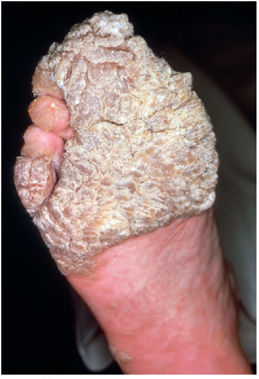
Features in Immunosuppressed Patients
- Lesions are multiple, coalescent, and extensive (see image below)
- Warts may have atypical morphology
- HPV in HIV patients: >90% of MSM have anal HPV; up to 50% have high-grade AIN; 30-50x increased risk of anal cancer
Treatment of HPV/Warts
Principle
Treatment is aimed at destruction of infected tissue; no treatment eradicates latent HPV, explaining high recurrence rates. In immunocompetent patients, up to two-thirds of warts resolve spontaneously within 2 years.
Destructive / Physical Methods
- Cryotherapy (liquid nitrogen): first-line for most warts; repeat every 2-3 weeks; painful
- Salicylic acid (topical): 10-40% preparations applied daily; effective for common and plantar warts; takes months
- Electrosurgery / laser: for refractory or large lesions
- Surgical excision: reserved for large, refractory lesions; risk of scarring
Immunotherapy
- Topical sensitizers (DNCB, squaric acid dibutylester, DPCP): applied topically; sensitize patient, then induce local immune response; 60-80% cure rates; useful for recalcitrant warts
- Intralesional Candida antigen: 0.1 mL injected into 1-3 lesions every 3-4 weeks; up to 80% cure rate; may cause cytokine-mediated systemic reactions (fever, chills, myalgia 6-8 hours post-injection)
Antiproliferative Agents
- Bleomycin (1 U/mL, intralesional): reserved for recalcitrant adult warts; ~90% response with 2 treatments; side effects include local pain, Raynaud's phenomenon of digits, rarely digital necrosis
- 5-FU (topical or intralesional): variable results
Topical Immunomodulators
- Imiquimod: approved for genital warts; significantly less effective than cryotherapy or topical immunotherapy for common/plantar warts; two large RCTs showed no difference from placebo for non-genital warts
- Podophyllotoxin: for genital warts (patient-applied)
- Sinecatechins: green tea extract, approved for external genital warts
For Genital Warts Specifically
- Patient-applied: imiquimod cream (3x/week), podophyllotoxin 0.5% (twice daily x 3 days/week)
- Provider-applied: cryotherapy, trichloroacetic acid (TCA), surgical removal, laser
Vaccination (Prevention)
- 9-valent HPV vaccine (Gardasil-9): covers HPV 6, 11, 16, 18, 31, 33, 45, 52, 58
- Recommended at age 11-12 (up to age 45 in some guidelines)
- Immunogenic and well-tolerated even in HIV-positive individuals
- VLPs from L1 protein are the vaccine antigens
Molluscum Contagiosum - Etiopathogenesis, Clinical Features, and Treatment
Etiopathogenesis
The Virus
Molluscum contagiosum (MC) is caused by Molluscum Contagiosum Virus (MCV), a member of the Poxviridae family. There are 4 types (MCV-1 to MCV-4):
- MCV-1: most common worldwide; virtually all infections in young children
- MCV-2: majority of infections (60%) in HIV-infected patients
- No difference in anatomic site of isolation based on type (unlike HSV)
Epidemiology - Three High-Risk Groups
- Young children (peak age 1-4 years)
- Sexually active young adults (age 20-29)
- Immunosuppressed individuals (especially HIV/AIDS)
Transmission: primarily by direct skin-to-skin contact, especially with wet skin; also via fomites (shared towels, swim equipment); autoinoculation is common.
Immune Evasion Mechanism
- MCV encodes an IL-18-binding protein that blocks the host's Th1 immune response by reducing local IFN-γ production
- MCV also inhibits NF-κB activation (via MC005 protein targeting NEMO-regulated IKK activation), preventing activation of innate immunity
- This allows MCV to persist in the epidermis without triggering a significant inflammatory response until the lesions eventually resolve
Histopathology
- Lesions primarily affect follicular epithelium
- Acanthotic, cup-shaped structure
- Henderson-Paterson bodies (molluscum bodies): large eosinophilic (early) then basophilic (late) cytoplasmic inclusion bodies in prickle cells; the bulk compresses the nucleus to the periphery
- Each lobule empties into a central crater (forming the umbilication)
- Inflammatory changes are slight or absent in uncomplicated lesions; resolving/inflamed lesions show dense lymphocytic and neutrophilic infiltrate
Clinical Features
Typical Presentation
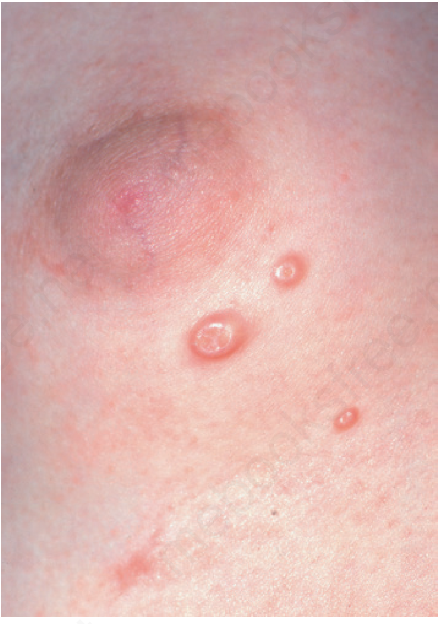
- Small, smooth-surfaced, firm, dome-shaped, pearly papule averaging 3-5 mm in diameter
- Central umbilication (central white core) - pathognomonic feature
- Side-lighting highlights the central opening; light cryotherapy highlights the umbilication
- May range from <1 mm (early) to >1 cm ("giant" cystic lesions)
By Patient Group
Young children:
- Usually generalized; few to >100 lesions
- Common sites: axillae, inguinal areas, popliteal/antecubital fossae, face
- Genital lesions as part of wider distribution in 10% (isolated genital MC in a child requires consideration of sexual abuse, though it is usually not from sexual contact)
- Children with atopic dermatitis (AD): 4x more likely to have >50 lesions; prolonged course; lesions tend to be confined to dermatitic skin
Adults (sexually transmitted):
- Lower abdomen, upper thighs, perineum, penile shaft in men
- Pubic hair removal (shaving, waxing) is a risk factor
- Mucosal involvement is rare
HIV/AIDS patients:
- Lesions favor the face (cheeks, neck, eyelids) and genitalia
- May develop larger (>1 cm), coalescent, verrucous, and widespread lesions
- Present in 10-30% of AIDS patients not receiving ART (virtually always when CD4 <100)
- MCV-2 predominates in this group
Inflammatory Reactions
- Molluscum dermatitis (40% of children): eczematous eruption surrounding individual lesions; more common in atopic children
- Inflamed MC (20%): erythema, swelling, sometimes pustulation of individual lesions; often heralds resolution - the "BOTE sign" (Beginning Of The End)
- Rupture into dermis: intense suppurative reaction resembling abscess or cellulitis
- Rarely: erythema annulare centrifugum, Gianotti-Crosti type reaction
Diagnosis
- Clinical diagnosis in most cases based on central umbilication
- Confirmation: express the pasty core between two microscope slides, stain with Wright, Giemsa, or Gram stain - shows Henderson-Paterson bodies
- Dermoscopy useful
- DDx: Cryptococcus, Histoplasmosis, Coccidioidomycosis (in immunosuppressed), keratoacanthoma, desmoplastic trichilemmoma, BCC
- Important to differentiate from condyloma acuminata in children
Treatment
Treatment Decision
Treatment is based on the clinical setting and immune status of the patient.
In immunocompetent children with numerous lesions: watchful waiting is reasonable since lesions spontaneously resolve. Individual lesions last 2-4 months; total duration of infection is typically 12-18 months. Aggressive treatment may be emotionally traumatic and may cause scarring.
Physical/Destructive Methods
| Method | Details |
|---|---|
| Curettage | Core removal; very effective; use lidocaine-prilocaine cream (EMLA) 1 hour before |
| Cryotherapy | Effective; use caution in dark-skinned patients (risk of dyspigmentation) |
| Cantharidin | Applied to lesion by wooden tip of cotton swab; left on 1-6 hours; ~20 lesions per session; high satisfaction rate; avoid on face and perineum in children |
| Trichloroacetic acid (TCA) | 3.5%-100%; applied to individual lesions |
| Needling/nicking | Nicking the opening with a blade/needle, then comedone extraction |
At-Home Therapies
- Pressing tape against lesion repeatedly, or continuous application of surgical tape daily for 16 weeks
- Topical tretinoin: applied with Q-tip to individual lesions; useful especially for facial lesions
Topical Agents - Limited Evidence
- Imiquimod: NOT recommended - two large RCTs showed no benefit over placebo
- Podophyllotoxin: similarly not effective for MC (failure to respond to these agents should prompt reconsideration of the diagnosis)
- 10% Potassium hydroxide (KOH): some evidence of efficacy
- Oral cimetidine: some immunomodulatory benefit, especially in atopic patients
For Genital MC in Adults
- Cryotherapy or curettage: very effective
- Screen for coexistent STIs; examine sexual partners
For Immunosuppressed/HIV Patients
- Primary treatment: antiretroviral therapy (ART) - results in dramatic resolution (though delayed 6-8 months)
- Topical 1-3% cidofovir or systemic IV cidofovir for refractory cases
- TCA peels (>35%) or daily 5-FU applications to point of erosion for widespread lesions
- Topical tretinoin nightly at highest tolerated concentration - reduces rate of new lesion appearance
- Shaving with blade razors should be discontinued in beard-area MC
Quick Comparison Summary
| Feature | HPV / Warts | Molluscum Contagiosum |
|---|---|---|
| Virus | Papillomaviridae, dsDNA | Poxviridae, dsDNA |
| Types | 150+ types | MCV 1-4 |
| Lesion | Verrucous/hyperkeratotic papule, interrupts skin lines | Dome-shaped, pearly, umbilicated papule |
| Hallmark | Koilcytes on histology | Henderson-Paterson bodies |
| Age peak | 5-20 yrs (non-genital); sexually active adults (genital) | 1-4 yrs children; 20-29 sexually active adults |
| Oncogenic risk | Yes (HPV 16/18 - cervical, anal, penile cancer) | No |
| Spontaneous resolution | Common in children (up to 2/3 within 2 yrs) | Common; 12-18 months typically |
| 1st-line Rx | Cryotherapy, salicylic acid | Cantharidin, curettage, watchful waiting |
| In HIV | Extensive, recalcitrant, accelerated malignancy | Widespread facial/genital lesions, MCV-2 predominant |
| Prevention | HPV vaccine (9-valent) | No vaccine; hygiene measures |
Sources: Andrews' Diseases of the Skin, 12e; Dermatology 2-Volume Set, 5e; Andrews' Diseases of the Skin E-Book, Clinical Dermatology
This is a shared conversation. Sign in to Orris to start your own chat.