
Face muscles
Reading File
The face muscles (muscles of facial expression) are a group of skeletal muscles in the superficial fascia of the face, most inserting into the skin rather than bone.
Searching sources...
Finding Sources
Finding Sources
Reading File
Reading File
Loading Image
Loading Image
Reading File
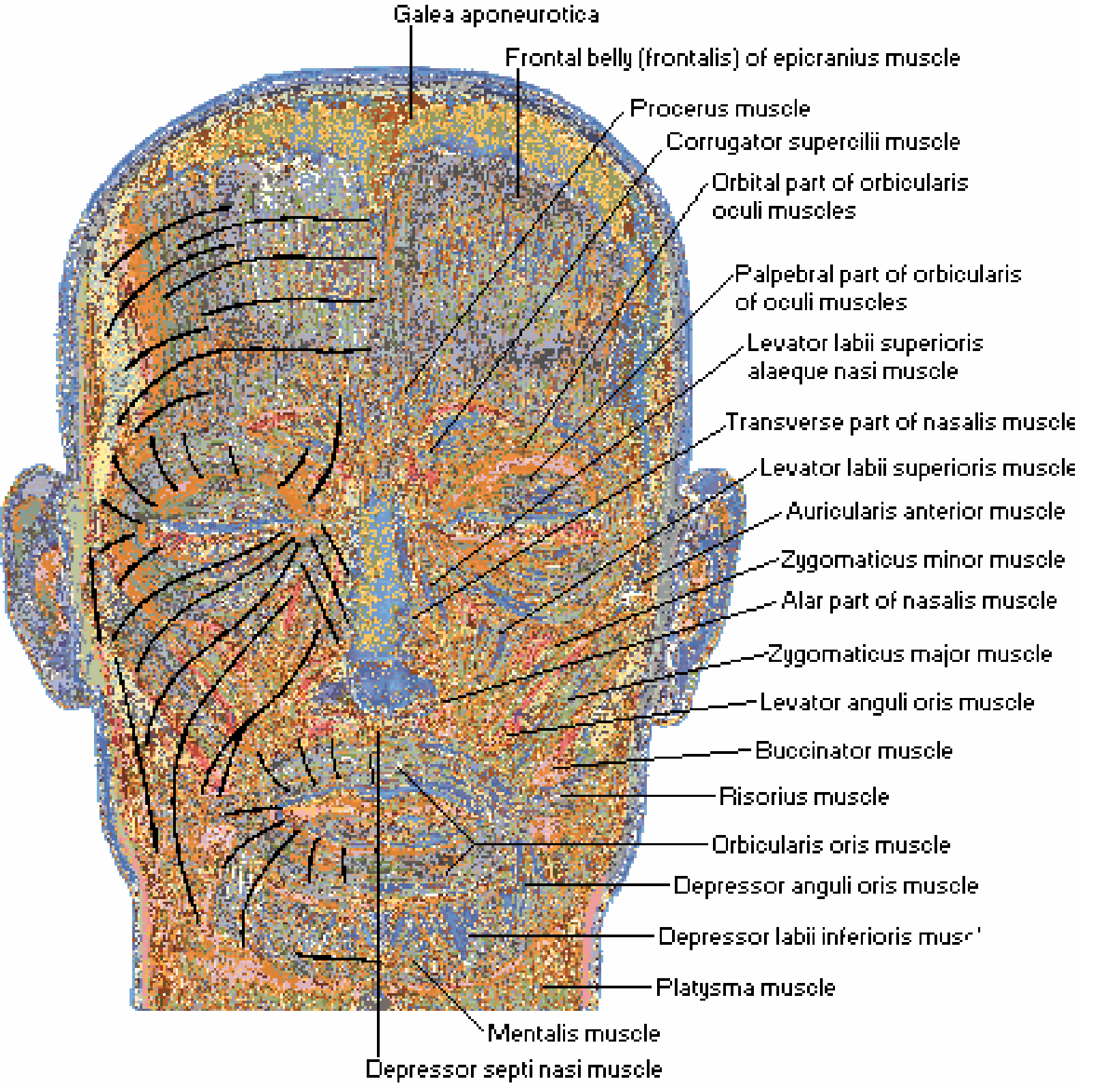
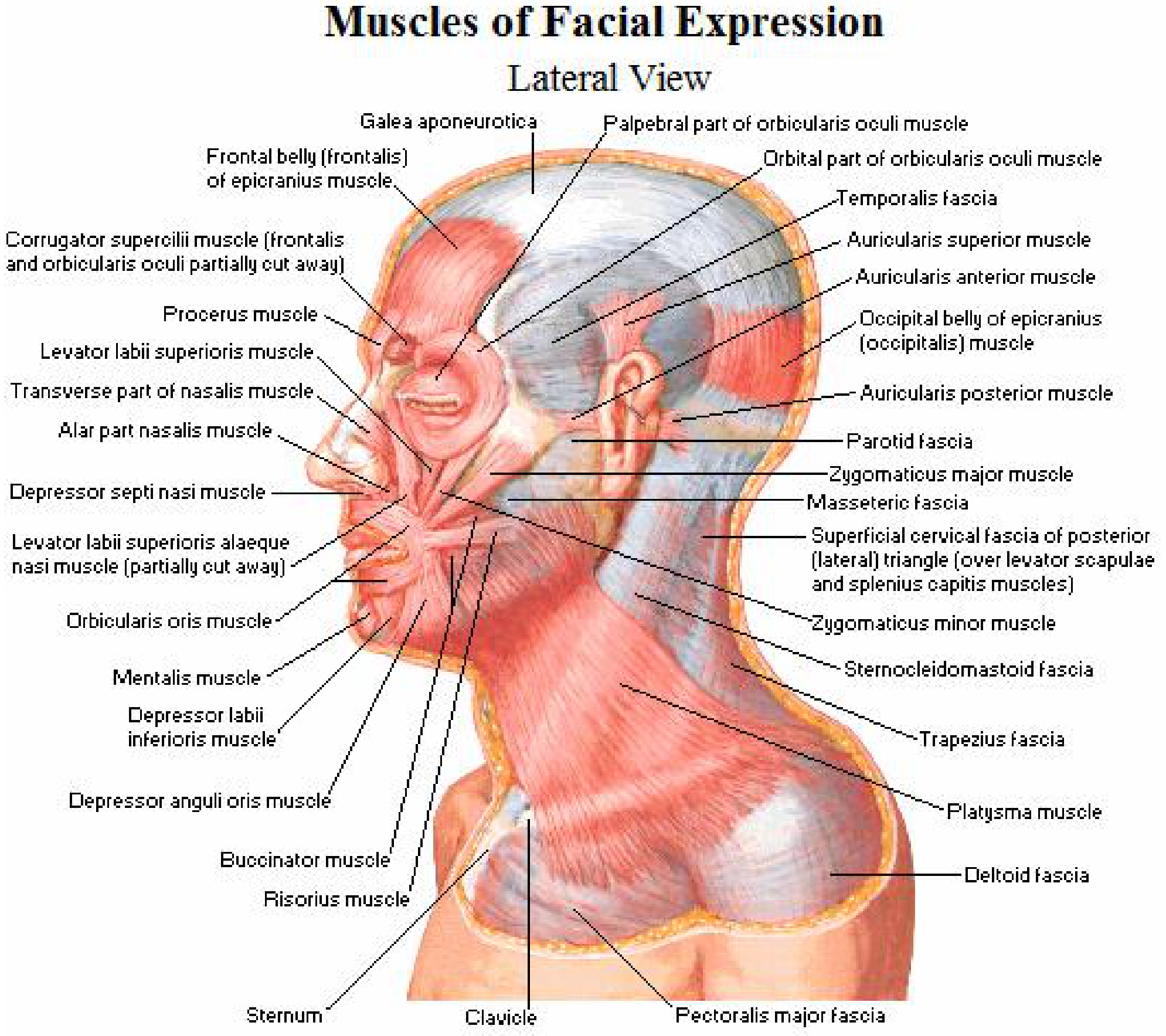
Here is a complete overview of the muscles of facial expression, drawn from Netter's Atlas of Human Anatomy and Dermatology 2-Volume Set 5e.
Muscles of Facial Expression
All muscles of facial expression share three key features:
- They arise from the second branchial arch
- All are innervated by cranial nerve VII (facial nerve)
- They insert into skin (not bone-to-bone), which is why they produce movement of the overlying skin
They are embedded within the SMAS (superficial musculoaponeurotic system), and their interaction with the SMAS allows fluidity of facial movement.
Anterior View (Netter's Atlas)
Lateral View (Netter's Atlas)
Muscle Groups by Region
Scalp & Forehead
| Muscle | Nerve Branch | Action | Clinical Note |
|---|---|---|---|
| Frontalis (anterior epicranius) | Temporal | Raises eyebrows; horizontal forehead lines | Loss = inability to raise brow; only 15% have cross-innervation |
| Occipitalis (posterior epicranius) | Posterior auricular | Pulls scalp posteriorly | Humans lack voluntary control of posterior belly |
The two bellies are connected by the galea aponeurotica. Horizontal forehead lines run perpendicular to frontalis fiber direction.
Periorbital (Around the Eye)
| Muscle | Nerve Branch | Action |
|---|---|---|
| Orbicularis oculi | Temporal & zygomatic | Closes eyelid (palpebral part = gentle blink; orbital part = forced closure) |
| Corrugator supercilii | Temporal | Draws brows medially and downward - the "scowl" muscle |
| Procerus | Temporal | Draws brow skin inferiorly; transverse nasal root lines |
The orbicularis oculi attaches at the medial and lateral canthal tendons and encircles the orbit. It is intertwined with the procerus, corrugator supercilii, and frontalis.
Nasal
| Muscle | Nerve Branch | Action |
|---|---|---|
| Nasalis (transverse + alar parts) | Zygomatic/buccal | Transverse part compresses nostrils; alar part dilates nares |
| Depressor septi nasi | Buccal | Pulls nasal tip downward |
| Levator labii superioris alaeque nasi | Buccal | Elevates upper lip and dilates nostril |
Perioral (Around the Mouth) - Elevators
| Muscle | Nerve Branch | Action |
|---|---|---|
| Orbicularis oris | Buccal & marginal mandibular | Closes and purses lips |
| Levator labii superioris | Zygomatic/buccal | Elevates upper lip |
| Zygomaticus major | Zygomatic/buccal | Draws angle of mouth upward and laterally (smiling) |
| Zygomaticus minor | Zygomatic/buccal | Elevates upper lip |
| Levator anguli oris | Buccal | Elevates angle of mouth |
| Risorius | Buccal/marginal mandibular | Retracts angle of mouth (grinning) |
Perioral - Depressors
| Muscle | Nerve Branch | Action |
|---|---|---|
| Depressor anguli oris (triangularis) | Marginal mandibular | Pulls angle of mouth downward |
| Depressor labii inferioris (quadratus) | Marginal mandibular | Pulls lower lip downward and laterally |
| Mentalis | Marginal mandibular | Raises and protrudes lower lip; chin wrinkling |
The mentalis has two bellies; the distance between them determines whether a "chin dimple" is present - a normal anatomic variant.
Cheek
| Muscle | Nerve Branch | Action |
|---|---|---|
| Buccinator | Buccal | Compresses cheek against teeth; aids chewing and blowing; interdigitates with orbicularis oris |
The parotid duct pierces the buccinator to enter the mouth at the second maxillary molar. The facial vascular bundle crosses its surface.
Neck (Extends onto Lower Face)
| Muscle | Nerve Branch | Action |
|---|---|---|
| Platysma | Cervical branch of CN VII | Tenses neck skin; draws lower lip and angle of mouth downward |
Periauricular
| Muscle | Nerve Branch | Action |
|---|---|---|
| Auricularis anterior | Temporal | Draws auricle forward |
| Auricularis superior | Temporal | Elevates auricle |
| Auricularis posterior | Posterior auricular | Draws auricle backward |
These are vestigial in humans and most people cannot voluntarily move them.
Wrinkle Lines
Wrinkle lines run perpendicular to the underlying muscle fiber direction - because repeated contraction creases the skin at 90° to the muscle pull. This is clinically important: elliptical incisions for skin tumor removal should conform to wrinkle line direction to minimize scarring.
Clinical Summary of CN VII Branch Loss
| Branch | Muscles Affected | Consequence |
|---|---|---|
| Temporal | Frontalis, corrugator, orbicularis oculi (upper) | Cannot raise eyebrow; incomplete eye closure (risk of corneal damage) |
| Zygomatic | Orbicularis oculi (lower), nasalis | Incomplete eye closure |
| Buccal | Buccinator, zygomaticus, levators, nasalis | Cannot smile; nasal collapse; difficulty eating |
| Marginal mandibular | Depressors, mentalis | Asymmetric mouth at rest and with movement |
| Cervical | Platysma | Platysmal banding |
- Dermatology 2-Volume Set 5e, Table 142.1
- Netter's Atlas of Human Anatomy, pp. 39-41
Motor points in face muscles that find out by stimulator
Motor points are the skin surface locations where electrical stimulation produces the strongest muscle contraction with the least current - they correspond to where the motor nerve enters the muscle belly.
Searching sources...
Finding Sources
Searching the Web
motor points facial muscles electrical stimulation map neuromuscular
Finding Sources
Reading Web Page
https://pmc.ncbi.nlm.nih.gov/articles/PMC11133044
Searching the Web
facial muscle motor points anatomical location diagram frontalis orbicularis zygomaticus buccinator electrode
Finding Sources
Here is a comprehensive guide to motor points of facial muscles as identified by electrical stimulator (pen electrode mapping).
What Is a Motor Point?
A motor point is the spot on the skin surface where electrical stimulation produces the maximum muscle contraction with the minimum current. It corresponds to the site where the motor nerve branch enters the muscle belly - the nerve has a lower threshold than muscle fiber, so stimulation at this point is most efficient.
How to Find It (Pen Electrode Technique)
- Apply low-intensity, low-frequency current via a pen (probe) electrode - the active/cathode electrode
- Systematically scan the skin surface over the target muscle
- Visually observe the muscle twitch response
- The point giving the strongest visible contraction at lowest current = motor point
- Mark it on the skin - this becomes the center of electrode placement
The inactive (anode/reference) electrode is placed at the nape of the neck or on a nearby tendon/fascia.
Motor Points of Facial Muscles - Organized by CN VII Branch
The face has no single universal motor point map (unlike limb muscles), because facial muscles are thin, overlapping, and the nerve branches are close to the surface. Stimulation targets the nerve branch entering each muscle belly.
Temporal Branch - Upper Face
| Muscle | Motor Point Location |
|---|---|
| Frontalis | Just above the eyebrow, midway between the pupil and midline; the nerve enters from the deep surface inferolaterally. Stimulate just above the brow arch. |
| Corrugator supercilii | Over the medial upper orbital rim, just above the nose bridge, between the two eyebrows |
| Orbicularis oculi (upper/orbital part) | Along the superior orbital rim, approximately 1 cm above the lateral canthus |
Zygomatic Branch - Mid Face (Eye & Cheek)
| Muscle | Motor Point Location |
|---|---|
| Orbicularis oculi (lower/palpebral part) | Inferior orbital rim, ~1 cm below the lateral canthus |
| Nasalis (transverse part) | Lateral wall of the nose, midway along the nasal bone/cartilage junction |
Buccal Branch - Mid/Lower Face
| Muscle | Motor Point Location |
|---|---|
| Procerus | Over the nasal root, midline between the eyes |
| Levator labii superioris alaeque nasi | Alongside the nose, at the level of the alar crease |
| Levator labii superioris | 1-2 cm below the infraorbital foramen, over the anterior cheek |
| Zygomaticus minor | Just medial to the zygoma, ~1-2 cm lateral to the nasolabial fold (upper portion) |
| Zygomaticus major | Along a line from the zygomatic arch to the corner of the mouth, approximately at its midpoint |
| Levator anguli oris | Just below the infraorbital foramen, slightly medial to zygomaticus major |
| Risorius | Over the cheek at the level of the mouth corner, running laterally |
| Buccinator | Central cheek, deep to the other muscles; stimulate at the mid-cheek just anterior to the masseter |
| Orbicularis oris | Perioral region: upper lip = philtrum area; lower lip = just below the vermilion border |
| Nasalis (alar part) | Over the alar cartilage/tip of nose |
Marginal Mandibular Branch - Lower Face / Chin
| Muscle | Motor Point Location |
|---|---|
| Depressor anguli oris | Just below and lateral to the corner of the mouth |
| Depressor labii inferioris | Below the lower lip, midway between the corner of the mouth and the chin midline |
| Mentalis | Over the chin prominence, slightly paramedian (one belly each side) |
Cervical Branch
| Muscle | Motor Point Location |
|---|---|
| Platysma | Along the anterior neck below the mandible; motor point is found in the mid-neck region over the anterior platysmal band |
Stimulation Parameters for Facial Muscles
| Parameter | Innervated muscle | Denervated muscle |
|---|---|---|
| Current type | Interrupted faradic (AC) | Interrupted galvanic (DC) |
| Pulse duration | 0.1-1 ms | ≥100 ms |
| Frequency | 30-50 Hz | ~30 per minute |
| Waveform | Biphasic | Monophasic DC, slowly rising |
| Electrode | Pen (active/cathode) on motor point | Pen (active) on motor point |
Key rule: Denervated muscle requires longer pulse durations (≥100 ms) and DC current because it cannot respond to the short pulses that innervated nerve fibers respond to.
Visual Reference
The lateral Netter's diagram from the previous answer shows the spatial relationships:
Clinical Notes
- In Bell's palsy and facial nerve palsy, motor point stimulation is used to maintain muscle bulk and prevent fibrosis during nerve recovery - per the IJSHR Bell's palsy NMES study.
- In synkinesis (post-reinnervation abnormal movement), electrical stimulation is contraindicated as it worsens abnormal co-contractions.
- The motor point may shift as muscle atrophies during denervation; repeat mapping is advisable in chronic palsy.
- Facial motor points are anatomically more variable than limb motor points due to thin, overlapping muscles - always confirm with pen electrode scanning rather than relying solely on fixed anatomical landmarks.
- Per the fNMES systematic review (PMC11133044), targeting the motor point reduces patient discomfort and maximizes contraction efficiency compared to arbitrary electrode placement.
This is a shared conversation. Sign in to Orris to start your own chat.