
/bestnotes on eye anatomy and physiology anatomy for 1st year mbbs student act as a professor and /teachme each and every point in /deepdive don't miss even a single point explain with the help of images Provide USMLE type questions on this topic with /3levels /doughtkiller.
Reading File
Finding Sources
Finding Sources
Finding Sources
Finding Sources
Finding Sources
Reading File
Reading File
Reading File
Reading File
Finding Sources
Reading File
Finding Sources
Reading File
Finding Sources
Loading Image
Loading Image
Loading Image
Loading Image
Loading Image
Now I have all the material I need. Let me compile the complete professor-style deep dive.---
👁️ EYE ANATOMY & PHYSIOLOGY — Professor's Deep Dive
For 1st Year MBBS | Best Notes Edition
"The eye is not just an organ of sight — it is a camera, a computer, and a chemical factory all in one. Master its structure and physiology will follow naturally."
🗺️ OVERVIEW FIRST: THE BIG PICTURE
The eyeball is a sphere about 2.5 cm in diameter, sitting in the bony orbit of the skull. Its job is to convert light energy (wavelengths 397–723 nm) into electrical signals (action potentials) sent to the cerebral cortex via the optic nerve (CN II).
Think of the eye as having three major concentric coats (tunics) and internal compartments filled with fluid:
| Layer | Components |
|---|---|
| Outer fibrous coat | Sclera (posterior 5/6) + Cornea (anterior 1/6) |
| Middle vascular coat (uvea) | Choroid + Ciliary body + Iris |
| Inner neural coat | Retina |
📍 IMAGE 1: Complete Cross-Section of the Eye
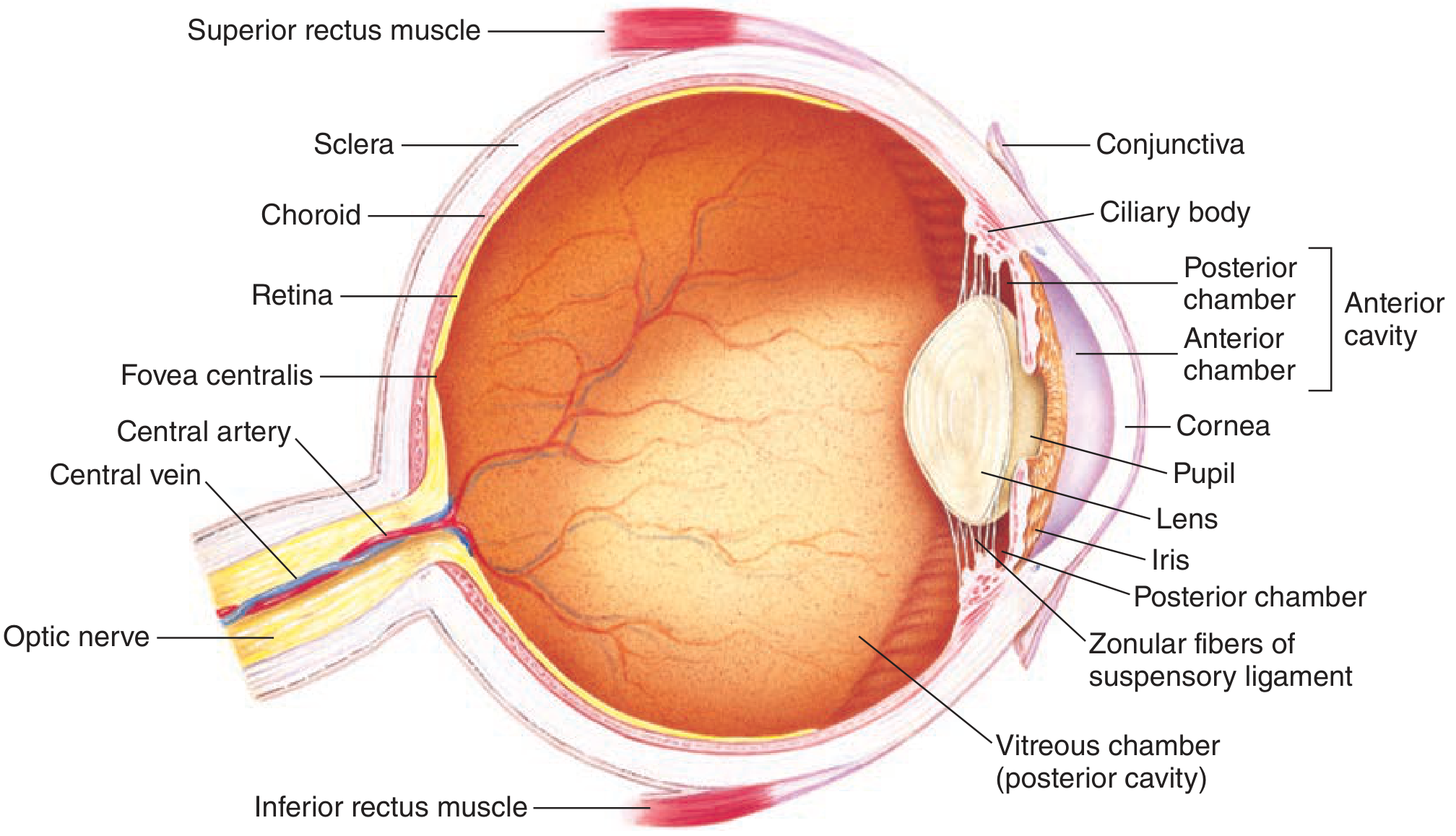
Fig. 10-1 — Full cross-section of the human eye. Study every label — each structure will be tested. (Ganong's Review of Medical Physiology, 26e)
📍 IMAGE 2: Gross Anatomy — External View
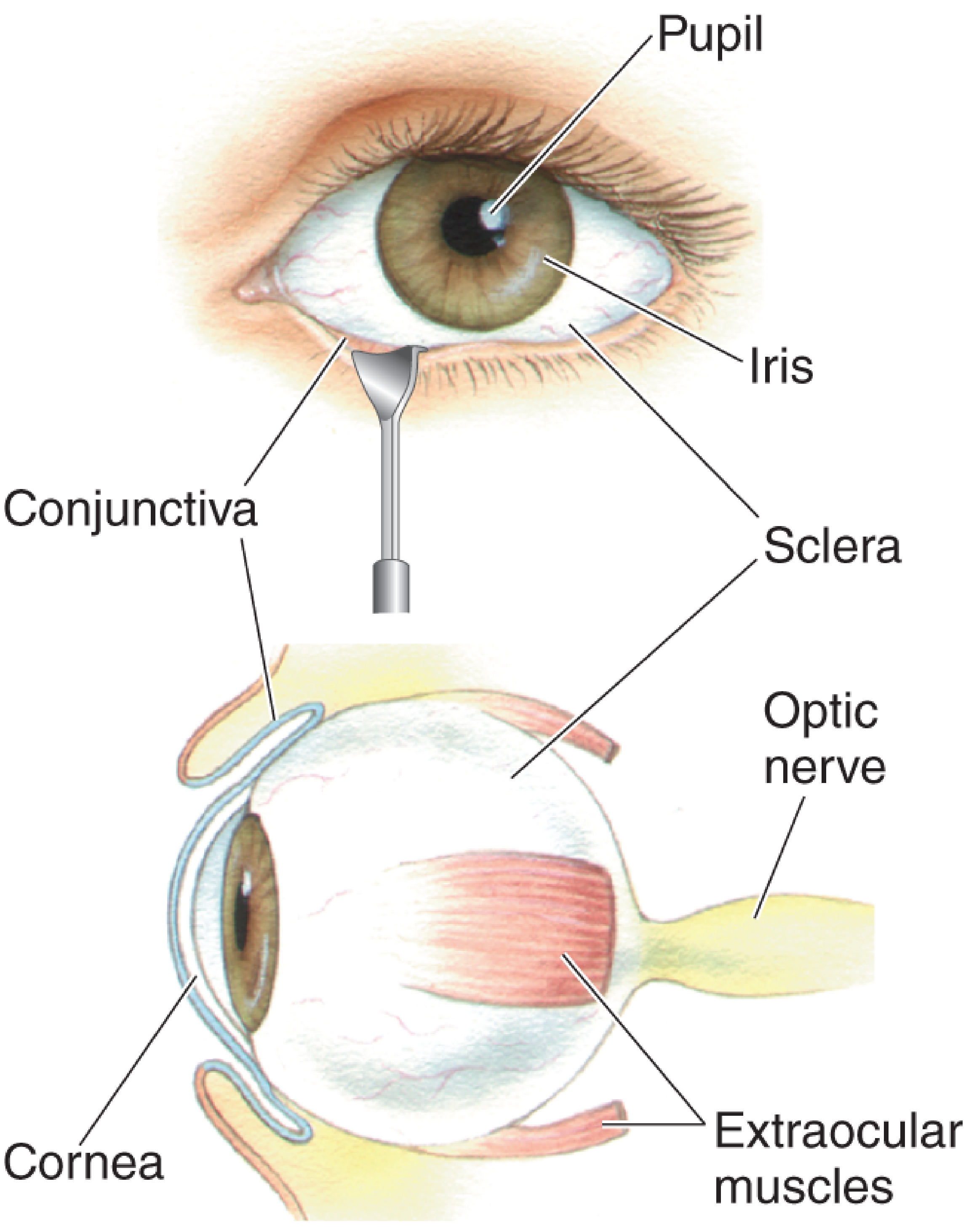
Fig. 9.4 — What you see when you look at someone's eye, and what lies behind it. (Neuroscience: Exploring the Brain, 5e)
PART 1 — THE OUTER FIBROUS COAT
1A. SCLERA
- The "white of the eye" — dense, avascular, opaque connective tissue
- Forms the posterior 5/6 of the eyeball
- Functions: structural protection, maintains shape, attachment point for the 6 extraocular muscles
- Continuous anteriorly with the cornea at the corneoscleral junction (limbus)
Professor's tip: The sclera is white because it contains densely packed collagen fibers in irregular arrangement — unlike the cornea where fibers are regular, allowing transparency.
1B. CORNEA
- The transparent anterior 1/6 of the outer coat
- Avascular — nourished entirely by aqueous humor (anteriorly) and tears (posteriorly)
- Sensitive to touch — rich sensory innervation by CN V1 (ophthalmic branch of trigeminal)
- Most powerful refracting surface of the eye (~43 diopters out of ~59 total diopters)
- Layers of cornea (from front to back): Epithelium → Bowman's layer → Stroma → Descemet's membrane → Endothelium
| Property | Value |
|---|---|
| Refractive power | ~43 diopters |
| Transparency mechanism | Regular collagen fibers + no blood vessels |
| Innervation | CN V1 (trigeminal, ophthalmic branch) |
| Nutrient source | Aqueous humor + tears |
Corneal reflex: Touch the cornea → blink. Afferent = CN V1; efferent = CN VII (facial). Test it in the exam!
1C. CONJUNCTIVA
- A clear mucous membrane covering the sclera and lining the inner eyelids
- Continuous with the corneal epithelium at the limbus
- Conjunctival goblet cells secrete mucus as part of the tear film
PART 2 — THE MIDDLE COAT: THE UVEA
The uvea has three components from back to front: Choroid → Ciliary body → Iris
2A. CHOROID
- Highly vascular, pigmented layer between sclera and retina
- Covers the posterior 2/3 of the eyeball
- Functions:
- Nourishes the outer retina (especially photoreceptors)
- Melanin absorbs stray light, preventing internal reflection (like the black inside a camera)
- Contains the choriocapillaris — a dense capillary network right next to the retina
2B. CILIARY BODY
- Located between the choroid and iris
- Contains two important things:
- Ciliary muscle — smooth muscle; controls lens shape for accommodation
- Ciliary processes — produce aqueous humor (by diffusion + active transport from plasma)
| Ciliary muscle type | Contraction effect | Vision |
|---|---|---|
| Circular (Müller's) | Relaxes zonular fibers → lens becomes rounded | Near vision |
| Longitudinal | Opens trabecular meshwork | Helps aqueous drainage |
Parasympathetic control (CN III): Ciliary muscle contraction → zonule fibers relax → lens rounds up → focus near (accommodation). Remember: "PARA = near, SYMPA = far"
2C. IRIS
- The colored, pigmented, circular diaphragm in front of the lens
- Has a central aperture = the pupil
- Contains two smooth muscles:
| Muscle | Action | Nerve supply | Effect |
|---|---|---|---|
| Sphincter pupillae (circular) | Constriction (miosis) | Parasympathetic (CN III) | Bright light, near vision |
| Dilator pupillae (radial) | Dilation (mydriasis) | Sympathetic (T1, superior cervical ganglion) | Darkness, fear |
- Pupil diameter changes produce up to a 16-fold change in light reaching the retina
- Uvea = iris + ciliary body + choroid (collective term — remember this!)
📍 IMAGE 3: The Eye in Cross-Section with Lens and Zonule Fibers
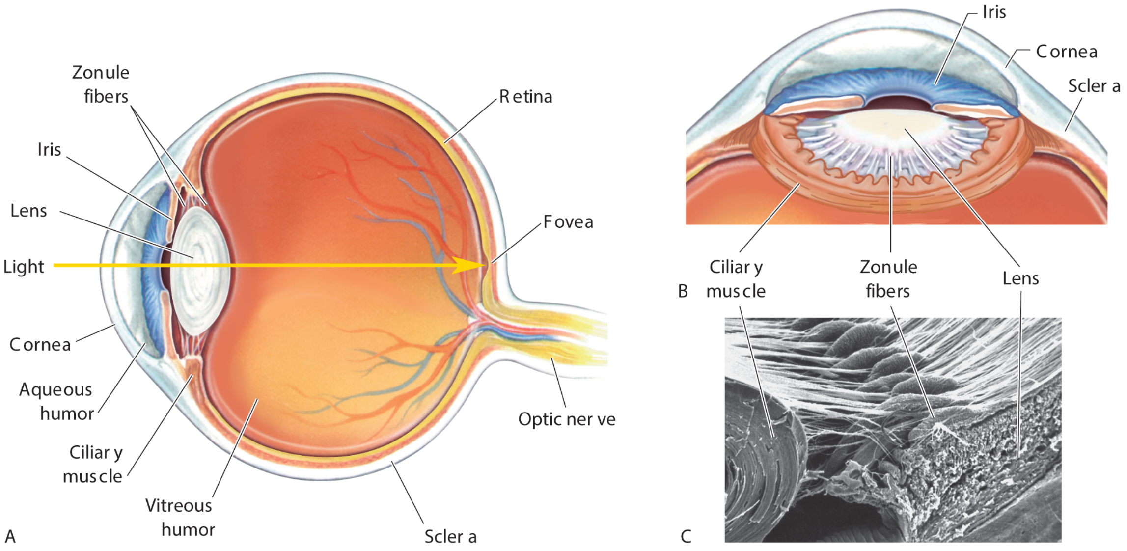
Fig. 9.6 — Cross-section with detailed lens-ciliary muscle-zonule relationship and scanning EM of zonule fibers (Neuroscience: Exploring the Brain, 5e)
PART 3 — THE LENS
- A biconvex, transparent, avascular, elastic structure
- Suspended by zonular fibers (suspensory ligament) anchored to the ciliary body
- Composed of: lens capsule → epithelium → lens fibers (elongated cells with no nucleus, packed with crystallin proteins)
Accommodation (Near Focus)
- Ciliary muscle contracts (parasympathetic, CN III)
- Ring of ciliary muscle gets smaller in diameter
- Tension on zonule fibers decreases
- Lens becomes rounder (more convex) due to inherent elasticity
- Refractive power increases → focus near objects
For far vision: ciliary muscle relaxes → zonule fibers tense → lens flattens → lower refractive power
Memory trick: Think of ciliary muscle as a hula hoop. When it contracts (gets smaller), the hoop presses inward, zonules go slack, lens pops outward like a ball.
Refractive Power
- Measured in diopters = 1/focal length (in meters)
- Total refractive power of the eye ~ 59 diopters
- Cornea contributes ~43 D (fixed)
- Lens contributes ~10-20 D (variable, for accommodation)
PART 4 — CHAMBERS AND FLUIDS
Anterior Cavity (between cornea and lens)
Divided into two sub-chambers:
| Chamber | Location | Fluid |
|---|---|---|
| Anterior chamber | Between cornea and iris | Aqueous humor |
| Posterior chamber | Between iris, zonule, and lens | Aqueous humor |
Vitreous Chamber (Posterior Cavity)
- Between lens and retina
- Filled with vitreous humor — clear, gelatinous (collagen + hyaluronic acid)
- Keeps the eyeball spherical
- Unlike aqueous, vitreous is NOT constantly replaced
AQUEOUS HUMOR — The Star Fluid
| Feature | Detail |
|---|---|
| Produced by | Ciliary body (active transport + diffusion from plasma) |
| Composition | Clear, protein-free, similar to plasma but NO proteins |
| Flow path | Posterior chamber → pupil → anterior chamber → trabecular meshwork → Canal of Schlemm → venous system |
| Function | Nourishes cornea and lens (avascular!), maintains IOP |
| Normal IOP | 10–20 mmHg |
Clinically gold: Obstruction of drainage → ↑ IOP → GLAUCOMA
- Open-angle: ↓ permeability of trabeculae into canal of Schlemm
- Closed-angle: Iris balloons forward, obliterates filtration angle
PART 5 — THE RETINA (The Neural Coat)
The retina is the most complex layer — it is actually brain tissue (outgrowth of the diencephalon during development). It lines the posterior 2/3 of the choroid.
5A. Two Layers of Retina
- Outer pigment epithelium (RPE) — single layer, absorbs stray light, recycles visual pigments, maintains photoreceptors
- Inner neural retina — contains photoreceptors and all the signal-processing neurons
5B. The 10 Layers of Retina (Inside to Outside)
From vitreous → choroid:
- Internal limiting membrane
- Nerve fiber layer (ganglion cell axons)
- Ganglion cell layer
- Inner plexiform layer
- Inner nuclear layer (bipolar, amacrine, horizontal cells)
- Outer plexiform layer
- Outer nuclear layer (photoreceptor nuclei)
- Outer limiting membrane
- Photoreceptor layer (rods and cones)
- Retinal pigment epithelium (RPE)
Professor's trick: "Girls Brow Beat Pretty Awesome Horizontal Oval Pupils" → Ganglion, Bipolar, (inner) Plexiform, Amacrine, Horizontal, Outer plexiform, Photoreceptors
Key concept: Light must pass through all the neural layers BEFORE hitting the photoreceptors — the retina is inverted (except at the fovea)!
📍 IMAGE 4: Retinal Layers and Photoreceptor Structure
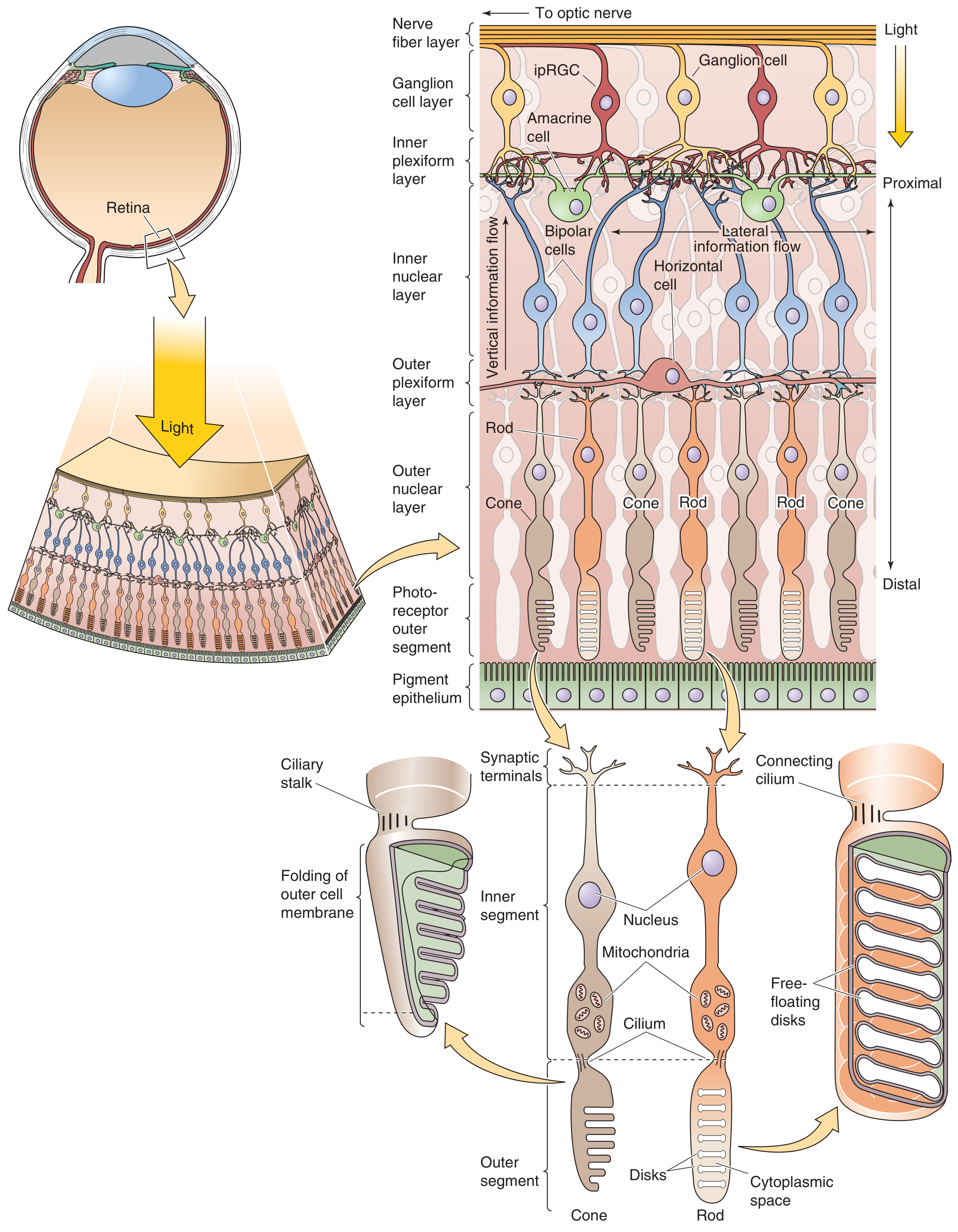
Fig. 15-9 — Neural circuits of the retina and photoreceptor ultrastructure. Note: light enters from the TOP (vitreous side) and reaches the photoreceptors at the BOTTOM. (Medical Physiology, Boron & Boulpaep)
5C. PHOTORECEPTORS: RODS vs CONES
| Feature | RODS | CONES |
|---|---|---|
| Number | ~120 million | ~6–7 million |
| Location | Peripheral retina | Concentrated at fovea |
| Vision type | Scotopic (dim/dark) | Photopic (bright/color) |
| Color vision | No (monochromatic) | Yes (3 types) |
| Acuity | Low | High |
| Photopigment | Rhodopsin (opsin + retinal) | Photopsin (3 types: S, M, L) |
| Convergence | High (many rods → 1 ganglion) | Low (1 cone → 1 bipolar → 1 ganglion at fovea) |
| Dark current | Yes | Yes |
- Rod:Cone ratio = approximately 16:1
- There is also a third photoreceptor type: intrinsically photosensitive retinal ganglion cells (ipRGC) — contain melanopsin, respond to blue light (~480 nm), regulate circadian rhythms and pupillary light reflex
5D. FOVEA CENTRALIS
- A small pit 300–700 μm in diameter in the center of the macula lutea
- Contains only cones (no rods), most densely packed (~0.05 μm between cones)
- The overlying retinal neurons are displaced laterally → light hits photoreceptors directly
- Highest visual acuity — the "HD camera" of the eye
- 1 cone → 1 bipolar → 1 ganglion cell at fovea (no convergence = maximum resolution)
5E. OPTIC DISC (BLIND SPOT)
- Where ganglion cell axons exit the eye to form the optic nerve
- No photoreceptors here → physiological blind spot
- ~15° medial to fovea (nasal side)
- Where the central retinal artery and vein enter/exit the eye
📍 IMAGE 5: The Retina as Seen Through an Ophthalmoscope
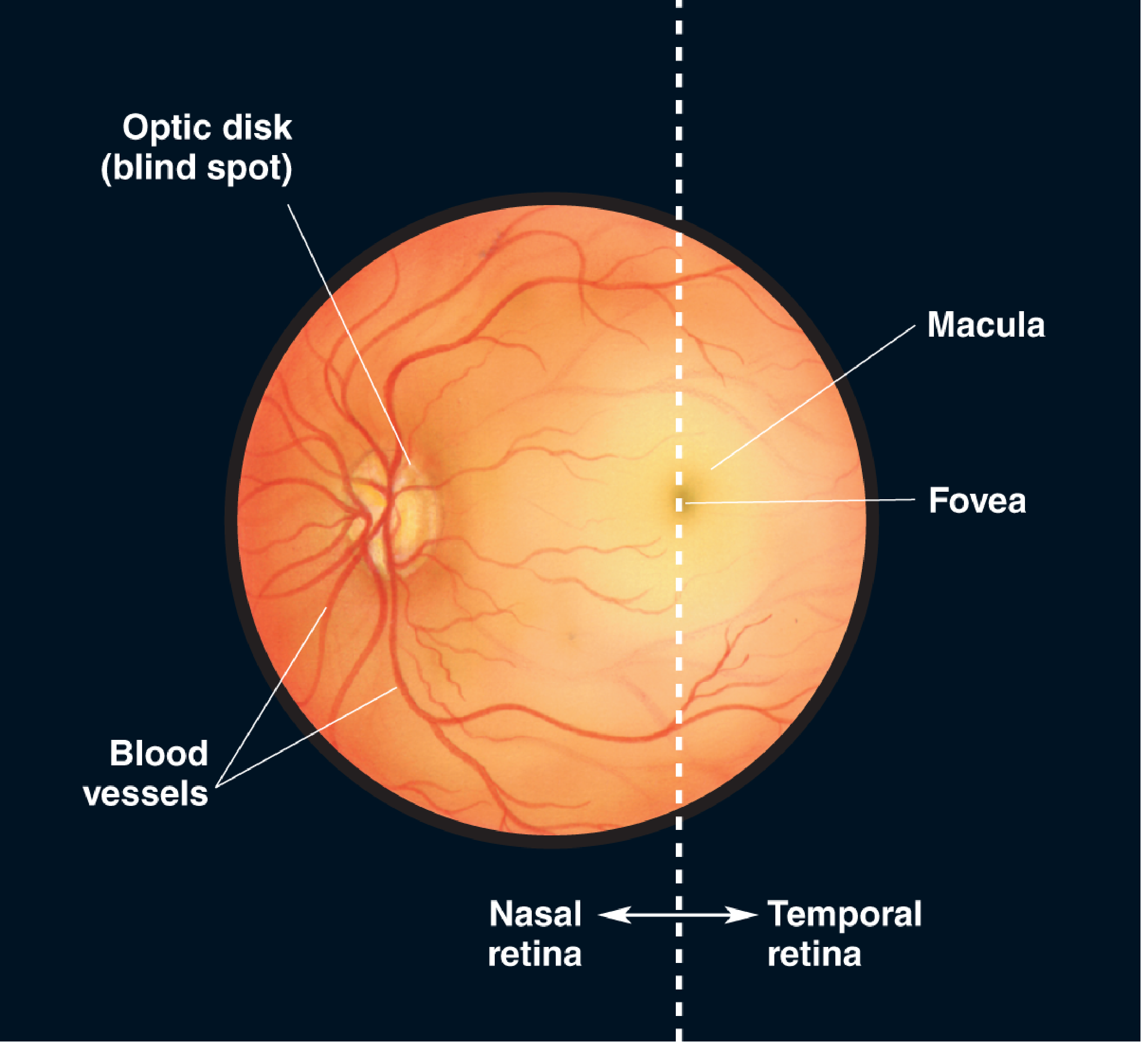
Fig. 9.5 — Ophthalmoscopic appearance of the fundus. The optic disc (blind spot) is to the nasal side; the fovea is at the center of the macula. (Neuroscience: Exploring the Brain, 5e)
PART 6 — PHOTOTRANSDUCTION: HOW LIGHT BECOMES ELECTRICITY
In the DARK (resting state)
- cGMP levels are high in the outer segment of photoreceptors
- cGMP keeps non-selective cation channels OPEN
- Na⁺ (90%) and Ca²⁺ (10%) flow IN → dark current
- Resting Vm ≈ -40 mV (partially depolarized)
- Glutamate is continuously released from the synaptic terminal
In LIGHT (activated state)
- Photon hits retinal (11-cis → all-trans retinal) on rhodopsin
- Rhodopsin activates transducin (G-protein)
- Transducin activates phosphodiesterase (PDE)
- PDE breaks down cGMP → 5'-GMP
- cGMP falls → cation channels CLOSE
- Na⁺ stops entering → cell hyperpolarizes (Vm → -70 mV)
- Glutamate release decreases
Key concept: Photoreceptors HYPERPOLARIZE (not depolarize) in response to light — the opposite of what most students expect!
Signal Cascade Magnification
- 1 photon → 1 rhodopsin → ~500 transducin → ~500 PDE → hydrolysis of ~10⁵ cGMP molecules
- Just 5–7 photons are enough to produce a sensation of light (Hecht et al., 1942)
PART 7 — PROTECTION AND SUPPORT STRUCTURES
The Lacrimal Apparatus
- Lacrimal gland (upper outer orbit) produces tears
- Tears course across the cornea → drain via lacrimal canaliculi → lacrimal sac → nasolacrimal duct → inferior meatus of the nose
- Tear film: 3 layers — Lipid (meibomian glands) + Aqueous (lacrimal gland) + Mucin (goblet cells)
Extraocular Muscles (6 muscles)
Inserted into the sclera, move the eyeball within the orbit:
| Muscle | Primary action | Nerve |
|---|---|---|
| Medial rectus | Adduction | CN III |
| Lateral rectus | Abduction | CN VI |
| Superior rectus | Elevation, adduction, intorsion | CN III |
| Inferior rectus | Depression, adduction, extorsion | CN III |
| Superior oblique | Depression, abduction, intorsion | CN IV |
| Inferior oblique | Elevation, abduction, extorsion | CN III |
Mnemonic: "LR6SO4" — Lateral Rectus = CN VI, Superior Oblique = CN IV, everything else = CN III
PART 8 — VISUAL PATHWAY (Brief Overview)
Retinal ganglion cells → Optic nerve (CN II) → Optic chiasm (nasal fibers cross) → Optic tract → Lateral geniculate nucleus (LGN) of thalamus → Optic radiation → Primary visual cortex (V1, calcarine sulcus, occipital lobe)
| Location of lesion | Visual field defect |
|---|---|
| Optic nerve | Monocular blindness (ipsilateral) |
| Optic chiasm (center) | Bitemporal hemianopia |
| Optic tract | Contralateral homonymous hemianopia |
| Occipital cortex | Contralateral hemianopia with macular sparing |
PART 9 — REFRACTIVE ERRORS (Clinical Correlates)
| Condition | Defect | Correction |
|---|---|---|
| Myopia (nearsighted) | Eyeball too long / cornea too curved; image falls in front of retina | Concave lens |
| Hyperopia (farsighted) | Eyeball too short; image falls behind retina | Convex lens |
| Astigmatism | Irregular corneal curvature | Cylindrical lens |
| Presbyopia | Age-related loss of lens elasticity | Reading glasses (convex) |
SUMMARY TABLE: Key Numbers to Remember
| Parameter | Value |
|---|---|
| Diameter of eyeball | ~2.5 cm |
| Visible light wavelengths | 397–723 nm |
| Total refractive power of eye | ~59 diopters |
| Corneal refractive power | ~43 diopters |
| Normal IOP | 10–20 mmHg |
| Number of rods | ~120 million |
| Number of cones | ~6–7 million |
| Rod:Cone ratio | ~16:1 |
| Fovea diameter | 300–700 µm |
| Photons to perceive light | 5–7 |
| Dark current Vm | ~-40 mV |
| Light (hyperpolarized) Vm | ~-70 mV |
🧠 USMLE-STYLE QUESTIONS — 3 LEVELS / DOUGHTKILLER
🟢 LEVEL 1 — RECALL / FOUNDATION
Q1. The aqueous humor is produced by which structure and drains via which route?
- A) Iris → pupil → Schlemm's canal
- B) Ciliary body → posterior chamber → canal of Schlemm
- C) Choroid → vitreous → trabecular meshwork
- D) Lens epithelium → anterior chamber → nasolacrimal duct
- E) Retinal pigment epithelium → optic disc
✅ Answer: B - Aqueous humor is secreted by the ciliary body processes, flows from the posterior chamber through the pupil into the anterior chamber, then drains through the trabecular meshwork into the canal of Schlemm. (Ganong's, p.197)
Q2. A student covers one eye and notices a small area of visual field where she cannot detect any stimulus. This corresponds to which retinal structure?
- A) Fovea centralis
- B) Macula lutea
- C) Optic disc
- D) Ora serrata
- E) Limbus
✅ Answer: C - The optic disc (blind spot) has no photoreceptors because it is where ganglion cell axons exit as the optic nerve and where the central retinal vessels enter. (Neuroscience: Exploring the Brain, p.829)
Q3. Which of the following photoreceptors is responsible for high-acuity, color vision in bright light and is densely concentrated at the fovea?
- A) Rods
- B) ipRGCs
- C) Cones
- D) Bipolar cells
- E) Horizontal cells
✅ Answer: C - Cones mediate photopic (bright light) color vision, are concentrated at the fovea, and have a 1:1:1 ratio of cone:bipolar:ganglion cell at the fovea, giving maximum acuity. (Medical Physiology, p.543)
Q4. The uvea consists of which three structures?
- A) Cornea, sclera, and conjunctiva
- B) Retina, vitreous, and lens
- C) Iris, ciliary body, and choroid
- D) Iris, lens, and cornea
- E) Choroid, retina, and sclera
✅ Answer: C - The uvea is the middle vascular coat and consists of the iris (anterior), ciliary body (middle), and choroid (posterior). (Ganong's, p.197)
Q5. Contraction of the sphincter pupillae muscle produces which effect, and is mediated by which nerve?
- A) Mydriasis; sympathetic nerve
- B) Miosis; parasympathetic (CN III)
- C) Miosis; sympathetic nerve
- D) Mydriasis; CN VI
- E) Accommodation; CN IV
✅ Answer: B - The sphincter pupillae is a circular muscle controlled by parasympathetic fibers from CN III (via ciliary ganglion), causing pupillary constriction (miosis) in bright light. (Ganong's, p.197)
🟡 LEVEL 2 — APPLICATION / REASONING
Q6. A 55-year-old man presents with gradually worsening peripheral vision loss over 2 years. Tonometry reveals IOP of 28 mmHg. Gonioscopy shows an open angle with decreased permeability of the trabecular meshwork. What is the underlying pathophysiology?
- A) Forward ballooning of the iris obliterating the filtration angle
- B) Overproduction of vitreous humor increasing posterior pressure
- C) Decreased permeability through trabeculae reducing aqueous outflow
- D) Ciliary body atrophy reducing aqueous production
- E) Increased aqueous absorption through the conjunctiva
✅ Answer: C - This is open-angle glaucoma: aqueous humor is produced normally but cannot drain through the trabecular meshwork into the canal of Schlemm because of decreased permeability. This raises IOP, damaging the optic nerve and causing peripheral visual field loss. (Ganong's, Clinical Box 10-1, p.198)
Q7. During near vision accommodation, which sequence of events occurs?
- A) Ciliary muscle relaxes → zonule fibers tighten → lens flattens
- B) Ciliary muscle contracts → zonule fibers tighten → lens rounds
- C) Ciliary muscle contracts → zonule fibers slacken → lens becomes more convex
- D) Dilator pupillae contracts → zonule fibers tighten → lens flattens
- E) Sympathetic stimulation → ciliary muscle contracts → lens rounds
✅ Answer: C - Parasympathetic stimulation (CN III) causes ciliary muscle contraction, which decreases the diameter of the ciliary ring, slackening the zonule fibers. With tension released, the elastic lens rounds up (becomes more convex), increasing refractive power for near focus. (Neuroscience: Exploring the Brain, p.832)
Q8. A 30-year-old patient receives pilocarpine eye drops for glaucoma. You explain the mechanism. Pilocarpine is a cholinergic agonist. Which effect does it produce that helps reduce IOP?
- A) Dilates the pupil, increasing aqueous outflow
- B) Relaxes the ciliary muscle, reducing aqueous production
- C) Contracts the ciliary muscle, opening the trabecular meshwork and increasing aqueous outflow
- D) Blocks beta receptors on the ciliary epithelium, decreasing aqueous production
- E) Stimulates adrenergic receptors, constricting the choroidal vessels
✅ Answer: C - Pilocarpine is a muscarinic agonist that causes ciliary muscle contraction. The longitudinal ciliary muscle fibers attach to the trabecular meshwork; their contraction opens the spaces in the meshwork, increasing aqueous outflow and reducing IOP. (Ganong's, Therapeutic Highlights, p.198)
Q9. In the dark, a rod photoreceptor's resting membrane potential is approximately -40 mV. This depolarized state is maintained by which mechanism?
- A) K⁺ flowing into the outer segment through cGMP-gated channels
- B) Ca²⁺ pumped in by the Na-Ca exchanger
- C) Na⁺ (and Ca²⁺) flowing into the outer segment through cGMP-gated non-selective cation channels (dark current)
- D) Cl⁻ exiting the inner segment through light-activated channels
- E) Na⁺/K⁺ ATPase in the outer segment keeping Vm depolarized
✅ Answer: C - In the dark, high intracellular cGMP keeps non-selective cation channels open in the outer segment. Na⁺ (~90%) and Ca²⁺ (~10%) flow in continuously — the "dark current" — maintaining the partially depolarized resting potential of ~-40 mV. (Medical Physiology, p.545)
Q10. A patient suffers a stroke affecting the right optic tract. What visual field defect will be found on examination?
- A) Right monocular blindness
- B) Bitemporal hemianopia
- C) Right homonymous hemianopia
- D) Left homonymous hemianopia
- E) Right superior quadrantanopia only
✅ Answer: D - The right optic tract carries fibers from the temporal retina of the right eye AND the nasal retina of the left eye — both representing the LEFT visual field. Destruction of the right optic tract causes left homonymous hemianopia (loss of the left half of the visual field in both eyes).
🔴 LEVEL 3 — SYNTHESIS / DOUGHTKILLER (HARDEST)
Q11. A biochemist studies isolated rod photoreceptors. She adds a drug that prevents phosphodiesterase (PDE) activation. Compared to a normal rod exposed to light, what would happen in this rod when light is shone on it?
- A) cGMP breaks down normally; the cell hyperpolarizes as usual
- B) cGMP remains elevated; cation channels stay open; the cell does NOT hyperpolarize
- C) Rhodopsin cannot absorb the photon; no response occurs
- D) Transducin is over-activated; cell hyperpolarizes more than normal
- E) Ca²⁺ exits via Na-Ca exchanger, causing excessive hyperpolarization
✅ Answer: B - In the normal phototransduction cascade: photon → rhodopsin → transducin → PDE → cGMP breaks down → channels close → hyperpolarization. If PDE is blocked, cGMP does NOT break down, channels remain open, dark current persists, and the cell cannot hyperpolarize in response to light — no visual signal is generated. (Medical Physiology, p.545)
Q12. A 45-year-old presents with acute-onset severe eye pain, visual blurring, nausea, and a fixed, mid-dilated pupil. The cornea appears hazy. IOP is 55 mmHg. Which mechanism best explains the pupillary finding?
- A) Ciliary muscle spasm pulling the iris root anteriorly
- B) The iris is locked in mid-dilation because the swollen iris sphincter cannot overcome the mechanical pressure of a shallow anterior chamber and iris bowing
- C) Sympathetic over-stimulation dilating the pupil fully
- D) CN III palsy preventing parasympathetic input to sphincter pupillae
- E) Cocaine use blocking norepinephrine reuptake
✅ Answer: B - This is acute angle-closure glaucoma. The iris root balloons forward (bombe configuration), blocking aqueous flow from posterior to anterior chamber. The resulting pressure differential locks the iris in a mid-dilated, fixed, non-reactive position because the sphincter cannot overcome the mechanical block and elevated IOP. The corneal haze is from epithelial edema due to massively elevated IOP. (Ganong's, Clinical Box 10-1)
Q13. An experiment measures the response of a single foveal cone and a peripheral rod to identical dim flashes of light. The rod detects the flash; the cone does not. A second experiment uses the same cells but tests spatial resolution with two closely spaced dots. The cone can distinguish them as separate; the rod cannot. Which combination of structural properties best explains BOTH findings?
- A) Rods are larger than cones; cones have more rhodopsin
- B) Rods have high convergence (many → 1 ganglion cell) giving summation + sensitivity; cones have 1:1:1 wiring giving high acuity but low summation
- C) Cones contain rhodopsin while rods contain photopsin; rhodopsin is less sensitive
- D) Rods express cGMP-gated channels; cones do not; hence cones are less light-sensitive
- E) The fovea has no blood supply, reducing cone sensitivity but improving acuity
✅ Answer: B - Two properties explain both observations: (1) Peripheral rods converge many receptors onto one ganglion cell (spatial summation) → detects dim light but poor acuity; (2) Foveal cones have 1:1:1 wiring → each ganglion cell represents a tiny area → high acuity but no summation → requires more light. This is the classic sensitivity vs. resolution trade-off in the retina. (Medical Physiology, p.543)
Q14. A medical student studying color vision recalls that the three cone subtypes differ in their peak absorption wavelengths. A patient with a genetic absence of long-wavelength (L) cones is evaluated. Which statement best describes his color vision defect, and what is this condition called?
- A) Cannot distinguish blue from yellow; tritanopia
- B) Cannot distinguish red from green (specifically red appears dark/black); protanopia
- C) Cannot distinguish any colors; achromatopsia
- D) Cannot distinguish blue from green; deuteranopia
- E) Has normal color vision since two cone types remain
✅ Answer: B - Absence of L-cones (long-wavelength, red-sensitive) is called protanopia. The patient cannot distinguish red from green because red light stimulates mainly L-cones — without them, red objects appear dark. Two cone types remain (M and S) but red-green discrimination is lost. Deuteranopia = absent M-cones (also red-green confusion, but red appears normal brightness). Tritanopia = absent S-cones (blue-yellow confusion).
Q15. A neuroscience researcher cuts the optic nerve of one eye in an experimental model and then tests the pupillary light reflex. She shines light in the eye with the intact optic nerve. She then shines light in the eye with the severed optic nerve (swinging flashlight test). What will she find?
| Test | Direct reflex (light eye) | Consensual reflex (other eye) |
|---|---|---|
| Light → intact eye | ? | ? |
| Light → severed nerve eye | ? | ? |
- A) Both direct and consensual are present in all cases — the iris responds locally
- B) Light to intact eye: direct present, consensual present. Light to severed eye: direct absent, consensual absent
- C) Light to intact eye: direct present, consensual present. Light to severed eye: direct present, consensual absent
- D) Light to severed eye: direct absent, consensual present (paradoxical dilation)
- E) No reflex occurs in either eye because the efferent pathway is disrupted
✅ Answer: B - The afferent limb for both direct and consensual pupillary reflex is the optic nerve (CN II) from the illuminated eye. If you sever the optic nerve of eye X: light shone into eye X sends NO afferent signal to the Edinger-Westphal nucleus → neither eye's iris constricts (direct AND consensual are absent). Light shone into the intact eye: sends afferent signal normally → both pupils constrict (direct and consensual present). This is the basis of the relative afferent pupillary defect (RAPD) or Marcus Gunn pupil.
📝 QUICK REVISION MNEMONICS
| Mnemonic | What it covers |
|---|---|
| LR6SO4 | Lateral Rectus = CN VI, Superior Oblique = CN IV |
| "PARA = near, SYMPA = far" | Accommodation vs. sympathetic |
| "Dark = Depolarized, Light = Hyperpolarized" | Photoreceptor physiology (opposite of intuition!) |
| "Canal of Schlemm = Drain pipe" | Aqueous drainage; block it = glaucoma |
| "Fovea = Only cones, no rods, no blood vessels" | Maximum acuity zone |
| Uvea = ICI | Iris + Ciliary body + choroId (UVEA = ICI) |
| "RPE recycles retinal" | Pigment epithelium regenerates visual pigment |
Sources: Ganong's Review of Medical Physiology 26e | Neuroscience: Exploring the Brain 5e | Boron & Boulpaep Medical Physiology | Kanski's Clinical Ophthalmology 10e
This is a shared conversation. Sign in to Orris to start your own chat.