
Bilateral carotid bifurcation atherosclerosis
carotid bifurcation atherosclerosis plaque ultrasound
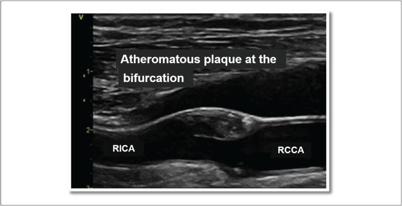
This diagnostic image is a B-mode ultrasound (sonography) of the carotid artery in a longitudinal view. It specifically highlights an atheromatous plaque located at the carotid bifurcation. The image shows the right common carotid artery (RCCA) branching into the right internal carotid artery (RICA). A prominent atherosclerotic plaque is visible as a heterogeneous, predominantly hypoechoic mass protruding from the arterial wall into the vessel lumen, causing significant luminal narrowing (stenosis). The normal arterial walls appear as hyperechoic, smooth linear structures, whereas the plaque exhibits an irregular surface and mixed echogenicity, suggesting a complex composition that may include lipids or necrotic tissue. This finding is clinically significant for evaluating cerebrovascular risk and identifying radiotherapy-induced arterial injuries or chronic atherosclerotic disease. The presence of the lesion at the bifurcation is a hallmark of typical carotid atherosclerosis.
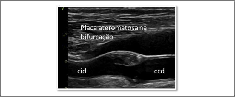
This diagnostic image is a B-mode ultrasound scan in longitudinal view of the right carotid artery. It demonstrates an atherosclerotic plaque located at the carotid bifurcation. The visual highlights a heterogeneous, eccentrically positioned plaque protruding into the vessel lumen from the posterior wall. The plaque exhibits mixed echogenicity, with hypoechoic areas suggesting a lipid-rich core or intraplaque hemorrhage, and hyperechoic regions likely representing fibrous tissue or focal calcification. Annotations include 'ccd' (common carotid artery right) and 'cid' (internal carotid artery right), indicating the anatomical transition zone. The image illustrates significant luminal narrowing, consistent with moderate carotid stenosis. This clinical photograph is used in cardiovascular medicine to assess stroke risk, evaluate intima-media thickness, and monitor progressive atherosclerosis or radiation-induced actinic arteritis.
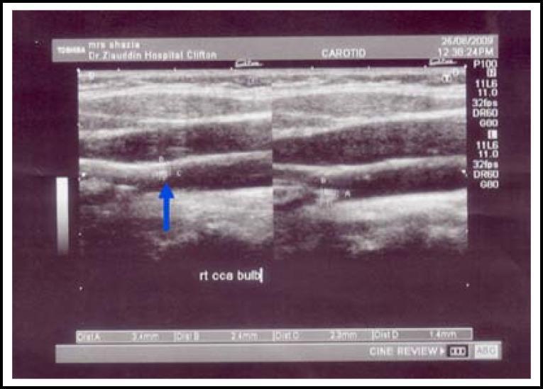
This diagnostic image is a B-mode ultrasound of the right carotid artery bifurcation and bulb in a longitudinal view. The image illustrates a clinical assessment of carotid intima-media thickness (CIMT) and the presence of atherosclerotic disease. The arterial walls show irregular echogenicity with visible hyperechoic foci indicative of calcified plaque formation along the vessel wall. Several calipers (labeled A, B, C, and D) demonstrate the measurement of the intima-media complex, with recorded values ranging from 1.4 mm to 2.4 mm, signifying pathological thickening. A blue arrow highlights a specific area of interest at the carotid bulb where the CIMT is increased. This visual serves as a primary diagnostic indicator for cardiovascular risk assessment in patients with hypercholesterolemia, demonstrating how ultrasound is used to identify early-stage atherosclerosis and structural vascular changes.
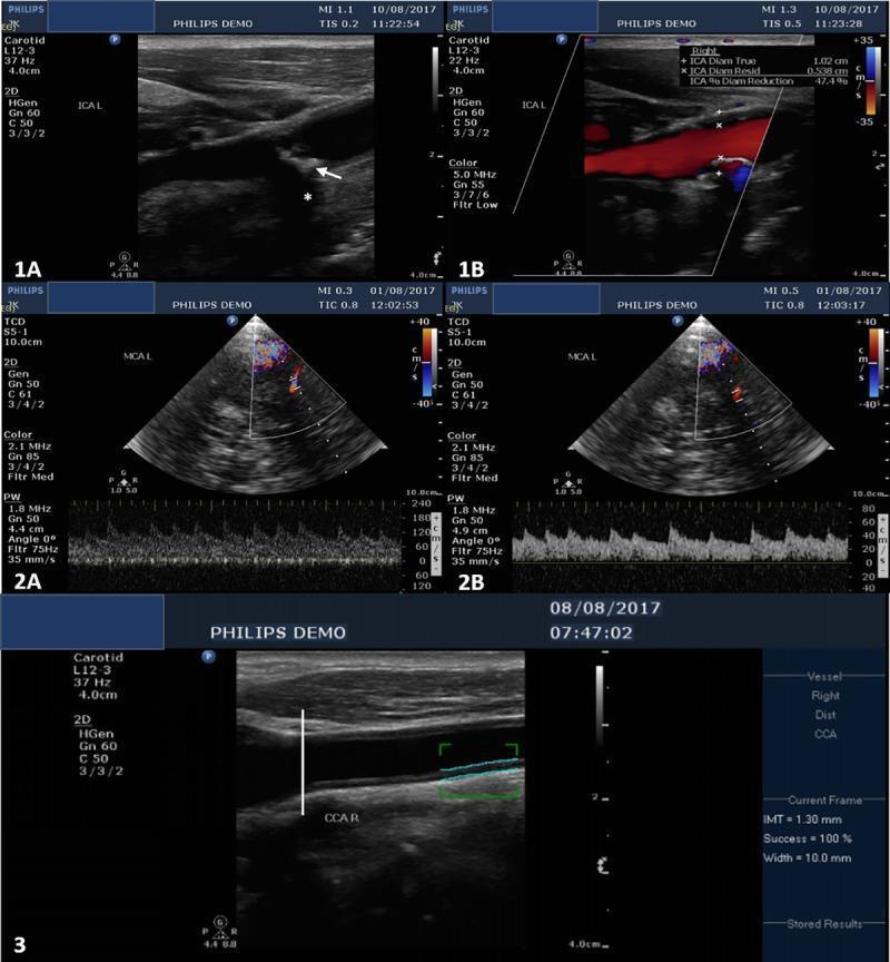
A composite diagnostic image set demonstrating extracranial and intracranial ultrasound imaging for atherosclerosis assessment. Panels 1A and 1B show B-mode and color Doppler longitudinal sections of the left carotid bulb, respectively. 1A identifies an isoechoic regular plaque (white arrow) with focal calcification causing characteristic acoustic shadowing (white asterisk). 1B illustrates a 47% diameter reduction due to this plaque. Panels 2A and 2B display Transcranial Color-Coded Duplex (TCCD) and Pulse Wave Doppler spectra of the left middle cerebral artery (MCA). 2A shows a stenotic segment with increased Peak Systolic Velocity (PSV) of 187 cm/s and Mean Flow Velocity (MFV) of 130 cm/s, while 2B shows a prestenotic segment with lower velocities, yielding a stenotic/prestenotic ratio of 3.25, consistent with >70% stenosis. Panel 3 demonstrates the measurement of carotid intima-media thickness (IMT) in the common carotid artery (CCA) using B-mode ultrasound. The measurement (1.30 mm) is taken in a longitudinal plane 10-30 mm proximal to the carotid bifurcation, indicated by electronic calipers (blue lines). These images serve as educational examples for vascular neurology and radiology.
carotid bifurcation atherosclerosis stenosis
Bilateral Carotid Bifurcation Atherosclerosis
Anatomy and Why the Bifurcation Is Vulnerable
Pathophysiology
- Thromboembolism (primary mechanism): Platelet-fibrin material, soft plaque debris, or superimposed thrombus embolizes from the bifurcation lesion to distal intracranial arteries. This is by far the most frequent cause of carotid-related ischemia. - Adams and Victor's Principles of Neurology
- Hemodynamic compromise: Severe stenosis (>70%) reduces flow, particularly when collateral reserves are inadequate - this becomes especially relevant in bilateral disease, where the contralateral ICA cannot compensate.
- Ulceration / irregular surface
- Soft (lipid-rich / hypoechoic) plaque
- Intraplaque hemorrhage
- Overlying thrombus
Clinical Presentation
| Category | Features |
|---|---|
| Asymptomatic | Incidental bruit or imaging finding; ~2% annual stroke risk |
| TIA | Transient monocular blindness (amaurosis fugax) or hemispheric deficits (<24 h) |
| Stroke | Fixed neurological deficit; 13% annual recurrence risk after symptomatic stenosis |
| Crescendo TIA | Multiple rapid-fire attacks - urgent intervention needed |
Diagnosis and Imaging
- Plaque morphology (echogenicity, surface characteristics)
- Carotid intima-media thickness (CIMT) - a surrogate of subclinical atherosclerosis (CIMT >1.0 mm is abnormal; values of 1.4-2.4 mm indicate significant disease)
- Degree of luminal stenosis by Doppler velocimetry
Risk Stratification by Degree of Stenosis (NASCET Criteria)
| Stenosis | Category | Management |
|---|---|---|
| <30% | Low risk | Medical therapy only |
| 30-49% | Mild | Medical therapy |
| 50-69% | Moderate (symptomatic) | CEA offers modest benefit |
| ≥70% | Severe | Strong indication for revascularization if symptomatic |
| Near-occlusion / Occlusion | Special case | CEA benefit reduced; medical therapy |
Medical Management
- Antiplatelet therapy: Aspirin is standard. Clopidogrel or aspirin + dipyridamole as alternatives. Note: anticoagulation has not been directly compared to antiplatelet therapy for carotid disease
- High-intensity statins: Stabilize plaque, reduce stroke risk independent of LDL
- Blood pressure control
- Smoking cessation
- Glycemic control in diabetics - Harrison's Principles of Internal Medicine 22E (2025)
Revascularization Options
1. Carotid Endarterectomy (CEA)
- Stenosis ≥70%: 2-year ipsilateral stroke risk 26% (medical) vs. 9% (CEA) - an absolute risk reduction of 17% (65% relative risk reduction)
- Stenosis 50-70%: Significant but less robust benefit
- Stenosis <30%: Surgery causes harm (ECST)
- Benefit is greatest when CEA is performed within 2 weeks of symptom onset
- Benefit more pronounced in patients >75 years and in men vs. women
- Stenosis ≥60%: 5-year stroke risk 11% (medical) vs. 5.1-6.4% (CEA)
- Absolute risk reduction: ~5.4-5.9% over 5 years (only ~1.2% annually)
- Perioperative complication rate higher in women - benefit in asymptomatic women remains controversial
- For CEA to be justified: institutional perioperative stroke/death rate must be ≤6%
2. Transfemoral Carotid Artery Stenting (TFCAS)
3. Transcarotid Artery Revascularization (TCAR)
- Avoids the atherosclerotic aortic arch
- Lower learning curve (proficiency after ~10-15 procedures)
- Stroke rate 1.4% (ROADSTER I) vs. 4.1% for TFCAS in CREST
- Symptomatic with >50% stenosis, OR asymptomatic with >80% stenosis
- CCA diameter ≥6 mm, landing zone ≥5 cm
- Bilateral carotid artery stenosis is itself a listed anatomic criterion making a patient eligible for TCAR
Bilateral Disease: Special Considerations
- No contralateral flow compensation - hemodynamic ischemia risk is higher when both ICAs are severely stenosed
- Staged procedures - typically the more symptomatic or more severely stenosed side is treated first; operating on both simultaneously carries prohibitive risk
- Contralateral carotid occlusion and bilateral stenosis are explicit high-risk anatomic criteria justifying TCAR over TFCAS
- Clamping risk in CEA - cross-clamp tolerance may be reduced if the contralateral side is also severely diseased; shunting should be considered
- Bilateral disease is a strong indicator for intensive medical management on the untreated side while awaiting staged intervention
Prognosis
| Situation | Annual stroke risk |
|---|---|
| Asymptomatic stenosis (any degree) | ~2% per year |
| Symptomatic stenosis (untreated) | ~13% per year |
| Post-CEA (≥70% symptomatic) | ~4.5% per year (5-year data) |
| Post-TCAR | Very low (1.4% perioperative) |