
Gall bladder reffer pain
gallbladder referred pain diagram right shoulder scapula
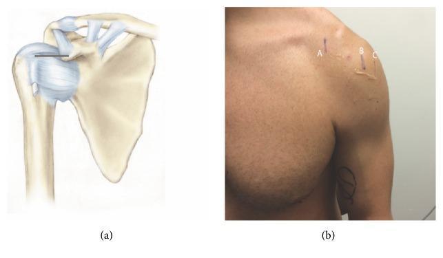
This composite educational graphic focuses on shoulder anatomy and surface landmark localization for clinical procedures. Figure (a) is an anatomical diagram of the right shoulder joint depicting the scapula, clavicle, and proximal humerus. It highlights the coracoclavicular and acromioclavicular ligaments, as well as the glenohumeral capsule. A horizontal black line illustrates the measurement distance between the coracoid process and the intertubercular (bicipital) groove. Figure (b) is a clinical photograph showing the anterior aspect of a human male's left shoulder with surface markings. Vertical skin markings labeled A, B, and C correspond to the underlying anatomy: (A) represents the coracoid process, (B) marks the intertubercular groove at 0° of external rotation, and (C) marks the same groove at 45° of external rotation. Visible ultrasound gel near markings B and C suggests that these points were localized using musculoskeletal ultrasound. This material is designed to teach clinical localization of the long head of the biceps tendon relative to fixed bony landmarks for diagnostic or therapeutic injections.
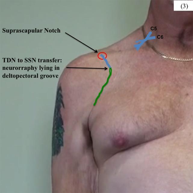
Anatomical Diagram and Clinical Overlay: This image depicts a surgical planning or post-operative schematic for a nerve transfer procedure on the right shoulder of a male patient. The visual combines a clinical photograph with color-coded overlays to illustrate the thoracodorsal nerve (TDN) to suprascapular nerve (SSN) transfer. Key anatomical landmarks are identified, including the cervical roots (C5 and C6) in the neck region and the suprascapular notch near the superior border of the scapula. A green line traces the distal path of the TDN terminal branches as they are mobilized superiorly from the axillary/lateral thoracic region toward the shoulder. A blue line represents the SSN, originating from the brachial plexus and traveling under the clavicle. The point of neurorrhaphy (nerve repair) is indicated where the green and blue lines meet within the deltopectoral groove, just inferior to the suprascapular notch. This diagram demonstrates the surgical management of an upper brachial plexus injury by utilizing a functional motor nerve donor (TDN) to reinnervate the target muscles of the suprascapular nerve.

This clinical photograph shows a posterior view of a patient's upper back and shoulders during a wall-push physical examination maneuver. The image demonstrates a clear case of right-sided scapular winging. Specifically, the right scapula is displaced laterally and posteriorly, with its medial border and inferior angle protruding prominently from the thoracic cage. This creates a visible asymmetric contour when compared to the neutral, stabilized left scapula. The right shoulder also appears slightly depressed. This clinical finding is often associated with neurological or muscular dysfunction, such as long thoracic nerve palsy affecting the serratus anterior muscle or injury to the spinal accessory nerve affecting the trapezius. The visual evidence of winging during forward flexion or a wall-push highlights the instability of the scapulothoracic joint, which is a key diagnostic sign in patients presenting with shoulder pain, weakness, or limited range of motion related to brachial neuritis or other neuromuscular conditions.
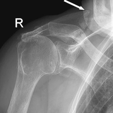
This anteroposterior (AP) X-ray of the right shoulder demonstrates an insufficiency fracture of the scapular spine, indicated by a white arrow. There is visible disruption of the cortical continuity of the scapula with associated periosteal reaction and hazy callus formation around the fracture site, though a clear fracture line remains, signifying a lack of complete bony consolidation. The image reveals generalized osteopenia with thinning of the cortical bone and decreased trabecular density, consistent with the patient's history of osteoporosis and chronic corticosteroid use. Additionally, there is superior migration of the humeral head relative to the glenoid, suggesting a chronic rotator cuff deficiency. The glenohumeral and acromioclavicular joints appear relatively preserved in terms of joint space. This imaging is clinically significant for evaluating non-traumatic or insufficiency fractures in elderly patients presenting with progressive shoulder pain and functional impairment.
Gallbladder Referred Pain
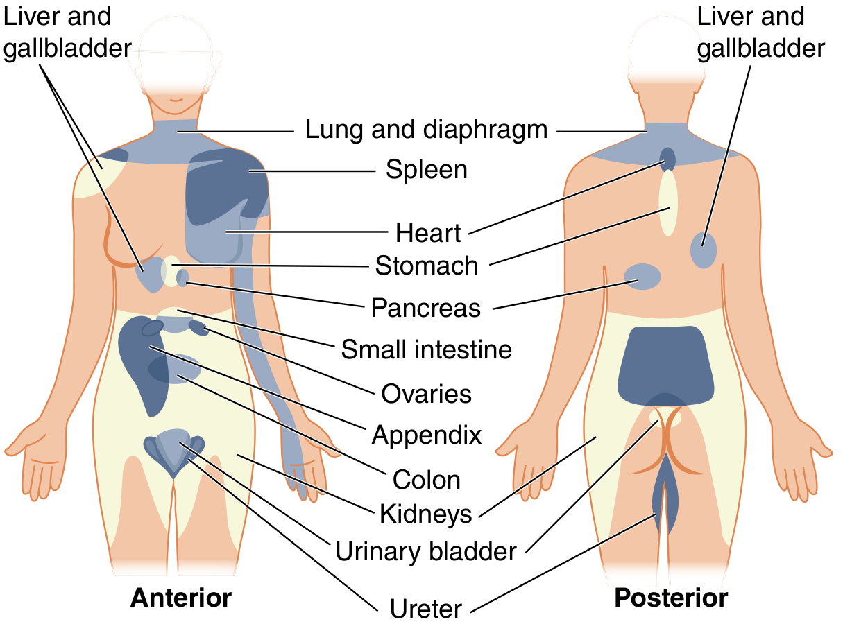
Where Does Gallbladder Pain Refer?
| Site | Direction |
|---|---|
| Right upper quadrant (RUQ) | Primary, local visceral pain |
| Right shoulder tip | Classic referred site (anterior) |
| Right infrascapular / interscapular region | Posterior referred site |
| Right side of neck (C3-C5 dermatomes) | Via phrenic nerve pathway |
Mechanism of Referred Pain
- Costanzo Physiology explains: "Referred pain is of visceral origin that is misperceived as pain arising from a somatic location... the dermatomal rule states that sites on the skin are innervated by nerves arising from the same spinal cord segments as those innervating the visceral organs." - Costanzo Physiology 7th Ed.
Nerve Pathway Details
- The gallbladder's pain fibers travel with sympathetic nerves and enter the spinal cord at T6-T9 (sometimes stated as T8-T11 for liver and gallbladder in Head zones)
- These converge on the same dorsal horn neurons as somatic fibres from the RUQ skin and right shoulder region
- Result: pain is referred to the epigastrium and RUQ anteriorly, and right infrascapular region posteriorly
- When gallbladder disease irritates the diaphragm (e.g., pericholecystic inflammation, bile leak, or a large distended gallbladder pressing upward), the phrenic nerve (C3-C5) is stimulated
- The phrenic nerve's somatic connections project pain to the right shoulder tip and right side of the neck - Sabiston Textbook of Surgery explains this as "referred pain to the shoulder because of the phrenic nerve's anatomic connections to the brachial plexus"
- The Color Atlas of Human Anatomy specifically names this: "The Eiselsberg phenomenon occurs on the right side of the shoulder as a 'false projection' - meaning that pain may radiate to the right shoulder due to disease of the liver or gallbladder. Pain sensations spread out in the dermatomes (C3-C5)." - Color Atlas of Human Anatomy, Vol. 1
- Compare: pancreatic disease refers to the left shoulder for the same reason
Head Zones (Dermatomal Map)
| Organ | Spinal Segments |
|---|---|
| Liver and gallbladder | T8-T11 |
| Heart | T3/4 |
| Stomach | T8 |
| Diaphragm | C4 |
| Pancreas | T5-T10 |
Clinical Summary
| Pain Type | Character | Location |
|---|---|---|
| True visceral | Dull, poorly localized, colicky | Epigastrium / RUQ |
| Parietal (peritonism) | Sharp, well-localized | RUQ (Murphy's sign) |
| Referred (somatic) | Aching | Right shoulder tip, right subscapular area |
| Diaphragmatic irritation | Sharp | Right shoulder tip, right neck (C3-C5) |