
All foraman of base of skull with content
Foramina of the Base of the Skull and Their Contents
Anterior Cranial Fossa
| Foramen | Contents |
|---|---|
| Foramen cecum | Emissary veins (connecting nasal cavity to superior sagittal sinus); may be absent |
| Foramina of cribriform plate | Olfactory nerve fibers (CN I) - 15-20 small foramina transmitting filaments from nasal mucosa to olfactory bulbs |
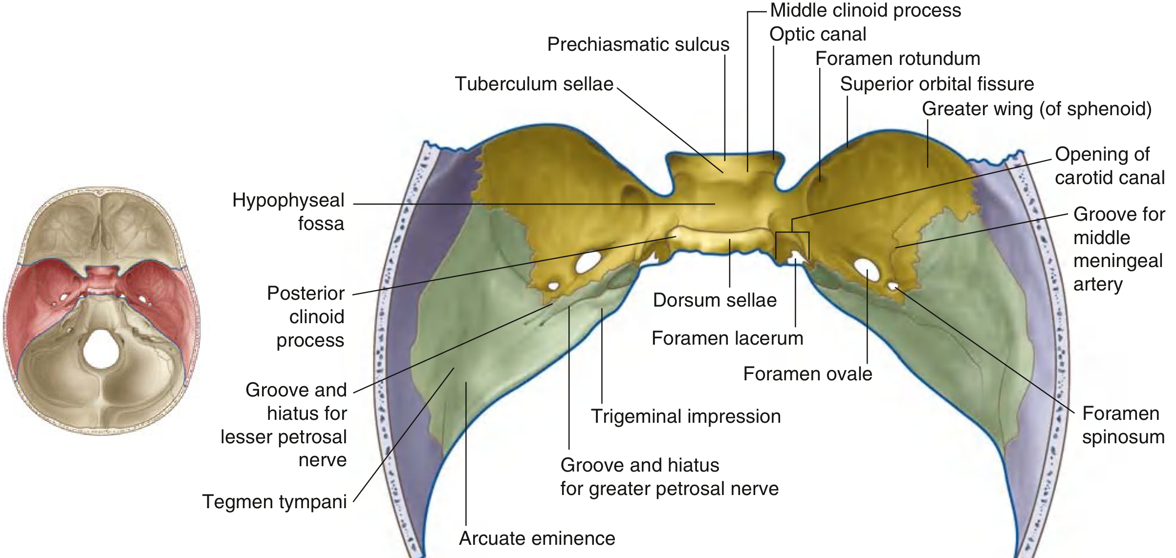
Middle Cranial Fossa
| Foramen | Contents |
|---|---|
| Optic canal | Optic nerve (CN II); ophthalmic artery |
| Superior orbital fissure | CN III (oculomotor), CN IV (trochlear), CN V1 (ophthalmic - frontal, lacrimal, nasociliary branches), CN VI (abducent); superior ophthalmic vein; inferior ophthalmic vein branch; orbital branch of middle meningeal artery |
| Foramen rotundum | CN V2 (maxillary nerve) - passes to pterygopalatine fossa |
| Foramen ovale | CN V3 (mandibular nerve); lesser petrosal nerve (from glossopharyngeal, IX); accessory middle meningeal artery (occasionally); emissary vein |
| Foramen spinosum | Middle meningeal artery and vein (meningeal branch); meningeal branch of mandibular nerve (nervus spinosus) |
| Foramen lacerum | Closed in life by fibrocartilaginous plug - no structures pass through completely; internal carotid artery traverses its upper part (exits canal, does not traverse); greater petrosal nerve; deep petrosal nerve; terminal branch of ascending pharyngeal artery; emissary vein |
| Carotid canal (opening) | Internal carotid artery (enters middle cranial fossa from below) |
| Hiatus for greater petrosal nerve | Greater petrosal nerve (CN VII branch) |
| Hiatus for lesser petrosal nerve | Lesser petrosal nerve (from tympanic plexus, CN IX) |
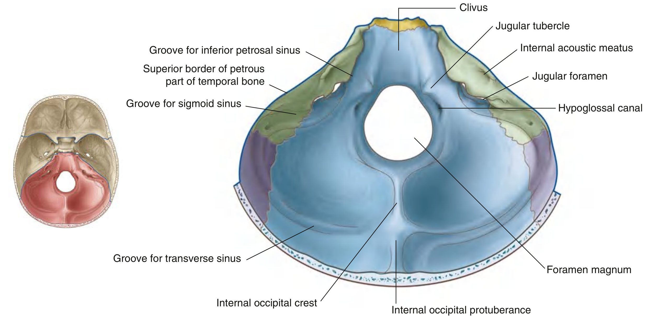
Posterior Cranial Fossa
| Foramen | Contents |
|---|---|
| Internal acoustic meatus (IAM) | CN VII (facial nerve + nervus intermedius); CN VIII (vestibulocochlear - superior vestibular nerve, inferior vestibular nerve, cochlear nerve); labyrinthine artery |
| Jugular foramen | Divided into three parts: Anterior (pars nervosa) - inferior petrosal sinus, CN IX (glossopharyngeal); Middle (pars vascularis) - CN X (vagus), CN XI (accessory); Posterior - internal jugular vein (continuation of sigmoid sinus), meningeal branches of occipital and ascending pharyngeal arteries, nodes of Krause |
| Hypoglossal canal | CN XII (hypoglossal nerve); meningeal branch of ascending pharyngeal artery; emissary vein; lymphatics |
| Foramen magnum | Medulla oblongata (continuing as spinal cord); spinal roots of CN XI (accessory nerve, ascending); vertebral arteries (bilateral); anterior spinal artery; posterior spinal arteries; meninges |
| Condylar canal | Emissary vein (when present - variable) |
Memory Aids
- Foramen lacerum - a common exam trap: it appears large but is plugged by cartilage in life; the internal carotid artery merely crosses its superior surface, it does not truly pass through it.
- Jugular foramen - carries the "9, 10, 11 + IJV" grouping (CN IX, X, XI + internal jugular vein).
- Foramen magnum - the largest foramen; carries the brainstem, vertebral arteries, and spinal accessory nerve.
- Superior orbital fissure - carries CN III, IV, V1, VI (all the "orbital movers" + ophthalmic division).
- The cribriform plate foramina are the smallest, yet the most numerous (15-20), carrying the olfactory nerve filaments.
Send me all Forman in one picture with content written below them
A detailed anatomical diagram of the base of the skull (internal view from above), showing all foramina clearly labeled with their contents listed below each label. The image should show a superior view of the skull base divided into three cranial fossae (anterior, middle, posterior), with the following foramina clearly marked and their contents written as small text annotations: ANTERIOR CRANIAL FOSSA: - Foramen cecum → Emissary veins to nasal cavity - Cribriform plate foramina → CN I (Olfactory nerve filaments) MIDDLE CRANIAL FOSSA: - Optic canal → CN II, Ophthalmic artery - Superior orbital fissure → CN III, IV, V1, VI; Ophthalmic veins - Foramen rotundum → CN V2 (Maxillary nerve) - Foramen ovale → CN V3, Lesser petrosal nerve, Accessory meningeal artery - Foramen spinosum → Middle meningeal artery & vein - Foramen lacerum → Closed by cartilage; ICA traverses; Greater & deep petrosal nerves - Carotid canal → Internal carotid artery POSTERIOR CRANIAL FOSSA: - Internal acoustic meatus → CN VII, CN VIII, Labyrinthine artery - Jugular foramen → CN IX, X, XI; Internal jugular vein; Inferior petrosal sinus - Hypoglossal canal → CN XII, Meningeal artery, Emissary vein - Foramen magnum → Medulla oblongata, Vertebral arteries, CN XI spinal roots, Spinal arteries - Condylar canal → Emissary vein The diagram should use a clean white background, with the three fossae shaded in different soft colors (light yellow for anterior, light green for middle, light blue for posterior), clear black labels with thin leader lines, and small descriptive text for contents. Medical textbook illustration style, high resolution, professional anatomy diagram.
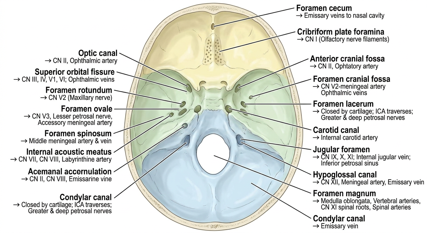
- Yellow - Anterior cranial fossa (Foramen cecum, Cribriform plate)
- Green - Middle cranial fossa (Optic canal, Superior orbital fissure, Foramen rotundum, Foramen ovale, Foramen spinosum, Foramen lacerum)
- Blue - Posterior cranial fossa (Internal acoustic meatus, Jugular foramen, Hypoglossal canal, Foramen magnum, Condylar canal)