
HEPATIC Cause of oedema
Hepatic Cause of Oedema
1. Hypoalbuminaemia → Reduced Plasma Oncotic Pressure
- Normal albumin synthesis: ~120 mg/kg/day; can double with low oncotic pressure, but a failing liver cannot compensate.
- Albumin is the major osmotically active intravascular colloid. When serum albumin falls, plasma colloid osmotic (oncotic) pressure drops.
- This allows fluid to leak out of capillaries into the interstitium, producing peripheral oedema (pitting, bilateral, dependent), and into the peritoneal cavity producing ascites.
"Hypoalbuminemia from decreased synthetic function in a cirrhotic liver results in reduced plasma oncotic pressure and contributes to the loss of fluid from the vascular compartment into the peritoneal cavity."
- Harrison's Principles of Internal Medicine, 22e
"Because albumin is the osmotically active intravascular colloid, hypoalbuminemia often results in edema."
- Henry's Clinical Diagnosis and Management by Laboratory Methods
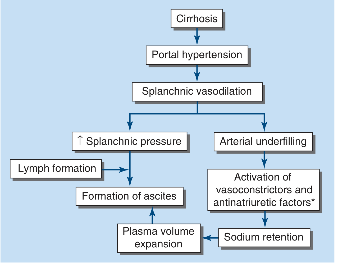
2. Portal Hypertension → Increased Hydrostatic Pressure
- The peritoneal cavity (ascites)
- The splanchnic interstitium
3. Splanchnic Vasodilation → Sodium and Water Retention
- Portal hypertension causes release of vasodilatory substances (especially nitric oxide) into the splanchnic circulation, causing splanchnic arterial vasodilation.
- This creates "arterial underfilling" - the systemic circulation senses reduced effective arterial blood volume.
- The body responds by activating the renin-angiotensin-aldosterone system (RAAS) and the sympathetic nervous system, resulting in avid renal sodium and water retention.
- This expands extracellular fluid volume, perpetuating peripheral oedema and ascites.
- The retained fluid continually leaks back out of the intravascular compartment, so the vascular "underfilling" sensation persists - creating a vicious cycle.
Summary Table
| Mechanism | Cause | Effect |
|---|---|---|
| Reduced albumin synthesis | Hepatocyte destruction | Low oncotic pressure → fluid leaks out |
| Portal hypertension | Sinusoidal fibrosis | Raised hydrostatic pressure → ascites, oedema |
| Splanchnic vasodilation + RAAS activation | Arterial underfilling | Na/water retention → extracellular fluid expansion |
| Increased hepatic lymph production | Obstruction of hepatic lymphatics | Ascites overflow |
Clinical Presentation
- Ascites (abdominal distension, shifting dullness, fluid thrill)
- Bilateral pitting peripheral oedema (ankles, legs)
- Hepatic hydrothorax (pleural effusion, usually right-sided, via diaphragmatic rent)
- Anasarca in severe cases
invesigaion correlaion
- Lab investigations used to diagnose hepatic oedema and what each finding correlates with clinically
- Correlation between specific investigation values and severity of oedema
What do you mean by 'investigation correlation' in the context of hepatic oedema?
boh
Investigations in Hepatic Oedema & Their Correlations
PART 1 - Lab Investigations & What Each Finding Means
A. Liver Function Tests (LFTs)
| Investigation | Finding in Hepatic Disease | Correlation with Oedema/Liver Failure |
|---|---|---|
| Serum Albumin | Low (<3.5 g/dL) | Most direct correlate of oedema - reduced oncotic pressure → fluid leaks into interstitium and peritoneal cavity |
| AST / ALT | Elevated (ALT > AST in viral; AST > ALT in alcoholic) | Hepatocyte necrosis/inflammation; severe elevation → synthetic failure → low albumin |
| Serum Bilirubin | Elevated | Reflects excretory failure; rising level in cirrhosis = decompensation |
| Alkaline Phosphatase (ALP) + GGT | Elevated in cholestatic disease | Biliary obstruction/cholestasis |
| Prothrombin Time / INR | Prolonged | Most sensitive synthetic function marker; rises within hours of major hepatic insult; elevated INR = severe failure |
| Platelet count | Low (thrombocytopaenia) | Hypersplenism from portal hypertension - correlates with severity of portal HTN |
| Serum Sodium | Hyponatraemia (dilutional) | Seen in advanced cirrhosis with ascites; RAAS activation causes water retention |
| Serum Creatinine / Urea | Raised | May indicate hepatorenal syndrome in decompensated cirrhosis |
"The initial evaluation of liver disease involves a battery of blood tests... assessing hepatic necroinflammation (aminotransferases), cholestatic biliary dysfunction (ALP, GGT), excretory function (bilirubin), and synthetic function (coagulation factors, albumin)."
- Goldman-Cecil Medicine
B. Serum Albumin - Key Correlations with Oedema Severity
| Serum Albumin Level | Interpretation | Clinical Oedema |
|---|---|---|
| 3.5 - 5.0 g/dL | Normal | No oedema from hypoalbuminaemia |
| 2.8 - 3.5 g/dL | Mild hypoalbuminaemia | Mild/early pitting oedema |
| <2.8 g/dL | Severe hypoalbuminaemia | Significant oedema + ascites likely |
| <2.0 g/dL | Critical | Anasarca; massive ascites; Child-Pugh score 3 |
PART 2 - Differentiating Hepatic vs Cardiac vs Renal Oedema
SAAG (Serum-Ascites Albumin Gradient)
| SAAG Value | Interpretation | Examples |
|---|---|---|
| ≥ 1.1 g/dL | Portal hypertension (transudate) | Cirrhosis, cardiac failure, Budd-Chiari |
| < 1.1 g/dL | Non-portal cause (exudate) | Malignancy, TB peritonitis, nephrotic syndrome |
"SAAG ≥1.1 g/dL has a diagnostic accuracy of 98% for portal hypertension-related ascites, compared to only 52-80% for other markers."
- Henry's Clinical Diagnosis and Management by Laboratory Methods
Comparative Table - Hepatic vs Cardiac vs Renal Oedema
| Feature | Hepatic | Cardiac | Renal (Nephrotic) |
|---|---|---|---|
| Serum albumin | Low (reduced synthesis) | Normal or mildly low | Very low (urinary loss) |
| Urinary protein | Normal/mild | Normal | >3.5 g/day (massive proteinuria) |
| AST/ALT | Elevated | Normal or mildly raised | Normal |
| INR/PT | Prolonged | Normal | Normal |
| Bilirubin | Elevated | Normal | Normal |
| BNP/NT-proBNP | Normal | Markedly elevated | Normal |
| Urine Na | Low (RAAS activation) | Low (RAAS activation) | Low (avid retention) |
| SAAG | ≥1.1 (portal HTN) | ≥1.1 (cardiac) | <1.1 |
| Ascitic fluid protein | Low (<2.5 g/dL) | Low-medium | - (rarely ascites) |
| USS abdomen | Cirrhotic liver, splenomegaly, varices | Enlarged heart, bilateral pleural effusions | Normal liver |
| ECG/Echo | Normal | LV dysfunction, valvular disease | Normal |
PART 3 - Severity Scoring: Child-Pugh Score
| Component | 1 point | 2 points | 3 points |
|---|---|---|---|
| Bilirubin | ≤2 mg/dL | 2-3 mg/dL | ≥3 mg/dL |
| Serum Albumin | ≥3.5 g/dL | 2.8-3.5 g/dL | ≤2.7 g/dL |
| INR | ≤1.6 | 1.7-2.2 | ≥2.3 |
| Ascites | None | Slight | Moderate-Massive |
| Encephalopathy | None | Grade 1-2 | Grade 3-4 |
| Class | A (5-6) = Compensated | B (7-9) = Moderate | C (10-15) = Decompensated |
- Sabiston Textbook of Surgery
The lower the albumin and the higher the bilirubin/INR, the more severe the synthetic failure - and the greater the degree of oedema and ascites.
Summary: Investigation Correlation at a Glance
Low Albumin  →  ↓ Oncotic pressure  →  Oedema / Ascites
High INR     →  Coagulation failure → Synthetic dysfunction
High Bilirubin → Excretory failure  → Severity of disease
High SAAG (≥1.1) → Portal hypertension → Confirms hepatic/cardiac cause
Thrombocytopaenia → Hypersplenism    → Portal HTN marker
Low serum Na → RAAS activation     → Severe decompensation
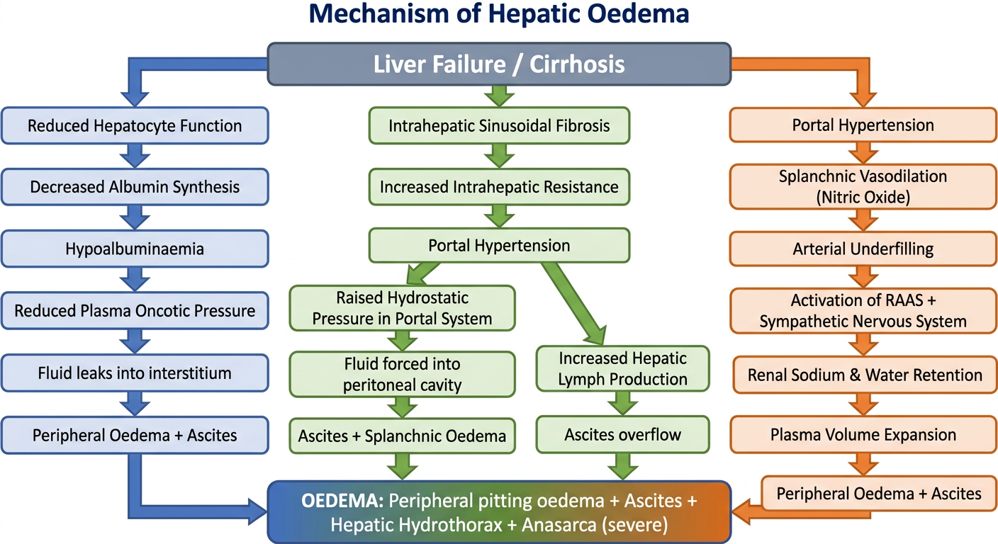
chart mechanism
A detailed medical flowchart diagram showing the mechanism of hepatic oedema. Title: "Mechanism of Hepatic Oedema". The chart has three parallel pathways all stemming from "Liver Failure / Cirrhosis" at the top center. Pathway 1 (left): Liver Failure / Cirrhosis → Reduced Hepatocyte Function → Decreased Albumin Synthesis → Hypoalbuminaemia → Reduced Plasma Oncotic Pressure → Fluid leaks into interstitium → Peripheral Oedema + Ascites Pathway 2 (center): Liver Failure / Cirrhosis → Intrahepatic Sinusoidal Fibrosis → Increased Intrahepatic Resistance → Portal Hypertension → Raised Hydrostatic Pressure in Portal System → Fluid forced into peritoneal cavity → Ascites + Splanchnic Oedema. Also from Portal Hypertension → Increased Hepatic Lymph Production → Ascites overflow Pathway 3 (right): Liver Failure / Cirrhosis → Portal Hypertension → Splanchnic Vasodilation (Nitric Oxide) → Arterial Underfilling → Activation of RAAS + Sympathetic Nervous System → Renal Sodium & Water Retention → Plasma Volume Expansion → Peripheral Oedema + Ascites At the bottom, all three pathways converge into a box: "OEDEMA: Peripheral pitting oedema + Ascites + Hepatic Hydrothorax + Anasarca (severe)" Use color coding: blue for pathway 1, green for pathway 2, orange for pathway 3. Clean clinical style with arrows, boxes, and clear labels. White background.
| Pathway | Key Step | End Result |
|---|---|---|
| Blue | ↓ Albumin synthesis → ↓ Oncotic pressure | Peripheral oedema + Ascites |
| Green | Portal hypertension → ↑ Hydrostatic pressure | Ascites + Splanchnic oedema |
| Orange | Splanchnic vasodilation → RAAS → Na/water retention | Peripheral oedema + Ascites |