
Umbilical hernia
umbilical hernia anatomy diagram
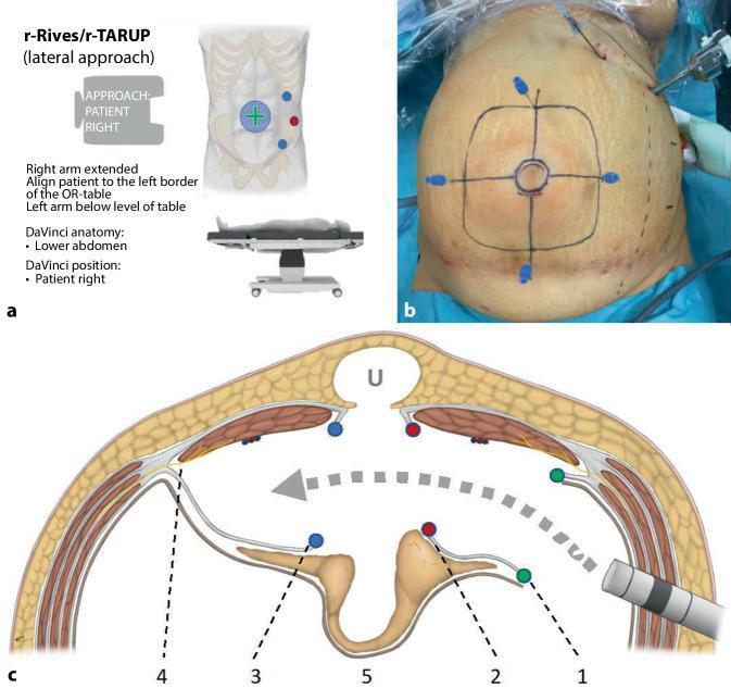
Educational graphic illustrating the surgical planning and anatomy for robotic transabdominal retromuscular umbilical prosthetic hernia repair (r-Rives or r-TARUP) via a left-lateral approach. Figure A shows an infographic detailing patient positioning on the operating table and port placement lateral to the left rectus muscle. Figure B is a clinical photograph of a patient's abdomen under pneumoperitoneum; ink markings delineate the umbilical hernia (central circle) and the anticipated mesh size (outer square). Transparietal needles are inserted at the square's perimeter to guide intra-abdominal preparation. Figure C is a cross-sectional anatomical diagram of the abdominal wall layers (skin, subcutaneous fat, rectus muscles, and posterior rectus sheaths). It maps the surgical dissection path (gray arrow) through the retrorectus space. Key steps are numbered: (1) green dots mark the lateral entry into the left posterior rectus sheath; (2) red dots signify the medial reopening of the sheath near the linea alba; (3) blue dots indicate the entry into the right posterior rectus sheath; (4) identifies lateral nerve preservation zones; (5) shows the mobilized hernia sac and peritoneal bridge. This content serves as a guide for surgical residents and fellows learning complex robotic ventral hernia repair techniques.
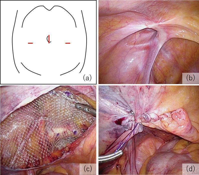
A multi-panel educational figure illustrating the key procedural steps of a Transabdominal Preperitoneal (TAPP) inguinal hernia repair. (a) Schematic diagram of the abdomen showing the laparoscopic port arrangement: a 12 mm umbilical port and two 5 mm lateral ports. (b) Intraoperative laparoscopic photograph showing the internal inguinal anatomy and the hernial orifice, classified as EHS-L2 (European Hernia Society classification for a medium indirect lateral hernia). (c) Surgical view demonstrating mesh fixation; a semi-transparent, macroporous polypropylene mesh is positioned in the preperitoneal space to cover the defect and is secured with visible surgical tacks. (d) Final procedural step showing the closure of the peritoneal flap using continuous suturing to exclude the mesh from the peritoneal cavity, preventing contact with abdominal viscera. The series emphasizes proper anatomical orientation, mesh placement, and peritoneal closure in minimally invasive hernia surgery.
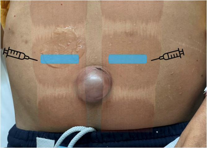
This clinical photograph displays a patient's abdomen in the supine position, featuring a large, prominent umbilical hernia. The hernia presents as a rounded, hemispherical protrusion several centimeters in diameter at the midline. The overlying skin appears tense and slightly erythematous, consistent with clinical incarceration. Superimposed on the image are educational annotations for a bilateral rectus sheath block (BRSB). Two horizontal blue boxes indicate the intended footprints for a linear ultrasound transducer, positioned transversely over the rectus abdominis muscles lateral to the umbilicus. Syringe icons illustrate the lateral-to-medial in-plane needle insertion technique. Also visible are vertical tan-colored markings on the skin following the abdominal wall anatomy, and ultrasound transmission gel applied to the right upper quadrant. The image serves as an educational guide for regional anesthesia landmarks and the clinical appearance of a complicated umbilical hernia.
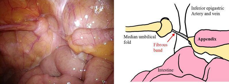
Laparoscopic view and corresponding schematic diagram of the right lower abdominal cavity demonstrating findings related to a de Garengeot hernia. The image shows the median umbilical fold incarcerated within the right femoral ring. A vermiform appendix is visible within the free abdominal cavity; notably, it exhibits a normal appearance with no signs of inflammation, enlargement, or hyperemia. A distinct fibrous band is seen connecting the tip of the appendix to the right femoral ring, indicating a prior or transient incarceration of the appendix. Surrounding anatomical landmarks include the inferior epigastric artery and vein, and loops of the small intestine. This visual illustrates a rare clinical scenario where the appendix is associated with a femoral hernia (de Garengeot hernia) but is found in a reduced state during surgical intervention. The diagram labels these structures to facilitate anatomical identification for educational purposes in general surgery and hernia repair training.
"umbilical hernia"[MeSH Terms] AND repair
Umbilical Hernia
Definition
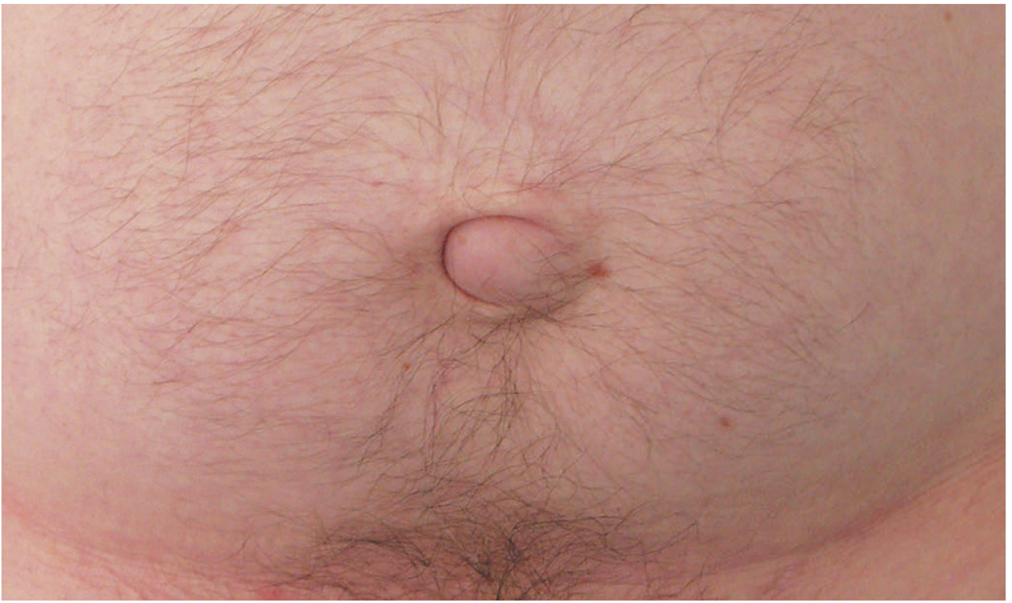
Classification / Types
| Type | Description |
|---|---|
| Exomphalos | Abdominal contents protrude into the umbilical cord, covered by a diaphanous membrane; a congenital anomaly |
| Congenital umbilical hernia | Passes through the centre of the umbilical scar; appears in the first few months of life; ~90% resolve spontaneously within 5 years |
| Acquired umbilical hernia (adult) | Protrudes through the umbilical scar in adult life; due to raised intra-abdominal pressure from pregnancy, ascites, ovarian cyst, fibroid, or bowel distension |
| Paraumbilical hernia | Most common acquired type; occurs just above (rarely below) the umbilicus between the two recti; affects obese, middle-aged women |
Embryology & Anatomy
Epidemiology
- Neonatal/infantile: Occurs in up to 10% of infants; higher incidence in premature babies; incidence in Black infants is up to 8 times higher than in White infants
- Adults: More common in overweight men (thinned midline raphe) and postpartum women; women are overall more frequently affected than men
- Predisposing conditions: pregnancy, obesity, liver cirrhosis with ascites, chronic raised intra-abdominal pressure
Clinical Features
- Appears within weeks of birth
- Often asymptomatic; increases in size on crying, assumes a classic conical shape
- Easily reducible; spontaneously reduces when the child lies down
- Definite cough impulse
- Contents usually small intestine (resonant to percussion)
- Strangulation is extremely uncommon below age 3
- The bulge is typically slightly to one side of the umbilical depression, creating a crescent-shaped appearance
- Pain is the dominant symptom (tissue tension, intermittent bowel obstruction)
- Often irreducible - contents (omentum or bowel) become adherent to the sac
- Sac neck is relatively narrow relative to sac size - prone to irreducibility, obstruction, and strangulation
- In large hernias, overlying skin may become very thin; ulceration can occur; spontaneous rupture is extremely rare
- In liver cirrhosis patients, the hernia can enlarge dramatically due to ascites
Investigations
- Diagnosis is usually clinical
- USS or CT may be used in obese patients or where contents are uncertain
- In cirrhotic patients: liver function tests, Child-Pugh scoring before any surgical planning
Treatment
Children
- Conservative (under 2 years, asymptomatic): Parental reassurance; 95% resolve spontaneously
- Surgical repair indicated if:
- Persists beyond age 2-5 years
- Hernia is very large or symptomatic
- Signs of incarceration (abdominal pain, bilious vomiting, tender hard umbilical mass) - mandates immediate exploration
Adults
- Hernia containing bowel - high strangulation risk; surgery should be strongly advised
- Symptomatic hernia
- Small asymptomatic hernias may be observed but tend to enlarge
| Defect Size | Technique |
|---|---|
| < 1 cm | Simple suture repair (no tension) or darn suture |
| Up to 2 cm | Mayo repair (transverse incision, sac opened and reduced, peritoneum closed, fascial edges overlapped - "waistcoat over trousers") with non-absorbable sutures |
| > 2 cm | Mesh repair strongly recommended; current evidence supports mesh even in smaller defects due to high recurrence risk with suture-only repair |
- Camera port + two lateral working ports
- Hernia contents reduced by traction/external pressure
- Falciform ligament and median umbilical fold taken down as needed
- A disc of non-adherent, intraperitoneal mesh placed on the undersurface of the abdominal wall, centred on the defect
- Fixed with staples, tacks, or sutures
- Advantages: secure repair, generous overlap, no damage to umbilicus
- Disadvantages: specialised mesh, risk of bowel adhesion, erosion, mesh pain
- Post-pregnancy: Exercise, weight loss, and increased abdominal tone first; surgery strongly avoided before or during pregnancy; many resolve postpartum
- Liver cirrhosis (Child's B/C): Extremely high operative mortality and morbidity; careful patient selection; hepatology support essential; fascial repair with fine continuous sutures to minimize post-op ascites leakage
Complications of Untreated Hernia
- Irreducibility (contents adhere to sac)
- Intestinal obstruction
- Strangulation (compromised blood supply) - more likely with narrow neck and small defect
- Skin ulceration / breakdown over large hernias
- Spontaneous rupture (rare)
Key Distinguishing Points - Summary Box
| Feature | Infantile | Adult/Paraumbilical |
|---|---|---|
| Age | Neonatal/early infancy | Middle age / postpartum |
| Sex | Equal | Females > Males |
| Spontaneous resolution | ~90-95% by age 5 | Does not resolve |
| Strangulation risk | Very low | Moderate to high |
| Management | Conservative first | Surgery advised |
| Surgical repair | Suture closure | Mesh preferred for > 2 cm |