
Explain gram negative and gram positive bacteria
gram positive gram negative bacteria cell wall structure comparison diagram
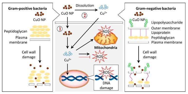
A pathophysiology diagram illustrating the antibacterial mechanisms of copper oxide nanoparticles (CuO NPs) on Gram-positive and Gram-negative bacteria. The left panel shows Gram-positive bacteria with a thick peptidoglycan layer above a plasma membrane. The right panel shows Gram-negative bacteria with a complex cell wall including lipopolysaccharides, an outer membrane, lipoproteins, and a thin peptidoglycan layer. Both panels demonstrate CuO NPs causing cell wall damage and lysis. The central panel details the intracellular bactericidal pathway labeled as mechanism 1 and 2. Mechanism 1 involves the internalization of CuO NPs into the cytosol, while mechanism 2 illustrates the dissolution of CuO NPs into copper ions (Cu2+) which then permeate the cell. Inside the cell, both Cu2+ ions and CuO NPs induce the production of reactive oxygen species (ROS). The resulting oxidative stress leads to mitochondrial disruption and DNA damage, ultimately resulting in cell death. This infographic highlights the role of nanoparticle morphology and ion release in targeting bacterial structural and metabolic integrity.
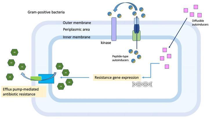
This pathophysiology diagram illustrates quorum-sensing-regulated resistance mechanisms in Gram-positive bacteria. The bacterial cell structure is depicted with three distinct layers: an inner membrane, a periplasmic area, and an outer membrane. The diagram highlights two primary signaling pathways involved in cell-to-cell communication. The first involves 'peptide-type autoinducers' (represented as blue/yellow spheres), which are processed and secreted via a transmembrane receptor and then detected by a membrane-spanning kinase. The second pathway involves 'diffusible autoinducers' (pink squares) that freely enter the cell. Both pathways converge to trigger 'resistance gene expression' at the DNA level. This genetic activation leads to physiological outcomes, specifically 'efflux pump-mediated antibiotic resistance,' where a transmembrane efflux pump actively transports antibiotic molecules (green hexagons) out of the intracellular space. This educational graphic demonstrates the interplay between bacterial signaling and multidrug resistance mechanisms.
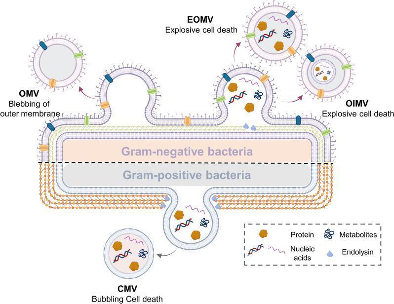
A medical microbiology diagram illustrating the biogenesis and classification of bacterial extracellular vesicles (BEVs) from Gram-negative and Gram-positive bacteria. The top half depicts a Gram-negative bacterium with its triple-layered cell envelope (outer membrane, periplasmic space, and cytoplasmic membrane). Three vesicle formation mechanisms are shown: blebbing of the outer membrane creating Outer Membrane Vesicles (OMV), and explosive cell death leading to Explosive Outer Membrane Vesicles (EOMV) and Outer-Inner Membrane Vesicles (OIMV). The bottom half represents a Gram-positive bacterium characterized by a thick peptidoglycan cell wall and a single cytoplasmic membrane. This section illustrates 'bubbling cell death' resulting in Cytoplasmic Membrane Vesicles (CMV). A legend identifies the internal cargo within these vesicles, including proteins (brown shapes), nucleic acids (wavy lines), metabolites (blue branched icons), and endolysin (light blue shapes). This pathophysiology diagram highlights the structural differences in bacterial envelopes and the various pathways—blebbing, explosive death, and bubbling—that contribute to BEV diversity and composition.
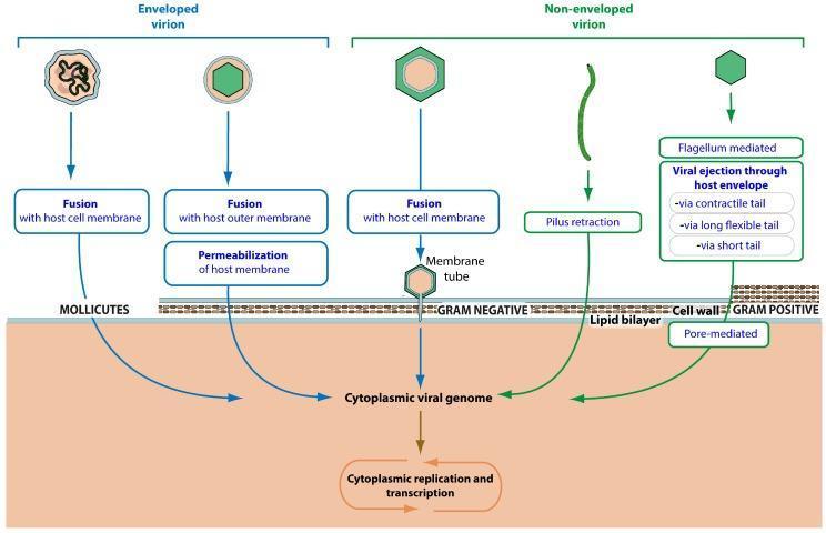
A scientific infographic illustrating the mechanisms of viral entry across different bacterial cell envelopes, focusing on enveloped and non-enveloped virions. The diagram is divided by host bacterial type: Mollicutes, Gram-negative, and Gram-positive bacteria. For enveloped virions infecting Mollicutes, the diagram shows fusion with the host cell membrane or host outer membrane, followed by membrane permeabilization. For Gram-negative bacteria, non-enveloped virions utilize mechanisms such as fusion with the cell membrane via a membrane tube or pilus retraction. For Gram-positive bacteria, entry is depicted as flagellum-mediated or viral ejection through the host envelope/cell wall using contractile, long flexible, or short tails, as well as pore-mediated entry. All pathways converge at the delivery of the 'Cytoplasmic viral genome' into the host cytoplasm, leading to 'Cytoplasmic replication and transcription.' The schematic effectively compares the structural challenges presented by different bacterial cell walls (e.g., lipid bilayers vs. thick peptidoglycan layers) and the corresponding viral penetration strategies.
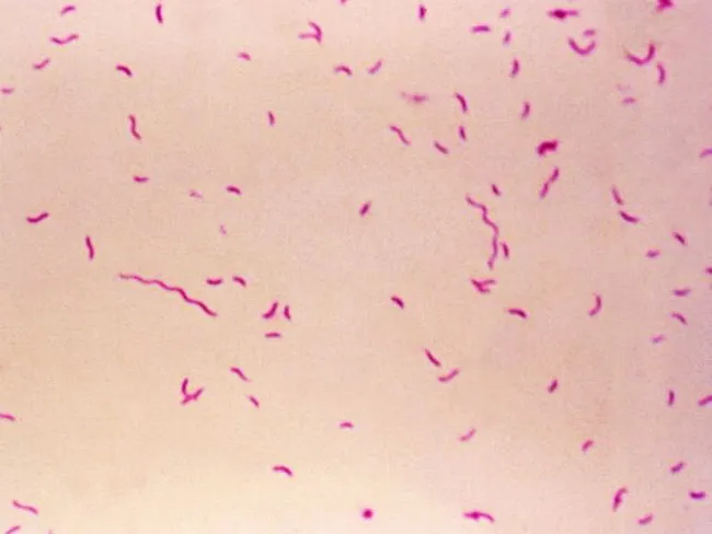
Bright-field light microscopy of a Gram-stained bacterial smear reveals numerous small, slender, curved Gram-negative rods with a characteristic gull-wing appearance consistent with Campylobacter species. The image captures bacteria oriented individually and in small clusters against a pale pink counterstain, illustrating Bacillary morphology typical of enteric pathogens. Gram staining shows purple/blue rods (crystal violet retained by cell wall) with a light pink background from the counterstain (safranin), enabling discrimination from Gram-positive organisms. The organisms appear slender and curved, with a single polar flagellum suggested by motility or alignment in the smear. Specimen type is a bacterial smear obtained from a gastrointestinal sample (feces or culture isolate), prepared for diagnostic microbiology. The imaging modality is bright-field microscopy at high magnification (approximately 1000x with oil immersion), following Gram staining to highlight bacterial cell wall structure. Clinically, detection of Campylobacter species supports infectious gastroenteritis; in the IPSID (immunoproliferative small intestinal disease) context, Campylobacter involvement has been proposed as a pathogenic trigger mirroring Helicobacter pylori's role in gastric MALT lymphoma. Differential diagnoses include Helicobacter, Vibrio, and other curved Gram-negative bacteria. This image serves educational and diagnostic utility for microbiology, clinical pathology, gastroenterology, and infectious disease research.
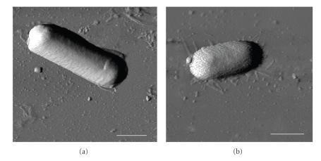
This diagnostic imaging set displays Atomic Force Microscopy (AFM) phase images of two distinct bacterial species, Bacillus cereus (a) and Escherichia coli (b), deposited on a mica surface. The images facilitate a morphological comparison between Gram-positive and Gram-negative bacteria at the nanometer scale. Panel (a) shows B. cereus as a distinctly elongated, rod-shaped cell with a relatively uniform, smooth surface texture and well-defined edges. In contrast, panel (b) depicts E. coli with a more compact, oval or capsule-shaped morphology. The E. coli cell exhibits a significantly rougher surface topography with visible granularity compared to the smoother envelope of B. cereus. The background depicts the mica substrate with faint thread-like structures visible around the E. coli cell, likely representing extracellular filaments or surface dehydration artifacts. A 1 μm scale bar is provided in both panels. These images are used in microbiology to study bacterial cell wall structural integrity, surface roughness (Rq), and biophysical responses to environmental conditions like relative humidity (RH).
Gram-Positive vs. Gram-Negative Bacteria
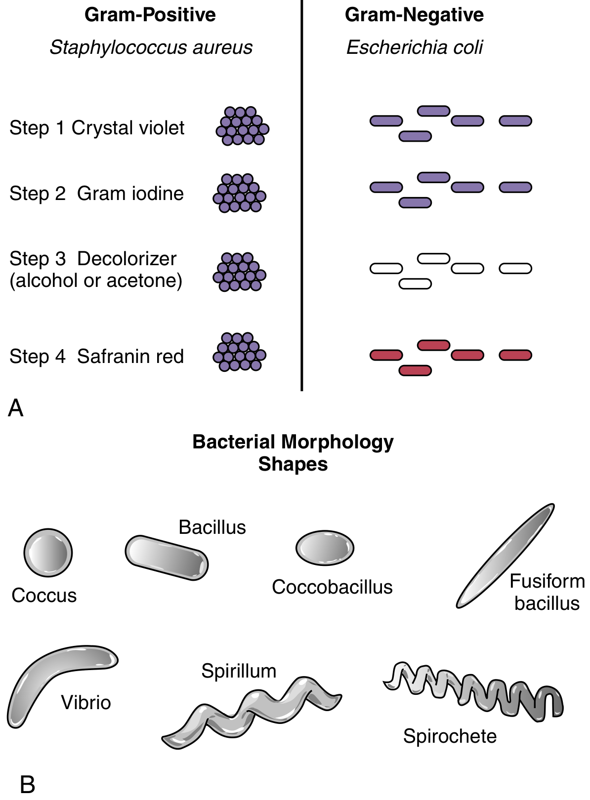
The Gram Stain - How It Works
| Step | Reagent | Purpose |
|---|---|---|
| 1 | Crystal violet | Primary stain - binds to peptidoglycan |
| 2 | Gram iodine | Mordant - causes crystal violet to precipitate inside the cell |
| 3 | Decolorizer (acetone-alcohol) | Washes out stain from gram-negative cells |
| 4 | Safranin (red counterstain) | Stains gram-negative cells pink/red |
Cell Wall Structure - The Core Difference
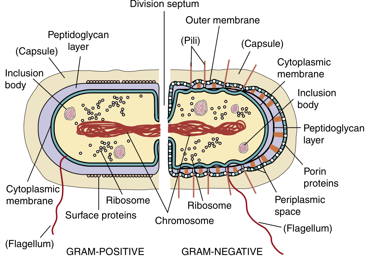
Gram-Positive Bacteria
- Peptidoglycan (multiple thick layers) - a meshlike exoskeleton; glycan chains cross-linked with short peptide bridges. Provides shape, protection from complement, and rigidity
- Teichoic acids - polyribitol phosphate or glycerol phosphate cross-linked to peptidoglycan; strengthens cell wall and sequesters calcium ions
- Lipoteichoic acids - lipid-linked teichoic acid anchored to the plasma membrane; activates innate host defenses
- Surface proteins - bound to peptidoglycan or teichoic acid; involved in immune evasion and adhesion
Gram-Negative Bacteria
- Thin peptidoglycan - single layer; provides shape but is insufficient to retain crystal violet
- Outer membrane (OM) - unique bilayer containing lipopolysaccharide (LPS) on the outer leaflet
- Lipopolysaccharide (LPS / endotoxin) - the most important virulence factor; activates the innate immune system via TLR-4, triggering fever, cytokine storm, and septic shock
- Periplasmic space - contains transport proteins, enzymes (including beta-lactamases responsible for antibiotic resistance), and biosynthetic machinery
- Porin proteins - channels in the outer membrane that allow small molecules (including some antibiotics) to enter
- Pili/fimbriae - adhesion structures for host-cell attachment
Head-to-Head Comparison Table
| Feature | Gram-Positive | Gram-Negative |
|---|---|---|
| Gram stain color | Purple | Pink/red |
| Peptidoglycan layer | Thick (150-500 Å) | Thin (single layer) |
| Outer membrane | Absent | Present |
| LPS (endotoxin) | Absent | Present (major virulence factor) |
| Teichoic acids | Present | Absent |
| Periplasmic space | Minimal | Present (contains beta-lactamases) |
| Porins | Absent | Present |
| Susceptibility to penicillin | Higher | Lower (OM is a barrier) |
| Susceptibility to lysozyme | Higher | Lower (OM protects) |
| Susceptibility to detergents | Lower | Higher (OM disrupted) |
Clinical Significance
Antibiotic targeting
- Beta-lactams (penicillins, cephalosporins) inhibit peptidoglycan synthesis (PBP binding). Gram-positive bacteria have more exposed peptidoglycan, making them more susceptible. Gram-negative bacteria have beta-lactamases in the periplasmic space that can destroy beta-lactams before they reach PBPs.
- Vancomycin works on gram-positive bacteria only (too large to cross the outer membrane of gram-negatives)
- Polymyxins target the outer membrane of gram-negative bacteria specifically
Sepsis and endotoxin
- Gram-negative sepsis is often more severe because LPS triggers massive TLR-4 activation, flooding the bloodstream with cytokines (TNF, IL-1, IL-6), causing fever, hypotension, and multi-organ failure
- Gram-positive bacteria lack LPS but peptidoglycan fragments can cause similar (though less potent) inflammatory cascades
Exceptions worth knowing
- Mycobacteria - have a unique waxy cell wall (mycolic acids); neither reliably gram-positive nor gram-negative - require Ziehl-Neelsen (acid-fast) stain
- Mycoplasma - no cell wall at all; cannot be Gram stained; naturally resistant to all beta-lactams
- Spirochetes (Treponema, Leptospira, Borrelia) - gram-negative structurally but stain poorly; identified by dark-field microscopy or serology