
Adenomatoid odontogenic tumour
"adenomatoid odontogenic tumor"[MeSH Terms]
Adenomatoid Odontogenic Tumour (AOT)
Overview
- Cummings Otolaryngology Head and Neck Surgery, p. 4050
- Scott-Brown's Otorhinolaryngology Head & Neck Surgery, p. 8950
Origin
The "Two-Thirds Rule" (Classic Mnemonic)
| Feature | Proportion |
|---|---|
| Female sex | 2/3 |
| Located in maxilla | 2/3 |
| Associated with impacted teeth (esp. canine) | 2/3 |
| Found in teenagers | 2/3 |
- Cummings, p. 4050
Clinical Features
- Slow-growing, painless, palpable bony-hard swelling
- Anterior maxilla is the dominant site
- Strong female predominance (M:F ratio ~1:1.9)
- Predominantly in the 2nd decade of life (most < 20 years)
- Occasionally arises peripherally within the gingiva (peripheral/extrafollicular variant)
Variants
| Type | Description |
|---|---|
| Follicular (most common) | Surrounds the crown of an unerupted tooth; resembles a dentigerous cyst radiographically |
| Extrafollicular / Periapical | Intraosseous but not associated with an impacted tooth; periapical or interdental location |
| Peripheral | Arises in the soft tissue of the gingiva, outside bone |
Radiographic Features
-
Unilocular radiolucency with a sharp, well-corticated to sclerotic border
-
Often encases the crown of an unerupted tooth (classically the maxillary canine), blending with the dental follicle - can mimic a dentigerous cyst
-
Key distinguishing feature from dentigerous cyst: presence of small discrete radiopaque foci (calcifications) within the lucency - seen in a significant minority of cases
-
Purely radiolucent lesions are most common, but this is among the classic "mixed radiolucent-radiopaque" jaw lesions
-
KJ Lee's Essential Otolaryngology, p. 4073-4075
Histopathological Features
- Spindle-shaped epithelial cells forming whorled masses or rosettes in a scant fibrous stroma
- Duct-like (adenomatoid) structures lined by cuboidal or columnar epithelial cells - the classic hallmark
- Eosinophilic amorphous material / amyloid-like droplets in the rosette/adenomatoid areas
- Mineralizations - not well organized; may resemble dentin, cementum, or abortive enamel
- Cystic spaces are common; the largest lumen is usually adjacent to the tooth crown
- Cummings, p. 4060-4062
- Scott-Brown's, p. 8953-8954
Treatment
- The associated impacted tooth can often be preserved for eventual eruption or managed orthodontically
- No recurrence is expected after enucleation
Prognosis
Key Differentials
| Feature | AOT | Dentigerous Cyst | Ameloblastoma |
|---|---|---|---|
| Age | < 20 years | Any | 30-60 years |
| Site | Anterior maxilla (canine) | Usually posterior mandible | Posterior mandible |
| Border | Well-corticated | Well-corticated | Can be scalloped |
| Calcifications | Often present | Absent | Absent |
| Recurrence | None | Rare | Common |
| Treatment | Enucleation | Enucleation | Wide resection |
Summary Points for Exams
- AOT = "two-thirds tumour" (female, maxilla, impacted canine, teenager)
- Formerly called adenoameloblastoma - misleading name, avoid
- Duct-like structures lined by columnar cells = histologic hallmark
- Mimics dentigerous cyst radiographically - calcifications help differentiate
- Treatment: simple enucleation; no recurrence
- Counts ~3% of odontogenic tumours
IHC markers
"adenomatoid odontogenic tumor" AND immunohistochemistry
PMID: 27541702
PMID: 38895097
adenomatoid odontogenic tumor IHC markers cytokeratin vimentin calretinin CD57
https://www.pathologyoutlines.com/topic/mandiblemaxillaadeno…
IHC Markers in Adenomatoid Odontogenic Tumour (AOT)
1. Epithelial / Structural Markers
| Marker | Expression | Notes |
|---|---|---|
| CK14 | Strongly positive (69% strong, 25% intermediate) | Most consistent epithelial marker; expressed in both follicular and extrafollicular variants; reflects dental lamina/stratum intermedium origin |
| CK19 | Positive | Expressed in more differentiated (columnar) cells; preameloblasts gradually replace CK14 with CK19 |
| Pan-cytokeratin (AE1/AE3) | Positive | Confirms epithelial nature of tumour cells |
| Vimentin | Focally positive | Co-expression with CK14 in spindle-shaped cells and cells around the duct-like structures; suggests epithelial-mesenchymal plasticity |
| Podoplanin (D2-40) | Positive | Expressed in odontogenic epithelium; supports origin from dental lamina |
2. Neural / Neuroectodermal Differentiation Markers
| Marker | Expression | Notes |
|---|---|---|
| CD57 (Leu-7 / HNK-1) | Positive | Reflects neuroectodermal/neural crest differentiation of odontogenic epithelium |
| Calretinin | Positive | Expressed in columnar cells of the duct-like structures; shared with ameloblastoma but weaker |
| Nestin | Positive | Neural stem cell marker; expressed in AOT epithelium; supports odontogenic origin; found in both the tumour cells and in the duct-like structures |
3. Ameloblastic / Odontogenic Differentiation Markers
| Marker | Expression | Notes |
|---|---|---|
| DSPP (Dentin Sialophosphoprotein) | Positive (variable) | Indicates dentinogenic differentiation; supports origin from the inner enamel epithelium |
| Amelogenin | Focally positive | Seen near calcified areas; represents abortive enamel matrix production |
| Sheathlin / Enamelin | Focally positive | Enamel matrix proteins detected near mineralised foci |
4. Proliferation & Tumour Suppressor Markers
| Marker | Expression | Notes |
|---|---|---|
| Ki-67 | Very low (<5%) | Confirms the extremely low mitotic activity and benign/hamartomatous nature |
| PCNA | Low expression | Parallels Ki-67 findings |
| p53 | Negative or minimal | Supports benign/non-aggressive behaviour |
| MDM2 | Low/variable | Regulator of p53; low expression consistent with benign biology |
| Cyclin D1 | Low expression | Cell cycle regulator; low compared to ameloblastoma |
| Bcl-2 | Positive | Anti-apoptotic; expressed in odontogenic epithelium; supports survival of quiescent cells |
5. Bone Resorption Markers
| Marker | Expression | Notes |
|---|---|---|
| RANKL | Expressed | Present in tumour epithelium; explains the capacity to displace (but not invade) bone |
| RANK | Present | Found in surrounding stromal/osteoclast precursor cells |
| OPG (Osteoprotegerin) | Present | The balance of RANKL/OPG is tilted toward limited resorption only - consistent with the non-aggressive expansile growth pattern |
6. Signalling Pathway Markers
| Marker | Expression | Notes |
|---|---|---|
| Beta-catenin | Cytoplasmic / membranous (not nuclear) | Nuclear translocation (indicative of active Wnt signalling) is absent or rare - unlike ameloblastoma where nuclear staining is seen |
| TGF-beta1 / Smad-2/3, Smad-4, Smad-1/5/8 | Expressed | Reflects TGF-beta signalling in epithelial differentiation |
| HGF / c-Met | Expressed | Hepatocyte growth factor axis; role in epithelial morphogenesis |
| FAK / Paxillin / PI3K | Lower expression than in ameloblastoma | Focal adhesion kinase signalling is present but at lower levels compared to ameloblastoma, correlating with non-invasive behaviour (Bello et al. 2021, PMID 32681423) |
| DNA methyltransferase | Expressed | Suggests epigenetic regulation of the tumour's differentiation state |
7. Stemness Markers
| Marker | Expression | Notes |
|---|---|---|
| SALL4 | Negative to weak (cytoplasmic only) | Unlike aggressive odontogenic tumours; cytoplasmic rather than nuclear expression likely represents an inactive/mutant form |
| Fascin | Weak or absent | A motility/invasion-related cytoskeletal protein; low expression is consistent with non-invasive behaviour (Kulkarni et al. 2022, PMID 38895097) |
8. Metallothionein & Matrix Metalloproteinases (MMPs)
| Marker | Expression |
|---|---|
| Metallothionein | Expressed in epithelial cells |
| MMPs | Low-level expression; significantly lower than ameloblastoma; consistent with lack of local invasion |
Summary Table (Quick Reference)
| Category | Positive | Negative / Low |
|---|---|---|
| Epithelial | CK14, CK19, pan-CK, podoplanin | - |
| Mesenchymal co-expression | Vimentin (focal) | SMA, desmin |
| Neural/odontogenic | CD57, calretinin, nestin | - |
| Odontogenic diff. | DSPP, amelogenin (focal) | - |
| Proliferation | Bcl-2 | Ki-67 (very low), p53 |
| Invasion/signalling | RANKL, TGF-beta | FAK/PI3K (low), fascin (low), nuclear beta-catenin |
Key Practical Takeaway
Difference between follicular and extrafollicular variant
"adenomatoid odontogenic tumor" AND (follicular OR extrafollicular OR variants)
PMID: 30256456
PMID: 39119433
adenomatoid odontogenic tumor follicular extrafollicular peripheral variant differences site age radiology
Follicular vs. Extrafollicular vs. Peripheral AOT
The Three Variants at a Glance
| Feature | Follicular (FAOT) | Extrafollicular (EAOT) | Peripheral |
|---|---|---|---|
| Proportion of all AOT | ~74% | ~21% | ~5% |
| Location | Inside bone; surrounds the crown of an unerupted (impacted) tooth | Inside bone; no association with the crown of an unerupted tooth; lies between or at the apex of erupted teeth | Outside bone; in the gingival soft tissue only |
| Tooth association | Strongly associated with impacted canine (~70% of FAOT involve the canine) | No impacted tooth; located periapically or interdentally among erupted teeth | No impacted tooth; arises from gingival remnants of dental lamina |
| Jaw preference | Maxilla >> mandible | Maxilla > mandible | Anterior jaw (maxilla or mandible) |
| Age | Classic teenage presentation | Same age range | Slightly broader |
| Sex | Female predominance | Female predominance | Female predominance |
| Radiographic appearance | Pericoronal unilocular radiolucency surrounding crown of unerupted tooth; mimics dentigerous cyst | Periapical/interdental unilocular radiolucency between roots or at apex of erupted teeth; mimics radicular cyst or lateral periodontal cyst | No bony lesion - soft tissue mass only (may show a shallow saucerization/cupping of underlying alveolar crest) |
| Key radiographic differential | Dentigerous cyst | Radicular cyst / Lateral periodontal cyst | Peripheral giant cell granuloma / Peripheral ossifying fibroma |
| Calcifications on X-ray | Present in a proportion - key distinguishing feature from dentigerous cyst | Present in some cases | May be visible on occlusal film |
| Histology | Identical across all three variants - duct-like structures, rosettes, spindle cell whorls, amyloid droplets, variable calcification | Same | Same |
| Capsule | Well-encapsulated | Well-encapsulated | No bony capsule; confined within gingival connective tissue |
| Treatment | Enucleation; impacted tooth can be preserved | Enucleation / curettage | Excision of the soft tissue mass |
| Recurrence | Essentially nil | Essentially nil | Essentially nil |
The Critical Clinical Distinction: Tooth Relationship
- Follicular - the tumour wraps around the crown of an unerupted tooth, just like a dentigerous cyst. The radiolucency is pericoronal. The most classic presentation is a teenager with a missing maxillary canine and a pericoronal lucency on OPG.
- Extrafollicular - sits between erupted teeth or at a tooth apex, with no impacted tooth involved. Can be mistaken for an apical/periapical cyst or lateral periodontal cyst. This variant is sometimes discovered incidentally.
- Peripheral - entirely in the soft tissue of the gingiva, no bone involvement. Diagnosed clinically as a gingival swelling and confirmed histologically.
Do the Variants Differ in Biology?
"No difference was found when comparing the clinical/radiological features of the follicular, extrafollicular, and peripheral variants."
- All three variants behave the same - benign, non-recurrent, conservatively managed
- IHC profiles (CK14, vimentin, Ki-67, etc.) are identical across variants
- Histopathology is identical - the variant classification is purely topographic, not biologic
- The impacted canine association (~70%) is specific to the follicular type, but even within FAOT, the canine is not always the associated tooth (incisors, premolars, molars are also reported)
Why Does the Distinction Matter Clinically?
| Reason | Relevance |
|---|---|
| Differential diagnosis | FAOT mimics a dentigerous cyst; EAOT mimics a radicular or lateral periodontal cyst; peripheral mimics a reactive gingival lesion - knowing the variant directs the correct differential |
| Pre-operative planning | FAOT may allow the associated impacted tooth to be preserved; EAOT and peripheral variants have no tooth management consideration |
| Radiographic recognition | Calcific foci within a pericoronal lucency (FAOT) or a periapical lucency without evidence of pulp pathology (EAOT) should raise suspicion for AOT |
| Avoiding over-treatment | EAOT near a tooth apex can be mistaken for a periapical cyst and lead to unnecessary root canal treatment or extraction |
Summary
Generate a comparison table/poster of AOT variants vs dentigerous cyst
A clean, professional medical education comparison poster titled "AOT Variants vs Dentigerous Cyst" for oral pathology students. The poster has a dark navy blue background with white and colored text. It contains a large comparison table with 5 columns: Feature, AOT Follicular, AOT Extrafollicular, AOT Peripheral, and Dentigerous Cyst. Each column header has a distinct color: teal for AOT Follicular, orange for AOT Extrafollicular, purple for AOT Peripheral, and red for Dentigerous Cyst. Rows include: Age (teens <20 / teens <20 / teens <20 / any age), Sex (female predominance all AOT / male slight predominance DC), Site (anterior maxilla / anterior maxilla periapical or interdental / gingival soft tissue / posterior mandible), Tooth association (wraps crown of unerupted tooth / no impacted tooth between erupted teeth / no tooth in soft tissue / wraps crown of unerupted tooth), Radiology (pericoronal unilocular lucency / periapical or interdental lucency / no bony lesion cupping of crest / pericoronal unilocular lucency), Calcifications (present key feature / present some cases / may be visible / absent), Key differential (dentigerous cyst / radicular cyst lateral periodontal cyst / reactive gingival lesion / AOT follicular), Lining epithelium (no true cyst lining duct-like structures / same / same / thin non-keratinized stratified squamous 2 cell layers), Treatment (enucleation preserve tooth / enucleation curettage / soft tissue excision / enucleation), Recurrence (nil / nil / nil / rare). Footer text: "All AOT variants: histologically identical. Biology is the same regardless of location." The table has alternating light and slightly darker row backgrounds for readability. A small tooth icon is in the top corner. The overall style is clean, modern, academic.
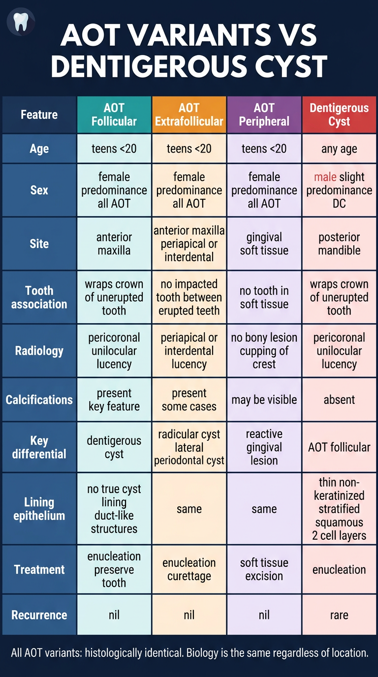
AOT Variants vs Dentigerous Cyst - Key Differences
| Feature | AOT Follicular | AOT Extrafollicular | AOT Peripheral | Dentigerous Cyst |
|---|---|---|---|---|
| Frequency | Most common AOT (~74%) | ~21% of AOT | ~5% of AOT | Most common developmental cyst |
| Age | Teens, <20 yrs | Teens, <20 yrs | Teens, <20 yrs | Any age (peak 2nd-3rd decade) |
| Sex | Female ++ | Female ++ | Female ++ | Male slight predominance |
| Site | Anterior maxilla | Anterior maxilla/mandible (periapical/interdental) | Gingival soft tissue only | Posterior mandible (3rd molar) |
| Tooth association | Surrounds crown of unerupted tooth | No impacted tooth; between erupted teeth | No tooth involvement | Surrounds crown of unerupted tooth |
| Radiology | Pericoronal unilocular lucency | Periapical/interdental lucency | No bony lesion (soft tissue only) | Pericoronal unilocular lucency |
| Calcifications | Present - key differentiating feature | Present in some | May be visible | Absent |
| Margins | Well-corticated/sclerotic | Well-corticated | - | Well-corticated |
| Lining | No true cyst lining; duct-like structures + rosettes | Same as follicular | Same | Thin non-keratinised stratified squamous epithelium (2-3 cell layers); reduced enamel epithelium origin |
| Capsule | Thick fibrous capsule; encapsulated | Encapsulated | Confined to gingival CT | Thin cyst wall |
| Key differential | Dentigerous cyst | Radicular cyst / Lateral periodontal cyst | Peripheral giant cell granuloma, pyogenic granuloma | AOT follicular |
| Treatment | Enucleation; tooth can be preserved | Enucleation/curettage | Soft tissue excision | Enucleation |
| Recurrence | Nil | Nil | Nil | Rare |