
Gram staining Microbiology and culture medium detailed explanation
Gram staining procedure bacteria purple pink
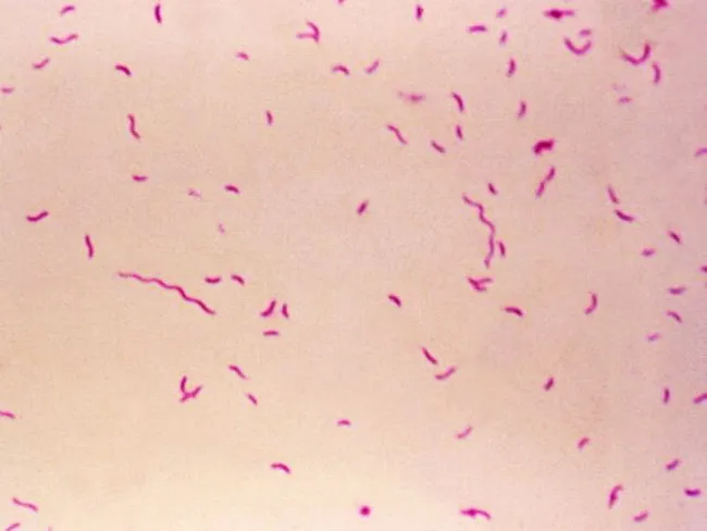
Bright-field light microscopy of a Gram-stained bacterial smear reveals numerous small, slender, curved Gram-negative rods with a characteristic gull-wing appearance consistent with Campylobacter species. The image captures bacteria oriented individually and in small clusters against a pale pink counterstain, illustrating Bacillary morphology typical of enteric pathogens. Gram staining shows purple/blue rods (crystal violet retained by cell wall) with a light pink background from the counterstain (safranin), enabling discrimination from Gram-positive organisms. The organisms appear slender and curved, with a single polar flagellum suggested by motility or alignment in the smear. Specimen type is a bacterial smear obtained from a gastrointestinal sample (feces or culture isolate), prepared for diagnostic microbiology. The imaging modality is bright-field microscopy at high magnification (approximately 1000x with oil immersion), following Gram staining to highlight bacterial cell wall structure. Clinically, detection of Campylobacter species supports infectious gastroenteritis; in the IPSID (immunoproliferative small intestinal disease) context, Campylobacter involvement has been proposed as a pathogenic trigger mirroring Helicobacter pylori's role in gastric MALT lymphoma. Differential diagnoses include Helicobacter, Vibrio, and other curved Gram-negative bacteria. This image serves educational and diagnostic utility for microbiology, clinical pathology, gastroenterology, and infectious disease research.
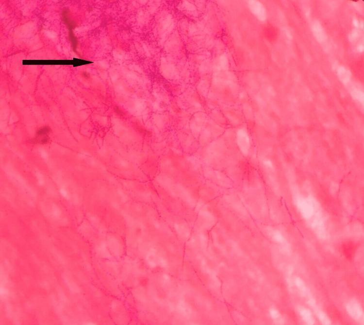
Diagnostic light microscopy image featuring a Gram stain of a clinical sputum sample at high magnification. The visual field demonstrates multiple Gram-positive (dark purple) microorganisms set against a pink-stained proteinaceous and cellular background. The pathogens exhibit a distinctive morphology characterized by long, slender, filamentous rods with frequent branching patterns. A characteristic 'beaded' appearance is evident along the length of the filaments, where the crystal violet stain appears irregularly distributed in alternating dark and light segments. These morphological features—Gram-positive staining, branching filaments, and beaded appearance—are classic diagnostic indicators for Nocardia species. This image serves as an educational resource for medical microbiology, illustrating the differentiation between typical pyogenic bacteria and branching actinomycetes in respiratory infections.
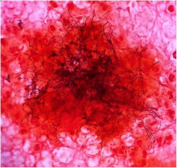
This light microscopy image demonstrates a Gram stain of an excised clinical sample, typically used in microbiology for diagnostic identification. The central focus shows a dense cluster of Gram-variable filamentous rods, characteristic of Actinomyces species. These organisms appear as dark, elongated, branching, thread-like structures. Some filaments exhibit a beaded appearance, staining more intensely (Gram-positive/purple) or less intensely (Gram-negative/pink) along their length. The background consists of host cellular debris and inflammatory cells, which are counterstained pink/red with safranin. These background elements include various eukaryotic cells and likely polymorphonuclear leukocytes. This visual is highly representative of Actinomycosis, demonstrating the classic 'sulfur granule' or 'tangled mass' morphology often seen in abscess aspirates or tissue samples. It serves as a critical educational tool for distinguishing filamentous bacteria from standard cocci or bacilli and highlights the importance of Gram stain variability in clinical diagnostics.
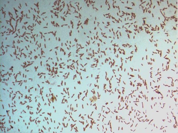
This diagnostic image shows a light microscopy view of a bacterial culture following Gram staining. The specimen displays numerous small, rod-shaped (bacilli) bacteria distributed across the field. The microorganisms exhibit a consistent pink-to-red hue, identifying them as Gram-negative. This staining characteristic indicates a cell wall structure with a thin peptidoglycan layer and an outer membrane that does not retain the crystal violet stain. The arrangement of the bacilli is predominantly individual or scattered, with occasional small clusters and pairs. Specifically identified as D. fastidiosa strain JC13T, this image serves as a primary microbiological reference for the morphological and staining properties of this novel genus within the family Erysipelotrichaceae. The visual demonstrates key diagnostic features including cell morphology, arrangement, and Gram reaction, which are essential for clinical microbiology classification and initial pathogen identification.
culture media types blood agar MacConkey selective differential bacteriology
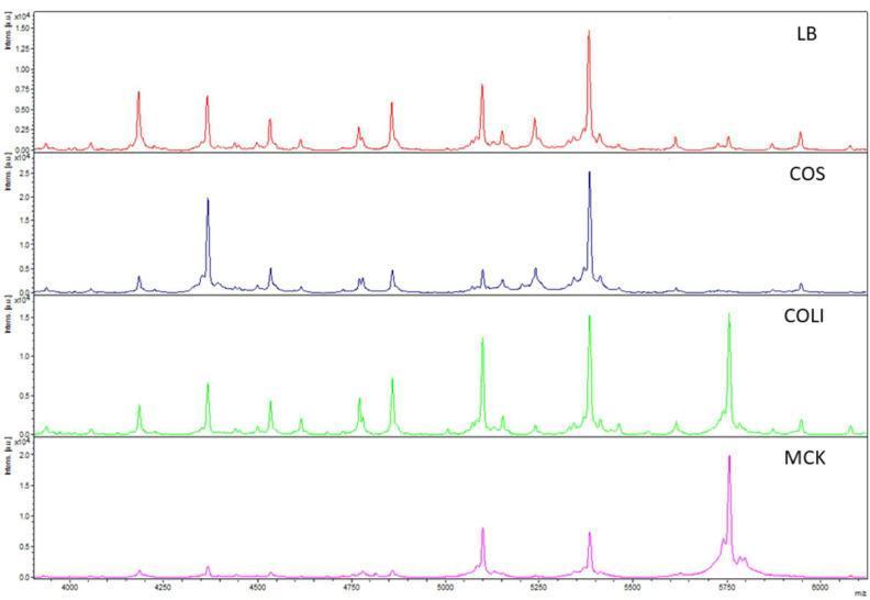
A multi-panel comparison chart of Matrix-Assisted Laser Desorption/Ionization Time-of-Flight (MALDI-TOF) mass spectra used in clinical microbiology. The image displays the spectral profiles of an Escherichia coli (E. coli) isolate grown on four different solid agar media: Luria Broth (LB, red), Columbia agar with sheep blood (COS, blue), Coliformen agar (COLI, green), and MacConkey agar (MCK, pink). The x-axis represents the mass-to-charge ratio (m/z) in the 4000–6000 range, while the y-axis indicates relative intensity. The LB spectrum shows the highest peak density and resolution, serving as the reference for reproducibility. In contrast, the COS, COLI, and MCK spectra demonstrate variations in peak presence and signal intensity, illustrating how different growth media influence the proteomic fingerprint of bacterial isolates. This diagnostic imaging comparison highlights the importance of standardized culture protocols for accurate microbial characterization and subspecies-level identification beyond simple species detection.
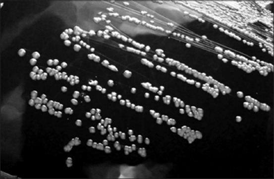
**Modality:** Microbiology culture photograph (grayscale). **Specimen:** *Corynebacterium pseudotuberculosis* (formerly *Corynebacterium ovis*) isolated on blood agar medium. **Morphological Features:** The image displays a streak plate demonstrating isolated bacterial colonies. The colonies are characterized by a distinct circular morphology with an entire margin. They exhibit a convex elevation and appear opaque with a grayish-white coloration against the dark background of the agar. The surface texture of the colonies appears slightly dry or waxy, which is characteristic of this genus. **Distribution:** Streaking pattern shows confluent growth in the primary sectors transitioning to well-defined, discrete individual colonies in the peripheral sectors, allowing for the observation of specific colony macro-morphology. **Diagnostic Context:** These visual features are consistent with the growth characteristics of *Corynebacterium* species, typically found in veterinary clinical microbiology samples. Key differentiating features include the specific convex profile and circular uniformity of the colonies on enriched media.
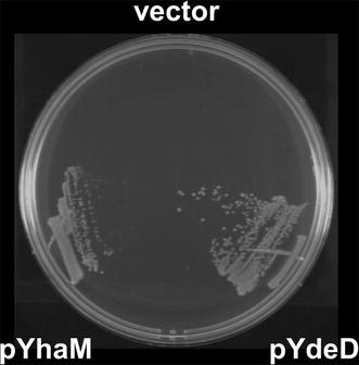
**Imaging Modality:** Microbiological culture photograph. **Specimen/Entity:** *Escherichia coli* (MG1655 strain) bacterial growth on a petri dish containing M9 minimal agar medium supplemented with 4 mM cysteine. **Experimental Setup:** The agar plate is divided into three distinct sectors for comparative growth analysis of different genetic constructs: 1. **Top (vector):** Empty vector control (pSTV29). 2. **Bottom Left (pYhaM):** *E. coli* expressing the *yhaM* gene. 3. **Bottom Right (pYdeD):** *E. coli* expressing the *ydeD* gene. **Characteristic Visual Features:** * **Differential Growth Patterns:** A marked difference in colony formation is visible between the sectors. * **Vector Control:** Shows complete inhibition of growth, indicating high sensitivity to the 4 mM cysteine concentration. * **pYhaM and pYdeD Sectors:** Both sectors demonstrate robust bacterial proliferation with visible streak lines and individual colony formation, indicating the conferral of cysteine resistance. * **Morphology:** Light-colored, opaque bacterial streaks and colonies against a dark background, typical of solid media cultivation. **Diagnostic Significance:** The image visually validates the functional role of YhaM and YdeD proteins in mitigating cysteine toxicity in *E. coli*, facilitating the study of sulfur metabolism and amino acid resistance mechanisms.
Gram Staining & Culture Media - Detailed Explanation
PART 1: GRAM STAINING
History & Significance
- Sherris & Ryan's Medical Microbiology, 8th ed., p. 129
- Jawetz, Melnick & Adelberg's Medical Microbiology, 28th ed., p. 44
The 4-Step Gram Staining Procedure
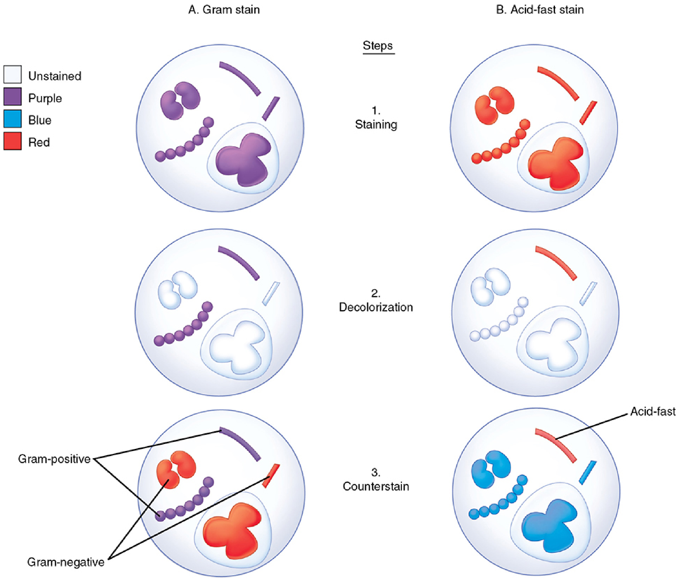
| Step | Reagent | What Happens |
|---|---|---|
| 1. Primary stain | Crystal violet (basic dye) | All bacteria stain blue-purple |
| 2. Mordant | Gram's iodine (iodine in KI) | Forms an insoluble crystal violet-iodine complex inside cells; acts as a fixative/mordant |
| 3. Decolorization | Acetone-alcohol or ethanol | Gram-positive cells retain the purple complex; Gram-negative cells lose it (outer membrane dissolves, thin peptidoglycan cannot hold the complex) |
| 4. Counterstain | Safranin (red dye) | Gram-negative cells (now colorless) take up red/pink color; Gram-positive cells remain purple |
"The iodine solution forms a mordanting action in which purple insoluble complexes are formed with ribonuclear protein in the cell. Gram-positive bacteria retain them; Gram-negative cells lose them through the outer membrane dissolved by solvent."
- Sherris & Ryan's Medical Microbiology, p. 129
Why the Differential Result? - The Cell Wall Basis
| Feature | Gram-Positive | Gram-Negative |
|---|---|---|
| Cell wall | Thick peptidoglycan layer (20-80 nm) | Thin peptidoglycan (2-7 nm) + outer membrane |
| Outer membrane | Absent | Present (lipopolysaccharide layer) |
| Decolorization | Crystal violet-iodine complex retained | Outer membrane dissolved by alcohol; complex washed out |
| Final color | Purple / Violet | Red / Pink |
"An intact cell wall is necessary for a positive reaction, and Gram-positive bacteria may fail to retain the stain if the organisms are old, dead, or damaged by antimicrobial agents."
- Sherris & Ryan's Medical Microbiology, p. 129
"An organism that is potentially Gram-positive may appear so only under a particular set of environmental conditions and in a young culture."
- Jawetz, Melnick & Adelberg, p. 44
Specimen Preparation for Gram Staining
- Place the most purulent or bloody portion of specimen on a clean microscopic slide - create both thick and thin areas
- For sterile body fluids (e.g., CSF): centrifuge to concentrate the specimen 10-100x before smearing
- Air dry, then fix with methanol or gentle heat
- Apply staining reagents: crystal violet → Gram iodine → decolorizer → safranin
Microscopic Examination
- Low-power objective first: look for large structures (nematode larvae, fungal forms, microcolonies)
- Oil immersion (1000x) for bacterial identification
- Important threshold: 10^5 organisms/mL must be present to see one organism per oil immersion field
- Evaluate: size, shape, Gram reaction, arrangement (pairs, chains, clusters)
"Reporting the presence of gram-positive cocci in pairs that resemble S. pneumoniae is more helpful than simply reporting gram-positive cocci in chains."
- Henry's, p. 1298
Clinical Significance of Gram Stain
| Gram Stain Finding | Likely Pathogen | Clinical Context |
|---|---|---|
| Gram-positive diplococci | Streptococcus pneumoniae | Lobar pneumonia in quality sputum |
| Small pleomorphic Gram-negative rods | Haemophilus influenzae | Respiratory infections |
| Gram-positive cocci in clusters | Staphylococcus aureus | Wound infections, sepsis |
| Gram-negative rods | Enterobacteriaceae | UTI, abdominal infection |
| Gram-positive rods | Bacillus, Listeria, Clostridia | Various infections |
"Done well the Gram-stained smear can achieve sensitivities of 70% to 80% and specificities of 90% to 95% for pneumococcal and H. influenzae pneumonia, respectively."
- Sherris & Ryan's Medical Microbiology, p. 130
Organisms That Do NOT Gram Stain Well (Gram Stain Limitations)
| Organism | Reason |
|---|---|
| Mycobacterium spp. | Waxy mycolic acid coat - requires Acid-Fast Stain (Ziehl-Neelsen) |
| Treponema pallidum | Too thin (~0.2 μm) - requires dark-field microscopy |
| Mycoplasma | No cell wall at all - cannot stain |
| Chlamydia, Rickettsia | Obligate intracellular - not detected in smears |
| Legionella | Stains poorly; requires silver stain or culture on special media |
| Fungi | May be seen but better visualized with KOH or calcofluor white |
Gram Stain Images
PART 2: CULTURE MEDIA
What is Culture Media?
- Biology of the organism (nutritional needs, oxygen requirements, temperature)
- Site of infection and specimen type
- Patient's immune response (which may suppress pathogen numbers)
- Quality of the culture media
Physical Forms of Culture Media
| Form | Description | Examples |
|---|---|---|
| Solid (agar) | Agar polysaccharide solidifies medium; isolated colonies visible | Blood agar, MacConkey agar |
| Liquid (broth) | No solidifying agent; allows growth of small numbers of organisms | Thioglycolate broth, enrichment broths |
| Semi-solid | Low agar concentration; used for motility testing | Motility test medium |
The 4 Main Types of Culture Media
"Culture media can be subdivided into four general categories: (1) enriched nonselective media, (2) selective media, (3) differential media, and (4) specialized media."
- Medical Microbiology 9e, p. 37
1. Enriched / Nonselective Media
| Medium | Composition | Use |
|---|---|---|
| Blood agar | Basal medium (tryptic soy, BHI, Brucella base) + 5% sheep/horse blood | General recovery of bacteria and fungi; demonstrates hemolysis (alpha, beta, gamma) |
| Chocolate agar | Blood agar heated to ~80°C - blood turns brown/chocolate colored | Fastidious organisms: Haemophilus spp., Neisseria gonorrhoeae, N. meningitidis (releases hemin and NAD from lysed RBCs) |
| Mueller-Hinton agar | Beef extract, casein hydrolysate, starch, divalent cations | Standard medium for antibiotic susceptibility testing (Kirby-Bauer disk diffusion) |
| Thioglycolate broth | Casein digest, glucose, yeast extract, cysteine, sodium thioglycolate | Enrichment broth for aerobic and anaerobic bacteria; sodium thioglycolate scavenges oxygen |
| Sabouraud dextrose agar | Casein/animal tissue digest + glucose (low pH) | Recovery and isolation of fungi; low pH and antibiotics suppress bacteria |
2. Selective Media
| Medium | Selective For | Inhibitory Agents |
|---|---|---|
| MacConkey agar | Gram-negative bacteria | Bile salts + crystal violet (inhibit Gram-positives) |
| Mannitol salt agar | Staphylococci | 7.5% NaCl (inhibits most bacteria) |
| Lowenstein-Jensen (LJ) medium | Mycobacterium spp. | Malachite green; egg-based |
| Middlebrook agar | Mycobacterium spp. | Various antibiotics |
| Campylobacter selective agar | Campylobacter spp. | Antibiotics (cefoperazone, vancomycin) + microaerophilic atmosphere at 42°C |
| TCBS agar | Vibrio spp. | Bile salts, thiosulfate, citrate |
| Thayer-Martin agar | Neisseria gonorrhoeae | VCN antibiotics (vancomycin, colistin, nystatin) |
3. Differential Media
| Medium | Differentiates | Principle |
|---|---|---|
| MacConkey agar | Lactose fermenters vs. non-fermenters | Contains lactose + neutral red pH indicator; fermenters produce acid → pink/red colonies; non-fermenters → colorless colonies |
| Mannitol salt agar | S. aureus vs. other staphylococci | Mannitol + phenol red; S. aureus ferments mannitol → yellow colonies |
| XLD agar (Xylose-lysine-deoxycholate) | Salmonella vs. Shigella in stool | Salmonella produces H₂S → black-centered colonies |
| EMB agar (Eosin methylene blue) | Gram-negatives; E. coli vs. others | E. coli produces characteristic metallic green sheen due to acid production |
| CHROMAgar | Multiple organisms | Chromogenic enzyme substrates produce colored colonies for different species |
4. Specialized / Enrichment Media
| Medium | Purpose |
|---|---|
| Selenite broth | Enrichment for Salmonella and Shigella from stool |
| GN broth (Gram-negative broth) | Enteric pathogens from feces |
| BCYE agar (Buffered charcoal yeast extract) | Legionella and Nocardia (requires iron + L-cysteine) |
| Cystine-tellurite agar | Corynebacterium diphtheriae |
| CCFA (Cycloserine-cefoxitin fructose agar) | Clostridioides difficile |
| Charcoal blood agar | Bordetella pertussis |
"Legionella was never grown in culture until it was recognized that recovery required using media supplemented with iron and l-cysteine. Campylobacter was not recovered in stool specimens until highly selective media were incubated at 42°C in a microaerophilic atmosphere."
- Medical Microbiology 9e, p. 37
Culture Media Summary Table
| Type | Media (Examples) | Purpose |
|---|---|---|
| Nonselective | Blood agar, Chocolate agar, Mueller-Hinton, Thioglycolate broth, Sabouraud dextrose agar | Recovery of bacteria and fungi |
| Selective, differential | MacConkey, Mannitol salt, XLD, Lowenstein-Jensen, Middlebrook, CHROMAgar | Isolate specific organisms; differentiate species |
| Specialized / Enrichment | BCYE, CCFA, Cystine-tellurite, Charcoal blood agar, Selenite broth | Recovery of fastidious or low-number organisms |
Key Concepts: Gram Stain + Culture Together
- A positive Gram stain with negative culture may indicate: organisms requiring specialized media, prior antibiotic administration, or unusual culture conditions needed
- A positive Gram stain finding guides initial empiric therapy AND the choice of which culture media to inoculate
- Example: Many Gram-negative coccobacilli in respiratory specimen → add chocolate agar to recover Haemophilus spp. that would be missed on blood agar alone
- The Gram stain also helps assess specimen quality: many squamous epithelial cells indicate oral contamination; many PMNs + bacteria = significant infection
"Demonstration of many bacteria on Gram stain that do not grow out in culture may indicate unusual organisms that require more specialized media or culture conditions."
- Henry's Clinical Diagnosis, p. 1298
Sources
- Jawetz, Melnick & Adelberg's Medical Microbiology, 28th Edition, pp. 44-45
- Sherris & Ryan's Medical Microbiology, 8th Edition, pp. 129-131
- Medical Microbiology, 9th Edition (Murray et al.), pp. 37-39
- Henry's Clinical Diagnosis and Management by Laboratory Methods, pp. 1297-1299