


IMG_2524.jpg
+2 more
What is this hyperpigmentation?? Reason ??
Reading File
analyse_media
analyse_media
Finding Sources
Finding Sources
Reading File
Finding Sources
Loading Image
Finding Sources
Finding Sources
Reading File
Finding Sources
I now have very comprehensive information. Let me compile the full answer.
Clinical Analysis: Facial Hyperpigmentation
What the Images Show
The three images show a South Asian/Indian female patient (confirmed by the clinical setting with gloved hands, hair cap, and gold jewelry) with:
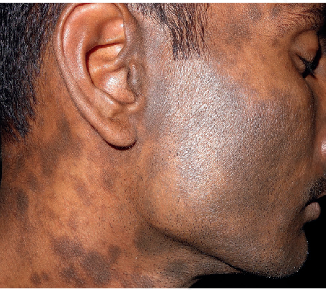
- Bilateral, diffuse, mottled slate-gray to dark brownish-gray macules and patches over the temples, lateral forehead, zygomatic/malar cheeks, and preauricular regions
- Ill-defined, irregular borders that fade gradually into normal skin
- Smooth skin surface - no scaling, induration, or scarring within the pigmented areas
- The grayish-blue tint strongly indicates dermal melanin deposition (melanin located in the dermis rather than the epidermis)
This matches almost exactly the textbook image of Lichen Planus Pigmentosus (LPP):
Most Likely Diagnosis: Lichen Planus Pigmentosus (LPP)
Why This Fits
| Feature | This Patient | LPP Typical Profile |
|---|---|---|
| Skin phototype | Dark (III-V) | III-V (Indian, Middle Eastern, Latin American) |
| Distribution | Temples, malar, preauricular | Forehead, temples, face, neck |
| Color | Slate-gray/blue-brown | Gray-brown to dark brown |
| Borders | Ill-defined | Irregularly shaped, ill-defined |
| Texture | Smooth, no scale | Usually asymptomatic, no papules |
| Surface | Intact | No induration or ulceration |
According to Andrews' Diseases of the Skin and Dermatology 2-Volume Set 5e:
"Lichen planus pigmentosus (LPP) is an uncommon variant of lichen planus that most often affects young to middle-aged adults with skin phototypes III-V, including those from India, Latin America, and the Middle East. It presents as irregularly shaped or oval, brown to gray-brown macules and patches that favor sun-exposed areas (especially the forehead, temples, and neck)."
Why Does It Happen? - Pathogenesis
LPP is a lichenoid interface dermatitis. The sequence of events:
- Immune-mediated basal layer damage - CD8+ T-lymphocytes attack basal keratinocytes via an interface/lichenoid reaction
- Keratinocyte apoptosis - basal cells undergo programmed cell death (civatte bodies)
- Melanin incontinence - melanin from damaged basal keratinocytes "falls" into the dermis
- Dermal melanophage accumulation - macrophages in the upper dermis engulf the melanin
- The deep location of melanin gives the characteristic slate-gray/blue-gray hue (the Tyndall effect - deeper pigment appears blue-gray rather than brown)
Triggering/Exacerbating Factors
- UV/sun exposure - the photo-distributed variant (LP actinicus) responds to sun protection with gradual fading. Very common in the Indian subcontinent
- Cosmetics/fragrances - some overlap with Riehl's melanosis (pigmented contact dermatitis)
- Thyroid disease (particularly hypothyroidism) - one study showed an association
- Hormonal factors - possible exacerbation reported
- Genetic predisposition - persons from India, Middle East, and Central America are susceptible regardless of sun exposure
Differential Diagnoses to Consider
| Condition | How to Differentiate |
|---|---|
| Melasma | More brown, less gray; symmetric; strongly hormonal/photo-driven; epidermal type responds to Wood's lamp; common in women |
| Riehl's melanosis (Pigmented contact dermatitis) | History of contact with cosmetics/fragrances; may have earlier inflammatory phase; histology shows lichenoid infiltrate |
| Erythema dyschromicum perstans (Ashy dermatosis) | Active lesions have erythematous border; similar slate-gray; seen in Latin Americans |
| Acquired bilateral nevus of Ota (Horii nevus) | Asian women 4th-5th decade; discrete macules; more zygomatic |
| Postinflammatory hyperpigmentation | History of preceding inflammatory lesion (acne, eczema, etc.) |
Management Outline
- Sun protection (broad-spectrum SPF 50+) - essential, especially if photoexacerbated variant
- Topical depigmenting agents: hydroquinone 2-4%, azelaic acid, kojic acid, tranexamic acid
- Topical calcineurin inhibitors (tacrolimus, pimecrolimus) - address the lichenoid inflammation
- Chemical peels (superficial glycolic/lactic acid)
- Laser therapy (Q-switched Nd:YAG, picosecond lasers) - used when dermal melanin is the primary component
- Address trigger: rule out contact allergens, evaluate thyroid function (TSH)
Important note: This is a clinical impression based on visual analysis. A skin biopsy remains the gold standard for confirming LPP - histology shows vacuolar degeneration of the basal layer, prominent dermal melanophages, and a lichenoid lymphocytic infiltrate.
Sources: Andrews' Diseases of the Skin (Clinical Dermatology) p. 2818-2832 | Dermatology 2-Volume Set 5e p. 1343 | Fitzpatrick's Dermatology Vol 1-2
This is a shared conversation. Sign in to Orris to start your own chat.