
Pneumonia case study
What kind of pneumonia case study would you like? · What's this for?
"community-acquired pneumonia" AND management
chest X-ray lobar consolidation community-acquired pneumonia
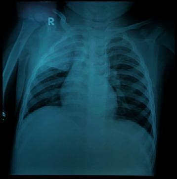
This diagnostic image is a posterior-anterior (PA) pediatric chest X-ray. The primary finding is a dense, homogeneous opacity located in the right upper lobe, characterized by a well-defined inferior border along the minor fissure, which is highly consistent with lobar consolidation. Bilateral perihilar and parenchymal spotting (reticulonodular opacities) are also visible, suggesting an associated bronchopneumonia component. The cardiac silhouette and mediastinal structures appear within normal limits for a pediatric patient, though the bony structures of the rib cage and vertebrae are visible with moderate clarity. This imaging demonstrates typical radiological signs of community-acquired or atypical pneumonia, such as right superior lobar pneumonia. Clinically, this serves as an educational example of differentiating focal lobar consolidation from generalized patchy bronchopneumonia patterns in a pediatric context.
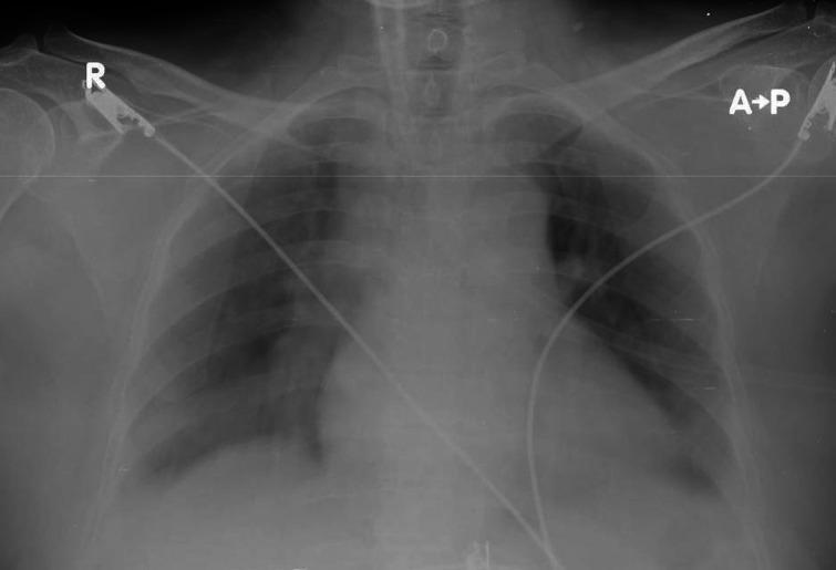
This diagnostic image is an anteroposterior (AP) chest X-ray of an adult patient, as indicated by the 'A-P' and 'R' (right side) labels. The radiograph demonstrates bilateral increased opacities with a lobar distribution, particularly prominent in the lower lung zones, consistent with consolidation seen in community-acquired pneumonia. The pulmonary vasculature appears congested, and there is a mild blurring of the cardiac borders. Several medical monitoring devices are visible: electrocardiogram (ECG) leads and wires are seen crossing the chest field, entering near the clavicular regions and extending toward the lower torso. The trachea is midline, and the osseous structures including the ribs and clavicles appear intact. This imaging is used to illustrate the radiographic presentation of lobar pneumonia and the presence of typical clinical monitoring equipment in a preoperative or intensive care setting.
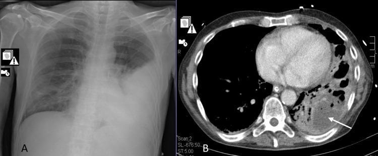
The composite image displays two diagnostic modalities for the chest. Image A is a frontal chest X-ray (radiography) demonstrating extensive opacification and consolidation in the left lower lung field, consistent with lobar pneumonia. The heart border is obscured, and the left costophrenic angle is obliterated. Image B is an axial computed tomography (CT) scan of the chest at the level of the heart, showing the evolution of the pathology. It reveals a large area of consolidation in the left posterior lung containing multiple low-density, hypodense regions. A white arrow points to a well-defined pulmonary abscess and surrounding cavitation within the necrotizing lung tissue. The right lung remains relatively clear on both modalities. These images illustrate the clinical progression from initial community-acquired pneumonia to necrotizing pneumonia and abscess formation, a significant complication often associated with specific bacterial pathogens or co-infections.
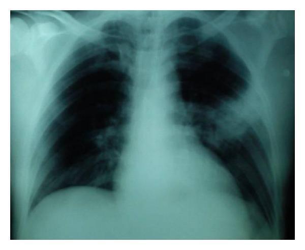
This diagnostic image is a posterior-anterior (PA) chest X-ray illustrating a significant pulmonary pathology. The primary finding is a dense, relatively homogeneous opacity located in the left lung field, consistent with lobar consolidation. This finding is characteristic of community-acquired pneumonia, specifically involving the left lung. In contrast, the right lung field appears clear with normal bronchovascular markings and no evidence of infiltrates or pleural effusion. The mediastinal shadow, including heart size and position, appears within normal limits. The costophrenic angles are sharp, and the diaphragmatic contours are intact. This radiograph serves as a classic educational example of pulmonary consolidation, highlighting the visual differences between air-filled lung parenchyma and fluid or cell-filled alveoli as seen in infectious pneumonia.
Case Study: Community-Acquired Pneumonia (CAP)
Patient Presentation
Diagnostic Workup
- CBC: WBC 18,200/µL with left shift
- BUN: 32 mg/dL (11.4 mmol/L)
- Chest X-ray: Right lower lobe consolidation, no effusion
- Blood cultures x2, sputum Gram stain/culture, urinary pneumococcal and Legionella antigen sent
- ABG: pH 7.34, PaO2 58 mmHg on room air, lactate 2.8 mmol/L
Severity Assessment: CURB-65
| Criterion | Present in this patient? |
|---|---|
| Confusion | Yes (1) |
| Urea > 19 mg/dL (7 mmol/L) | Yes, 32 mg/dL (1) |
| Respiratory rate ≥ 30/min | Borderline at 28 - scored 0 here, but trending up |
| BP (systolic <90 or diastolic ≤60) | Yes, 98/62 (1) |
| Age ≥ 65 | Yes, age 68 (1) |
Diagnosis
Management
- Site of care: ICU admission given severe CAP criteria and hemodynamic instability.
- Empiric antibiotics - started within the first hour, before culture results, per Goldman-Cecil Medicine (p. 3693-3697):
- For severe inpatient CAP: beta-lactam (ceftriaxone or ampicillin-sulbactam) PLUS a macrolide (azithromycin), OR beta-lactam plus a respiratory fluoroquinolone (levofloxacin/moxifloxacin) - Fishman's Pulmonary Diseases, Table 128-6.
- Add MRSA coverage (vancomycin or linezolid) and Pseudomonas coverage (piperacillin-tazobactam, cefepime, or meropenem) if risk factors are present (prior MRSA/Pseudomonas isolation, recent hospitalization, structural lung disease such as this patient's COPD).
- Cover Legionella empirically with the macrolide/fluoroquinolone, especially since he has multiple comorbidities and moderate-severe disease - Goldman-Cecil, "Alternative Regimens," p. 3942.
- Supportive care: Supplemental oxygen to target SpO2 ≥90-92%, IV fluids for hypotension, VTE prophylaxis, glycemic control, early mobilization once stable.
- De-escalate antibiotics once culture/antigen results return and the patient is clinically improving (typically 48-72h), narrowing to the identified pathogen.
- Duration: Minimum 5 days, extending if slow to respond, bacteremic, or if Pseudomonas/MRSA/less common organisms are confirmed.
Expected Course & Teaching Points
- Why this case is "severe": Hypotension, altered mentation, and hypoxemia in an elderly patient with comorbidities are red flags separating this from routine outpatient CAP.
- Common pathogens: Streptococcus pneumoniae remains the leading cause of CAP requiring hospitalization; atypicals (Mycoplasma, Chlamydophila, Legionella) and viruses (influenza) are also frequent, which is why empiric coverage is broad until a pathogen is confirmed.
- Pitfall to avoid: Relying on CURB-65 alone can underestimate severity in obese patients or those with certain comorbidities - clinical gestalt and ATS/IDSA major/minor criteria should always be layered on top of the score.
- Complication to watch for: Failure to improve by 72 hours should prompt evaluation for empyema, lung abscess, or resistant/unusual organisms.