
Malignancy in gyncelogy
Reading File
The topic is broad. Let me clarify before diving into a full research effort.
Asking for Preferences
Which area of gynecologic malignancy would you like to focus on? · What level of detail do you need?
Finding Sources
Finding Sources
Finding Sources
Berek & Novak's Gynecology is the primary gynaecology reference. Let me read the key sections for all cancers in parallel.
Finding Sources
Reading File
Reading File
Good. Now let me read detailed content from the core Berek & Novak's sections on each cancer type.
Reading File
Reading File
Let me get the key clinical content from the main Berek & Novak gynecologic cancer chapters.
Reading File
Finding Sources
Finding Sources
Finding Sources
Reading File
Finding Sources
Reading File
Finding Sources
Reading File
Reading File
I now have excellent clinical content. Let me read the vulvar cancer section and get a bit more on cervical cancer staging/treatment, then synthesize.
Reading File
Reading File
Reading File
Searching Images
gynecologic cancer cervical ovarian endometrial staging diagram
Here is a comprehensive detailed clinical overview of malignancy in gynaecology.
Malignancy in Gynaecology — Comprehensive Clinical Overview
1. CERVICAL CANCER
Epidemiology & Risk Factors
Cervical cancer is the 3rd most common gynecologic malignancy in the United States (behind uterine corpus and ovary) and the 2nd most common cause of cancer death in women in developing countries. Mean age at diagnosis: 47 years (bimodal peaks: 35–39 and 60–64 years).
Key risk factors:
- HPV infection (the causal agent) — HPV 16 & 18 account for ~70% of cases; confers a 50-fold increased risk for precancerous lesions
- Early sexual intercourse (< 16 years)
- Multiple sexual partners
- Cigarette smoking
- High parity
- Chronic immune suppression (HIV)
- Low socioeconomic status / limited access to screening
- Long-term OCP use (weak cofactor; ~1.9× RR with ≥5 years)
- Chlamydia trachomatis and herpes simplex as cofactors
Pathology
| Type | Features |
|---|---|
| Squamous cell carcinoma (~70%) | Large cell keratinizing (keratin pearls), large cell non-keratinizing, or small cell (worst prognosis) |
| Adenocarcinoma (~25%, rising incidence) | Glandular origin; harder to detect on Pap smear |
| Small-cell anaplastic / neuroendocrine | Rare; most aggressive; resembles oat-cell lung cancer |
| Adenosquamous, clear cell, glassy cell | Uncommon variants |
Staging (FIGO 2018 — updated, allows imaging + pathology)
| Stage | Description |
|---|---|
| I | Confined strictly to cervix |
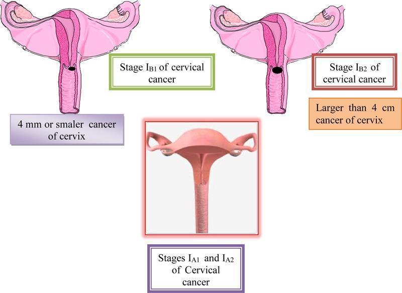
| IA1 | Stromal invasion ≤3 mm depth |
| IA2 | Invasion >3–5 mm |
| IB1 | Clinically visible ≤2 cm |
| IB2 | >2–4 cm |
| IB3 | >4 cm |
| II | Beyond cervix, not to pelvic wall/lower 1/3 vagina |
| IIA | No parametrial invasion |
| IIB | Parametrial invasion |
| IIIA | Lower 1/3 vagina involvement |
| IIIB | Pelvic wall and/or hydronephrosis |
| IIIC1 | Pelvic lymph node metastasis |
| IIIC2 | Para-aortic lymph node metastasis |
| IVA | Bladder/rectum mucosa invasion |
| IVB | Distant metastasis |
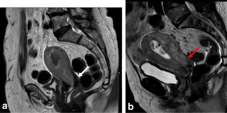
Imaging note: MRI is superior for parametrial disease detection. PET-CT has best sensitivity/specificity for lymph node metastases.
Treatment
| Stage | Treatment |
|---|---|
| Stage IA1 | Cone biopsy (fertility-preserving) or simple hysterectomy |
| Stage IA2–IB1 | Radical hysterectomy + pelvic lymph node dissection OR radiation |
| Stage IB2–IIA | Radical hysterectomy + PLND OR chemoradiation |
| Stage IIB–IVA | Concurrent chemoradiation (cisplatin-based) + brachytherapy — standard of care |
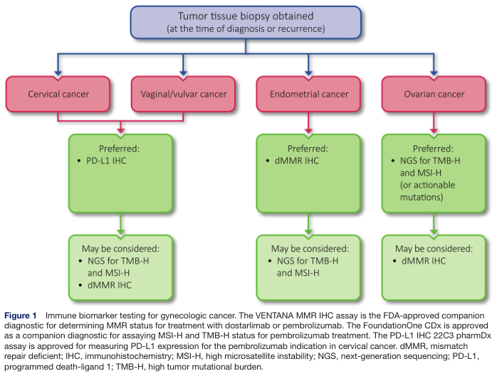
| Stage IVB / recurrent | Palliative systemic therapy; pembrolizumab (PD-L1+) approved |
Robotic/laparoscopic radical hysterectomy is now used at experienced centers; note that recent data (LACC trial) showed worse oncologic outcomes with MIS for IB1 cervical cancer compared to open surgery — open radical hysterectomy remains preferred.
2. ENDOMETRIAL (UTERINE CORPUS) CANCER
Epidemiology
The most common gynecologic malignancy in the United States. Average age: 60 years; 75% occur in women >50 years. Overall 5-year survival: ~75%.
Risk Factors
Core concept: prolonged unopposed estrogen stimulation
| Risk Factor | Notes |
|---|---|
| Obesity | Fat → aromatization of androgens → estrone; major risk factor |
| Nulliparity | Fewer progesterone-dominant cycles |
| Late menopause | Prolonged estrogen exposure |
| Anovulatory cycles (PCOS) | Unopposed estrogen |
| Exogenous unopposed estrogen | 4–8× increased risk |
| Tamoxifen use | Acts as uterine estrogen agonist |
| Lynch syndrome (HNPCC) | MLH1, MSH2, MSH6 mutations → 40–60% lifetime risk |
| Diabetes, hypertension | Associated (likely via obesity pathway) |
Protective: Combined OCP use, progestins, IUD use, smoking (paradoxically reduces estrogen)
Precursor: Endometrial Hyperplasia
| Type | Progression to Cancer |
|---|---|
| Simple without atypia | 1% |
| Complex without atypia | 3% |
| Simple with atypia | 8% |
| Complex with atypia | 29% |
Pathology (Type I vs Type II)
| Feature | Type I | Type II |
|---|---|---|
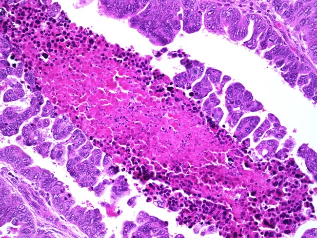
| Histology | Endometrioid | Serous, clear cell |
| Estrogen-related | Yes | No |
| Background | Hyperplasia | Atrophic endometrium |
| Grade | Low (G1–G2) | High (G3) |
| Prognosis | Better | Worse |
| Molecular | PTEN, KRAS, MLH1 | p53 mutation, ERBB2 |
| Proportion | ~80% of cases | <10% of cases, but >50% of deaths |
Clinical Features
- Postmenopausal vaginal bleeding — the cardinal symptom (>90% of cases)
- Pelvic pressure/discomfort with advanced disease
- < 5% are asymptomatic at diagnosis
- Any postmenopausal bleeding must be investigated regardless of quantity
Diagnosis
- Transvaginal ultrasound (TVUS) — endometrial thickness >4 mm in postmenopausal women warrants biopsy
- Office endometrial aspiration biopsy — first-line investigation
- Hysteroscopy + D&C — if biopsy inadequate or high clinical suspicion
Staging (FIGO — surgical)
| Stage | Description |
|---|---|
| I | Confined to uterus |
| IA | No or < ½ myometrial invasion |
| IB | ≥ ½ myometrial invasion |
| II | Cervical stromal invasion |
| IIIA | Uterine serosa / adnexa |
| IIIB | Vaginal/parametrial involvement |
| IIIC1 | Pelvic LN metastasis |
| IIIC2 | Para-aortic LN metastasis |
| IVA | Bladder / bowel mucosa invasion |
| IVB | Distant metastases |
Treatment
- Surgical staging — Total hysterectomy + bilateral salpingo-oophorectomy (BSO) + peritoneal cytology ± lymph node assessment (standard for most patients)
- Adjuvant radiotherapy — vaginal vault brachytherapy (reduces vaginal recurrence); external beam pelvic RT for high-risk features
- Chemotherapy — carboplatin + paclitaxel for advanced/recurrent disease; improves DFS
- Hormonal therapy — progestins for low-grade, hormone receptor–positive recurrent disease
- Immunotherapy — pembrolizumab + lenvatinib for advanced mismatch repair–proficient (pMMR) disease; single-agent pembrolizumab for dMMR/MSI-H
Key Prognostic Variables (adverse)
Advancing age | Non-endometrioid or grade 3 histology | Deep myometrial invasion | Lymphovascular space invasion | Large tumor size | Cervical extension | LN metastasis | Intraperitoneal spread
Uterine Sarcomas
Rare but highly malignant uterine tumors:
- Leiomyosarcoma — most common uterine sarcoma; arises from smooth muscle; worst prognosis
- Endometrial stromal sarcoma (ESS) — low-grade and high-grade subtypes
- Carcinosarcoma (MMMT) — mixed Müllerian tumor; behaves as high-grade carcinoma; treated with chemotherapy
- Adenosarcoma — low malignant potential
3. OVARIAN CANCER
Epidemiology
The 2nd most common gynecologic malignancy but the leading cause of gynecologic cancer death (because ~75% present at Stage III–IV). ~20,000 new cases/year in the US.
Risk Factors & Genetics
| Factor | Risk |
|---|---|
| BRCA1 mutation | 40–60% lifetime risk |
| BRCA2 mutation | 15–25% lifetime risk |
| Lynch syndrome | Increased risk (colorectal, endometrial, ovarian) |
| Nulliparity, early menarche, late menopause | Incessant ovulation theory |
| HRT (estrogen-only) | Modest increase |
| Protective: OCP use, breastfeeding, tubal ligation | Reduce ovulation and retrograde menstruation |
Pathology (Ovarian Epithelial Tumors — 90% of malignant tumors)
| Histotype | Features |
|---|---|
| High-grade serous carcinoma (HGSC) | Most common (~70%); TP53 mutation; often fallopian tube origin; BRCA-associated |
| Low-grade serous carcinoma | KRAS/BRAF mutations; indolent but chemoresistant |
| Endometrioid | ARID1A, PTEN mutations; endometriosis-related |
| Clear cell | Chemoresistant; frequent in Asia; endometriosis-related |
| Mucinous | Often borderline; intestinal metastasis in DDx |
| Brenner tumor | Transitional cell–like; usually benign |
Non-epithelial tumors:
- Germ cell tumors — dysgerminoma, yolk sac tumor, immature teratoma (young women; often curable)
- Sex cord-stromal tumors — granulosa cell tumor (estrogen-secreting), Sertoli-Leydig
Clinical Features
- Typically vague, nonspecific symptoms: abdominal bloating, early satiety, urinary urgency, pelvic/abdominal pain
- Advanced disease: ascites, palpable omental mass, pleural effusion
- CA-125 elevated in ~80% of epithelial ovarian cancer (not specific; also elevated in endometriosis, fibroids, PID)
Diagnosis
- TVUS + CA-125 (primary evaluation)
- CT chest/abdomen/pelvis for staging
- Tissue diagnosis at surgery (not routine pre-op biopsy)
FIGO Staging (2013)
| Stage | Description |
|---|---|
| IA | One ovary; capsule intact; no ascites |
| IB | Both ovaries; capsule intact |
| IC | One/both ovaries with surgical spill, ruptured capsule, or malignant ascites |
| IIA | Extension to uterus/tubes |
| IIB | Extension to other pelvic tissues |
| IIIA1 | Positive retroperitoneal LN only |
| IIIA2 | Microscopic peritoneal metastasis above pelvic brim |
| IIIB | Peritoneal metastasis ≤2 cm above brim |
| IIIC | Peritoneal metastasis >2 cm above brim |
| IVA | Pleural effusion with positive cytology |
| IVB | Parenchymal organ metastases |
Treatment
Primary treatment = cytoreductive surgery + platinum-based chemotherapy
Cytoreductive ("debulking") surgery goals:
- Complete cytoreduction (no macroscopic residual disease) = best survival (~30–40% disease-free at 5 years)
- Optimal cytoreduction: residual <1 cm
- Procedure: TAH + BSO + omentectomy + pelvic/para-aortic LN dissection + peritoneal biopsies
Chemotherapy:
- Carboplatin + paclitaxel (IV or intraperitoneal) — first-line for stage III–IV
- Bevacizumab (anti-VEGF) — added for high-risk stage III–IV; maintenance therapy
- PARP inhibitors (olaparib, niraparib, rucaparib) — maintenance therapy especially for BRCA-mutated disease; significantly improves PFS
Neoadjuvant chemotherapy (NACT):
- 3 cycles → interval debulking surgery → 3 more cycles
- Offered when upfront complete cytoreduction is unlikely or patient is high perioperative risk
- Non-inferior to primary surgery in RCTs
4. VULVAR CANCER
Epidemiology
Accounts for ~4% of all gynecologic malignancies. Predominantly affects older women (postmenopausal). Two distinct pathways:
| Pathway | Associated Condition | Age |
|---|---|---|
| HPV-related | Vulvar intraepithelial neoplasia (VIN), HPV 16/18 | Younger |
| Non-HPV | Lichen sclerosus → differentiated VIN | Older |
Pathology
- Squamous cell carcinoma (~90%) — most common
- Melanoma (~5%) — worst prognosis
- Bartholin gland carcinoma (adenocarcinoma / adenoid cystic)
- Basal cell carcinoma, verrucous carcinoma (low malignant potential)
- Rare: vulvar sarcoma, lymphoma, Merkel cell carcinoma
Routes of Spread
- Direct extension to vagina, urethra, anus
- Lymphatic → inguinofemoral LN → pelvic LN (key prognostic factor)
- Hematogenous — late event
Staging (FIGO — surgical)
| Stage | Description |
|---|---|
| I | Confined to vulva or perineum, node-negative |
| IA | ≤2 cm, stromal invasion ≤1 mm |
| IB | >2 cm or stromal invasion >1 mm |
| II | Adjacent perineal structures (lower 1/3 urethra, vagina, anus), node-negative |
| IIIA | 1 positive LN (≥5 mm) or 1–2 LN (<5 mm) |
| IIIB | ≥2 positive LN (≥5 mm) or ≥3 LN (<5 mm) |
| IIIC | Positive nodes with extracapsular spread |
| IVA | Upper urethra, bladder, rectum, pelvic bone, or fixed/ulcerated nodes |
| IVB | Any distant metastasis |
Treatment Principles
- Multidisciplinary and individualized approach
- Radical local excision — adequate for T1 and early T2 lesions (replaces radical vulvectomy)
- Inguinofemoral lymphadenectomy — always thorough when groin dissection is indicated
- Sentinel lymph node dissection — for unifocal tumors <4 cm with clinically negative nodes (experienced centers)
- Chemoradiation → then limited surgical resection — best for large T2 and T3 primary tumors
- Recurrent disease — pelvic exenteration for central recurrence; chemoradiation for previously unirradiated patients
Key rule: Vulvar lesions always require biopsy to avoid delay in diagnosis.
5. VAGINAL CANCER
Key Points
- Rare (<2% of gynecologic malignancies)
- 80–90% are squamous cell carcinoma
- Shares epidemiology with cervical cancer (HPV-related)
- Clear cell adenocarcinoma — associated with in utero DES exposure
- FIGO staging is clinical
- Radiation therapy is the mainstay of treatment for most patients
- Select patients with stage I disease can be managed with radical surgery
6. GESTATIONAL TROPHOBLASTIC NEOPLASIA (GTN)
A spectrum of tumors arising from trophoblastic tissue:
- Hydatidiform mole (complete and partial) — benign but with malignant potential
- Invasive mole
- Choriocarcinoma — highly malignant; hematogenous spread; excellent response to chemotherapy (MTX or actinomycin D)
- Placental site trophoblastic tumor (PSTT)
Beta-hCG is a sensitive tumor marker for monitoring. GTN is one of the most chemosensitive solid tumors, with near-100% cure rate for low-risk disease.
Summary Table
| Cancer | Most Common Type | Hallmark Presentation | Key Tumor Marker | Primary Treatment |
|---|---|---|---|---|
| Cervical | Squamous (~70%) | Abnormal vaginal bleeding/discharge | None (Pap + HPV test) | Surgery (early) / Chemoradiation (advanced) |
| Endometrial | Endometrioid | Postmenopausal bleeding | CA-125 (limited) | Hysterectomy + BSO ± adjuvant RT/chemo |
| Ovarian | HGSC | Vague abdominal symptoms, ascites | CA-125 | Cytoreduction + carboplatin/paclitaxel |
| Vulvar | SCC | Pruritus, visible vulvar lesion | None | Radical local excision ± groin dissection |
| Vaginal | SCC | Vaginal bleeding/discharge | None | Radiation |
| GTN | Choriocarcinoma | Post-molar/post-pregnancy | β-hCG | Chemotherapy (MTX/Act-D) |
*Sources: Berek & Novak's Gynecology (9781496380357); Goldman-Cecil Medicine (9780323930345); Schwartz's Principles of Surgery (9781259835346)*Your credits are exhausted. Your daily credits reset tomorrow, or you can purchase a top-up for more.
This is a shared conversation. Sign in to Orris to start your own chat.