
Microscopy of endometrial. Carcinoma
Reading File
This is a visual/pathology question - I need to search for endometrial carcinoma microscopy sections and images. Let me run parallel searches.
Finding Sources
Finding Sources
Excellent - I found rich content with microscopy images. Let me read the key sections from Robbins pathology books and fetch the images in parallel.
Reading File
Reading File
I have the text content. Now let me fetch the microscopy images and read the serous carcinoma morphology section from Robbins' Pathologic Basis of Disease.
Loading Image
Loading Image
Reading File
I now have excellent content and microscopy images. Let me fetch the remaining key images and the serous carcinoma morphology section.
Loading Image
Reading File
Now let me get the serous carcinoma microscopy images from Robbins' Pathologic Basis of Disease.
Finding Sources
Reading File
I now have all the content and images needed for a thorough answer. Let me also fetch the serous carcinoma image from the large Robbins textbook.
Finding Sources
I have comprehensive content. Now I'll compose the full answer.
Microscopy of Endometrial Carcinoma
Endometrial carcinoma has two main histologic types with distinct microscopic appearances: endometrioid carcinoma (80-85% of cases) and serous carcinoma (~15%). Their microscopy differs dramatically.
Endometrial Hyperplasia (Precursor Lesion)
Before invasion, the precursor stages are recognizable on H&E:
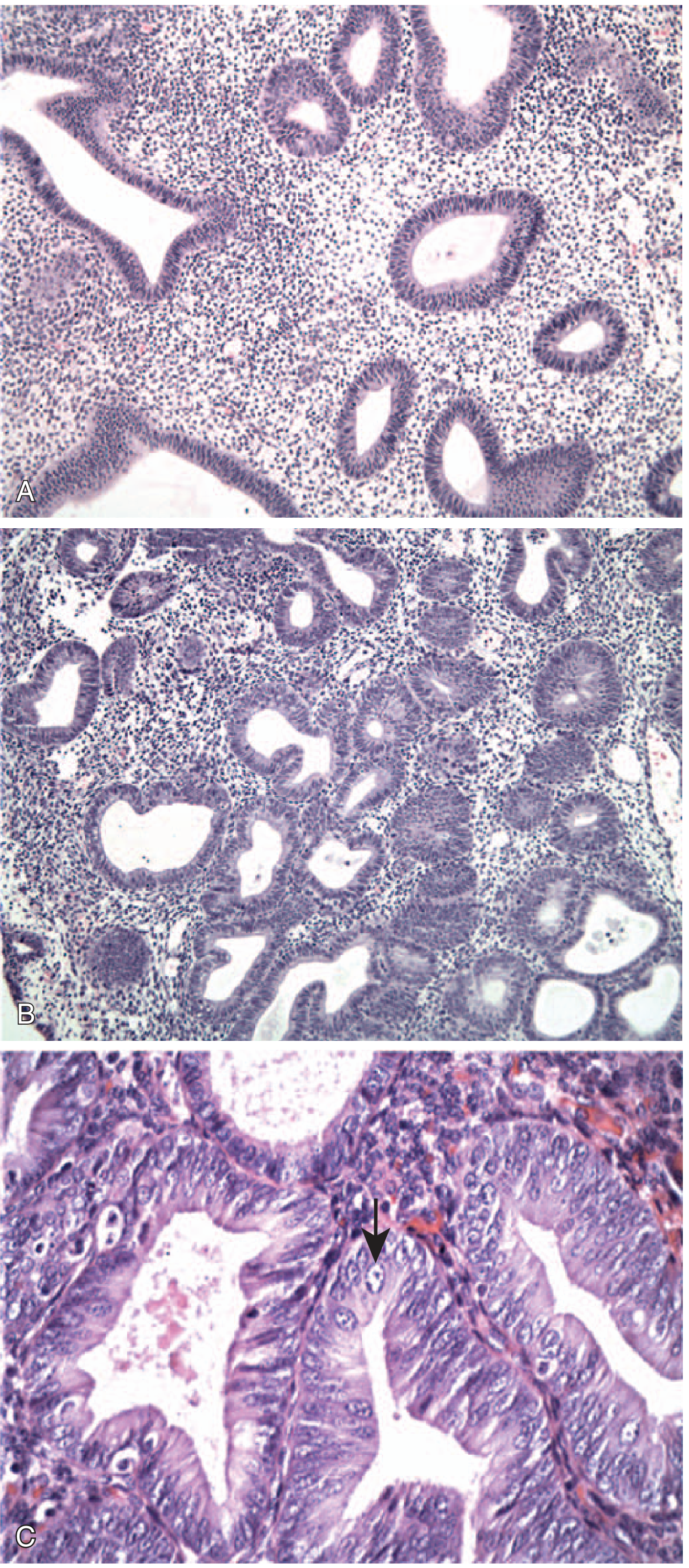
FIG. 17.11 - Endometrial hyperplasia. (A) Without atypia: mild glandular crowding and cystic dilation. (B) With atypia: glandular crowding + cellular atypia. (C) High magnification of atypical hyperplasia: rounded vesicular nuclei with prominent nucleoli (arrow). - Robbins & Kumar Basic Pathology
Endometrioid Carcinoma - Microscopic Features
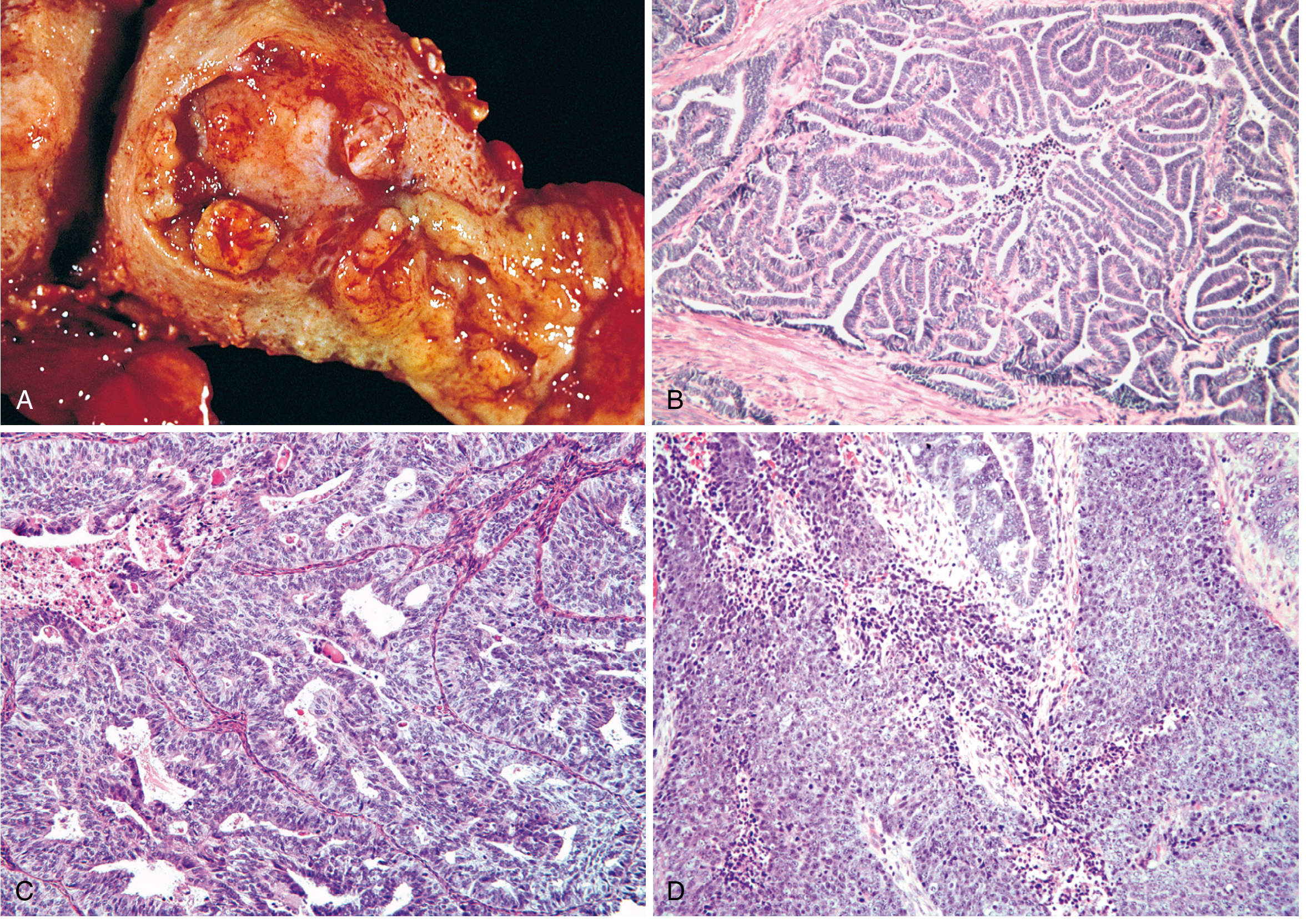
FIG. 22.24 - (A) Gross fungating mass. (B) Grade 1 well-differentiated endometrioid adenocarcinoma with preserved glandular architecture, back-to-back glands, no intervening stroma. (C) Grade 2 moderately differentiated with glandular architecture plus solid areas. (D) Grade 3 poorly differentiated with predominantly solid growth. - Robbins, Cotran & Kumar Pathologic Basis of Disease
Key microscopic features of endometrioid carcinoma:
Glandular architecture: The tumor closely mimics proliferative endometrium. Glands are crowded back-to-back with loss of intervening stroma ("gland-to-gland" apposition).
Grading (FIGO 1-3):
- Grade 1 (well differentiated): Almost entirely well-formed glands; minimal solid component (<5%). Cells show mild atypia, elongated nuclei.
- Grade 2 (moderately differentiated): Well-formed glands mixed with solid sheets that make up 6-50% of the tumor. Moderate cytologic atypia.
- Grade 3 (poorly differentiated): Predominantly solid growth pattern (>50% solid). Marked nuclear atypia, prominent nucleoli, frequent mitoses.
Distinguished from hyperplasia by:
- Desmoplastic stromal reaction around invading glands
- Confluent glandular growth without intervening stroma
- Papillary growth patterns
- Myometrial invasion
Squamous differentiation: Up to 20% of endometrioid carcinomas contain foci of squamous metaplasia. In low-grade tumors, the squamous elements appear benign (morules); in high-grade tumors they may be frankly malignant.
Tumor-infiltrating lymphocytes (TILs): Tumors with DNA mismatch repair defects (MLH1 loss by promoter methylation) or polymerase-epsilon (POLE) mutations often show abundant infiltrating T cells - an indicator of microsatellite instability.
Serous Endometrial Carcinoma - Microscopic Features
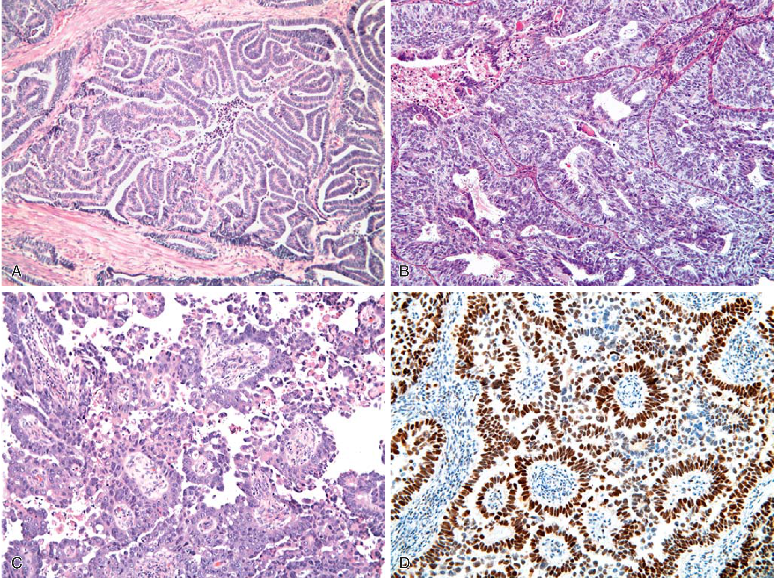
FIG. 17.12 - (A) Endometrioid type grade 1 with glandular infiltration into myometrium. (B) Grade 3 with solid growth. (C) Serous carcinoma with papilla formation and marked cytologic atypia. (D) Diffuse strong p53 IHC staining (brown nuclei) = TP53 mutation. - Robbins & Kumar Basic Pathology
Key microscopic features of serous carcinoma:
| Feature | Detail |
|---|---|
| Architecture | Papillary tufts and complex branching papillae; can also form glands |
| Cytologic atypia | Marked - high nuclear-to-cytoplasmic ratio, hyperchromasia, prominent nucleoli |
| Mitoses | Numerous, often atypical |
| Grade | Always high-grade by definition, regardless of architectural pattern |
| Precursor (SEIC) | Serous endometrial intraepithelial carcinoma - malignant cells confined to epithelial surface only, identical morphology |
| p53 IHC | Diffuse, strong nuclear positivity (mutant p53 accumulates) - key distinguishing feature |
Serous vs. endometrioid distinction: Both can form glands, but serous carcinoma shows far greater nuclear atypia. When architecture alone is ambiguous, p53 IHC helps - diffuse block positivity (or complete absence/"null pattern") indicates TP53 mutation.
Comparison Summary
| Feature | Endometrioid | Serous |
|---|---|---|
| Frequency | 80-85% | ~15% |
| Architecture | Glandular (back-to-back glands) | Papillary tufts, complex papillae |
| Cytologic atypia | Mild to moderate (G1-G2); severe (G3) | Always severe/high-grade |
| p53 IHC | Focal/weak (wild-type pattern) | Diffuse strong or null (mutant pattern) |
| Precursor | Atypical hyperplasia | Serous endometrial intraepithelial carcinoma (SEIC) |
| Molecular hallmark | PTEN, PIK3CA, KRAS mutations; MMR defects | TP53 mutations (>90%) |
| Associated setting | Estrogen excess (obesity, perimenopausal) | Endometrial atrophy (older postmenopausal) |
| Prognosis | Good (stage I: ~90% 5-year survival) | Poor (5-year survival 18-27%) |
Sources: Robbins & Kumar Basic Pathology (9780323790185); Robbins, Cotran & Kumar Pathologic Basis of Disease (9780443264528)
This is a shared conversation. Sign in to Orris to start your own chat.