
Molecular basis diagnostic marker and therapeutic role in glial tumours and gliomas md pathology exam 10 marks
Reading File
Finding Sources
Finding Sources
Finding Sources
Finding Sources
Searching PubMed
glioma molecular classification WHO
Finding Sources
Finding Sources
Finding Sources
Finding Sources
Finding Sources
Molecular Basis, Diagnostic Markers, and Therapeutic Role in Glial Tumours / Gliomas
(Pathology - 10 Marks)
1. Introduction
Gliomas are the most common primary CNS neoplasms, arising from glial cells (astrocytes, oligodendrocytes, ependymal cells). Since the WHO CNS Tumour Classification (5th edition, 2021), gliomas are no longer classified purely on histomorphology - molecular/genetic alterations now form the backbone of diagnosis, grading, prognostication, and treatment selection ("integrated diagnosis").
2. WHO Molecular Classification of Adult-Type Diffuse Gliomas
| Tumour | Key Molecular Signature |
|---|---|
| Astrocytoma, IDH-mutant | IDH1/IDH2 mutation, ATRX loss, TP53 mutation, no 1p/19q loss |
| Oligodendroglioma, IDH-mutant and 1p/19q-codeleted | IDH1/IDH2 mutation + 1p/19q co-deletion, TERT promoter mutation |
| Glioblastoma, IDH-wildtype (Grade 4) | IDH-wildtype + TERT promoter mutation, EGFR amplification, or +7/-10 copy number changes |
Pediatric-type diffuse gliomas are grouped separately (H3 K27-altered midline glioma, H3 G34-mutant hemispheric glioma, MAPK pathway-altered low-grade glioma) - Harrison's Principles of Internal Medicine 22E; GOLDMAN-CECIL Medicine.
3. Key Molecular Markers - Diagnostic Significance
a) IDH1/IDH2 mutations (Isocitrate Dehydrogenase)
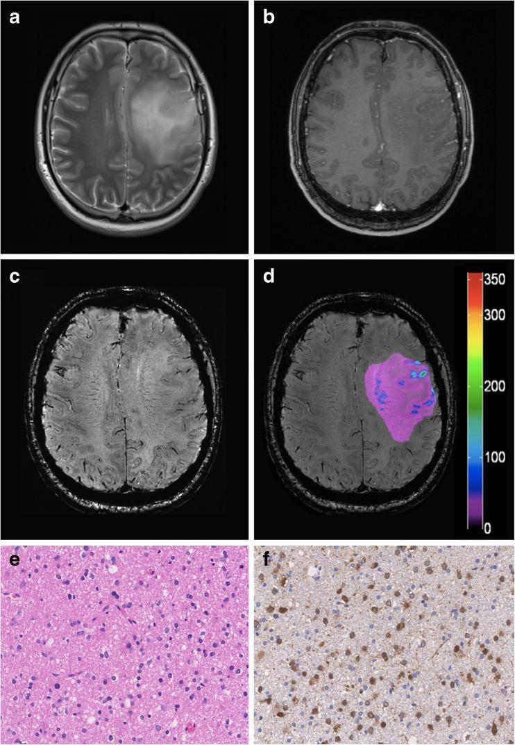
- Most common: IDH1 R132H (~90%); detected by immunohistochemistry (mutant-specific antibody) or sequencing if IHC-negative
- Early, defining event in diffuse gliomas - distinguishes IDH-mutant (younger patients, better prognosis) from IDH-wildtype tumours
- ~95% of glioblastomas are IDH-wildtype - Bradley and Daroff's Neurology in Clinical Practice; Grainger & Allison's Diagnostic Radiology
b) 1p/19q co-deletion
- Defining marker of oligodendroglioma (always occurs with IDH mutation)
- Best prognosis among lower-grade gliomas (mean survival ~8 years)
- Predicts increased chemosensitivity and radiosensitivity - Henry's Clinical Diagnosis and Management by Laboratory Methods
c) ATRX loss / TP53 mutation
- Loss of ATRX expression (by IHC) + TP53 mutation characterizes IDH-mutant astrocytomas (mutually exclusive with 1p/19q co-deletion)
d) TERT promoter mutation
- Found in IDH-wildtype glioblastoma and in IDH-mutant, 1p/19q-codeleted oligodendroglioma
- One of three genetic criteria (with EGFR amplification or +7/-10) sufficient to designate a histologically lower-grade astrocytoma as glioblastoma, Grade 4 - Robbins & Kumar Basic Pathology
e) EGFR amplification
- Present in ~40% of IDH-wildtype glioblastomas (chromosome 7)
- Along with PTEN mutation and CDKN2A deletion, drives growth factor signaling in primary (de novo) glioblastoma
f) MGMT promoter methylation
- O6-methylguanine-DNA methyltransferase is a DNA repair enzyme that removes alkylating adducts
- Promoter hypermethylation -> reduced MGMT expression -> tumor cells cannot repair temozolomide-induced DNA damage -> better response to alkylating chemotherapy
- Strongest predictive and prognostic biomarker in glioblastoma management - Harrison's Principles of Internal Medicine; Washington Manual of Medical Therapeutics
g) H3 K27M mutation (histone H3.3/H3.1)
- Defines diffuse midline glioma (e.g., DIPG - diffuse intrinsic pontine glioma), automatically assigned WHO Grade 4 regardless of histology
- Dismal prognosis; detected by mutant-specific IHC
h) BRAF alterations
- KIAA1549-BRAF fusion: characteristic of pilocytic astrocytoma (WHO Grade 1)
- BRAF V600E point mutation: seen in pleomorphic xanthoastrocytoma and a subset of low-grade gliomas - actionable with BRAF inhibitors
4. Therapeutic Role of Molecular Markers
| Marker | Therapeutic Implication |
|---|---|
| MGMT methylation | Predicts response to temozolomide (alkylating agent); unmethylated MGMT = temozolomide resistance, worse prognosis |
| IDH mutation | Better response to radiotherapy/chemotherapy; IDH inhibitors (e.g., vorasidenib) under trial for lower-grade IDH-mutant gliomas |
| 1p/19q co-deletion | Predicts favorable response to PCV chemotherapy (procarbazine, CCNU, vincristine) and radiotherapy in oligodendroglioma |
| EGFR amplification/BRAF V600E | Potential target for EGFR inhibitors / BRAF-MEK inhibitors (e.g., dabrafenib-trametinib in BRAF-mutant gliomas) |
| VEGF pathway (angiogenesis) | Bevacizumab (anti-VEGF monoclonal antibody) used in recurrent glioblastoma - improves progression-free survival and reduces edema/steroid requirement, though no overall survival benefit |
| H3 K27M | Guides prognosis; investigational targeted/epigenetic therapies (ONC201/dordaviprone) being studied |
Standard therapeutic paradigm for glioblastoma: maximal safe surgical resection -> concurrent radiotherapy + temozolomide -> adjuvant temozolomide for 6 months, with MGMT status guiding expected benefit (Washington Manual of Medical Therapeutics; Harrison's 22E).
5. Adverse Prognostic Factors in Glioblastoma
Older age, unmethylated MGMT promoter, poor Karnofsky performance status, unresectable tumor, IDH-wildtype status - Harrison's Principles of Internal Medicine 22E.
6. Summary Diagram (Exam Point Form)
- Classification: IDH status -> 1p/19q status -> grade
- Diagnostic IHC panel: IDH1-R132H, ATRX, p53, Ki-67, GFAP, H3K27M
- Molecular tests: FISH/PCR for 1p/19q, MGMT methylation assay (PCR/pyrosequencing), NGS panel for IDH1/2, TERT, EGFR, BRAF
- Prognostic use: IDH-mutant > IDH-wildtype; 1p/19q co-deleted = best prognosis
- Predictive/therapeutic use: MGMT methylation -> temozolomide response; BRAF/EGFR -> targeted therapy; VEGF -> bevacizumab
Recent evidence note: A 2025 CNS-guideline systematic review (PMID: 39747718) updates neuropathology recommendations for WHO grade II diffuse gliomas, and a 2024 systematic review (PMID: 39063221) discusses glioma stem cell pathways and targeted therapies - both consistent with, and extending, the textbook framework above without contradicting it.
Sources: Harrison's Principles of Internal Medicine 22E; Robbins & Kumar Basic Pathology; Bradley and Daroff's Neurology in Clinical Practice; Grainger & Allison's Diagnostic Radiology; GOLDMAN-CECIL Medicine; Washington Manual of Medical Therapeutics; Henry's Clinical Diagnosis and Management by Laboratory Methods; Adams and Victor's Principles of Neurology 12th ed.
Glioma Robbins vaded
Finding Sources
Finding Sources
Your credits are exhausted. You can purchase a top-up or subscribe to a plan to continue using Orris. If you're already subscribed, your daily credits will reset tomorrow.
Glioma Robbins based
Finding Sources
Finding Sources
Finding Sources
Reading File
Reading File
Reading File
Reading File
Gliomas - Robbins & Kumar Basic Pathology (Textbook-Based Notes)
Robbins organizes gliomas under the 2021 WHO classification, which incorporates molecular features (IDH mutation status, 1p/19q codeletion) into what was previously a purely histology-based scheme. Diffuse gliomas are split into adult-type and pediatric-type, and graded 1-4 by biologic behavior. Lower-grade tumors tend to progress to higher grade over time via clonal evolution.
Adult-type diffuse gliomas comprise three distinct entities, which together account for most malignant CNS tumors in adults:
1. Astrocytoma, IDH-Mutant
Epidemiology/Clinical: Arises from astrocytes, usually in the cerebral hemispheres; peak incidence 4th-6th decades. Presents with seizures, headache, and focal neurologic deficits.
Grading: WHO grades 2-4 (grade 1 is not used - all diffuse gliomas are considered malignant). Molecular features affect grading directly: homozygous deletion of CDKN2A/CDKN2B upgrades the tumor to grade 4 even if histology looks lower grade.
Pathogenesis: Driver mutations in IDH1 (or less often IDH2), plus frequent inactivating mutations in TP53 and ATRX.
Morphology:
- Grade 2-3: poorly defined, gray, infiltrative tumors that expand/distort brain without a discrete mass; well-differentiated cells with mild-moderate nuclear pleomorphism and a fibrillary background of GFAP-positive astrocytic processes; indistinct tumor-normal margin with infiltration many centimeters from the main lesion
- Grade 3: denser cellularity, more nuclear pleomorphism, mitoses present
- Grade 4: cellular crowding, cytologic atypia, increased proliferation, microvascular proliferation and/or necrosis - but (unlike IDH-wildtype glioblastoma) usually lacks large areas of central necrosis/hemorrhage
Clinical course: Can remain static for years before progressing. Median overall survival: >10 years (grade 2), 5-10 years (grade 3), ~3 years (grade 4).
2. Glioblastoma, IDH-Wild-Type
The most common malignant glioma - ~50% of all primary malignant adult brain tumors. Always WHO grade 4 (no lower-grade precursor); very poor prognosis.
Pathogenesis (hallmarks of cancer are acquired via):
- Evasion of senescence: TERT mutations or alternative telomere lengthening
- Escape from growth control: biallelic CDKN2A deletion (loss of p16)
- Growth factor signaling activation: EGFR or PDGFR amplification
- Resistance to apoptosis: TP53 mutation
- MGMT promoter methylation - downregulates a DNA repair enzyme and increases sensitivity to alkylating chemotherapy (temozolomide)
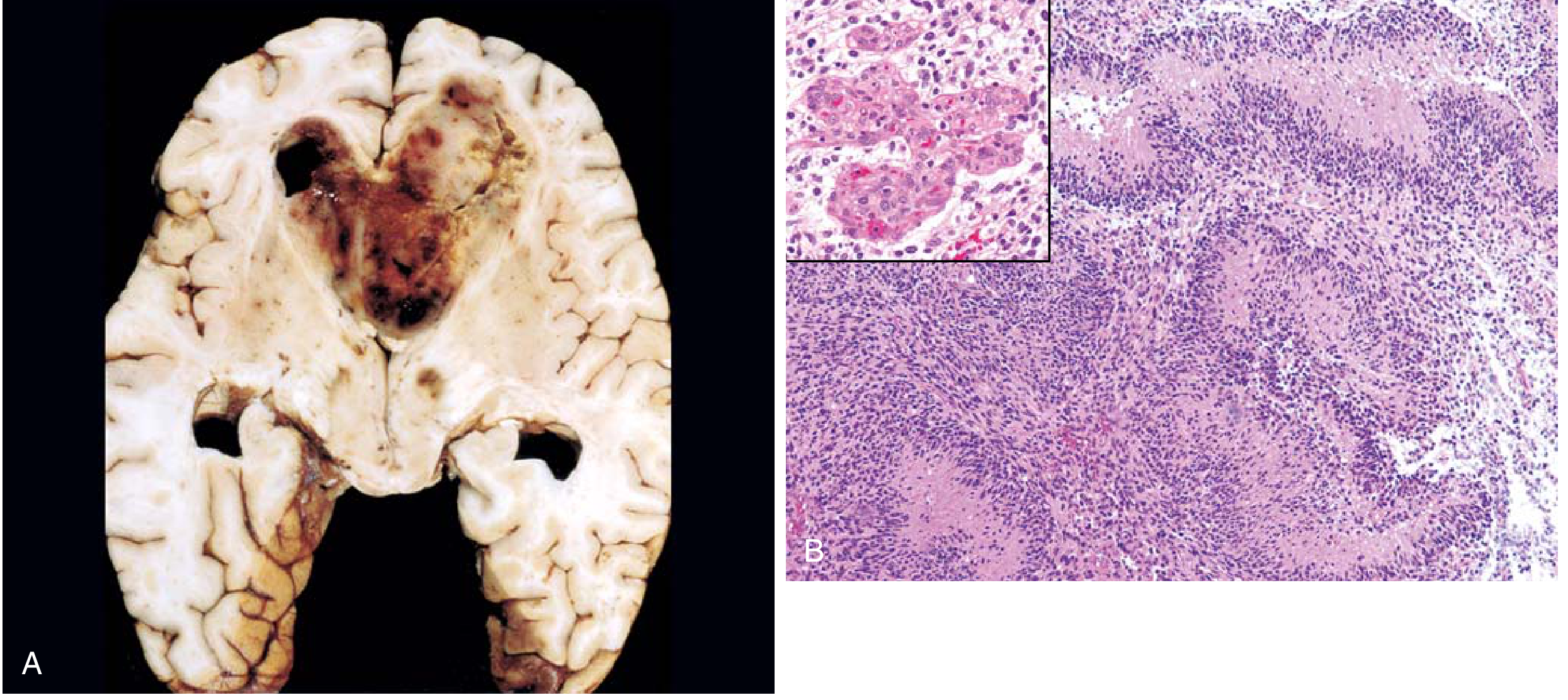
Morphology: Grossly heterogeneous - firm/white areas alongside soft/yellow necrotic zones, cystic degeneration, and hemorrhage. Microscopically: high cellularity, poorly differentiated pleomorphic cells, brisk mitoses, serpiginous "pseudopalisading" necrosis (tumor nuclei lined around anucleate necrotic zones), and microvascular proliferation. Molecular criteria (TERT promoter mutation, EGFR amplification, or +7/-10 chromosomal copy-number change) alone can assign grade 4 status even without necrosis or microvascular proliferation.
3. Oligodendroglioma, IDH-Mutant and 1p/19q-Codeleted
Composed of cells resembling oligodendrocytes; carries the best prognosis among diffuse glial tumors when corrected for grade. Accounts for 5-15% of gliomas, typically presenting in the 4th-5th decades with a history of seizures, most often in frontal/temporal lobes.
Pathogenesis: Codeletion of chromosomes 1p and 19q, always occurring together with an IDH1/IDH2 mutation. Most also carry TERT promoter mutations (increased telomerase activity).
Morphology (grade 2): Gelatinous gray infiltrative mass, often with cysts, hemorrhage, and calcification (up to 90% of tumors). Classic "fried egg" appearance - regular cells with spherical nuclei and finely granular chromatin surrounded by a clear cytoplasmic halo - plus a delicate "chicken-wire" capillary network. Low mitotic activity. Grade 3 shows higher cellularity, nuclear anaplasia, more mitoses, and often microvascular proliferation/necrosis.
Clinical course: Average survival 10-20 years (grade 2), 5-10 years (grade 3) with combined surgery, chemotherapy, and radiotherapy.
4. Pilocytic Astrocytoma (Circumscribed Glioma, WHO Grade 1)
Relatively benign, affecting children and young adults. Well-circumscribed (unlike diffuse gliomas), most commonly arising in the cerebellum, but also third ventricle, optic pathways, spinal cord, or cerebral hemispheres. Presents with mass effect, obstructive hydrocephalus, and raised intracranial pressure; often cystic. Because it is well circumscribed, it is usually curable with complete surgical resection. (Molecularly driven by KIAA1549-BRAF fusion, per Robbins' broader discussion.)
Quick Comparison Table
| Feature | Astrocytoma IDH-mut | Glioblastoma IDH-wt | Oligodendroglioma |
|---|---|---|---|
| IDH status | Mutant | Wild-type | Mutant |
| 1p/19q | Intact | Intact | Co-deleted |
| Other mutations | TP53, ATRX | EGFR/PDGFR amp, TERT, CDKN2A del | TERT promoter |
| Grade | 2-4 | Always 4 | 2-3 |
| Prognosis | Better (median >10 yr at grade 2) | Worst (poor survival) | Best among diffuse gliomas |
| Histology clue | Fibrillary, GFAP+ | Pseudopalisading necrosis, microvascular proliferation | "Fried egg" cells, "chicken-wire" capillaries |
Source: Robbins & Kumar, Basic Pathology, Ch. 21 (Gliomas), p. 858-861.
If you want, I can also pull the equivalent Robbins, Cotran & Kumar Pathologic Basis of Disease (the larger reference edition) coverage for comparison, or turn this into a condensed 10-mark exam answer format.
Combine both
Reading File
Reading File
Gliomas - Combined Robbins Coverage
(Robbins & Kumar Basic Pathology + Robbins, Cotran & Kumar Pathologic Basis of Disease)
Both Robbins texts anchor their discussion in the 2021 WHO Classification of CNS Tumors, which folds molecular markers (IDH mutation status, 1p/19q codeletion) into the histologic grading scheme. Gliomas are the most common primary intraparenchymal brain tumors and include astrocytomas, oligodendrogliomas, and ependymomas - thought to arise from multipotent progenitor cells that differentiate along a particular glial lineage, with characteristic anatomic and age predilections.
Diffusely infiltrating astrocytomas and glioblastomas were once viewed as one continuous low-to-high-grade spectrum. Both texts now agree they are two genetically and clinically distinct entities, separated primarily by IDH1/IDH2 mutation status.
1. Astrocytoma, IDH-Mutant
Epidemiology (combined):
- Basic Pathology: peak in 4th-6th decades, cerebral hemispheres
- Pathologic Basis of Disease adds precision: less common than glioblastoma; median age 38 years; cerebral hemispheres of young-to-middle-aged adults
Presentation: Seizures, headache, focal neurologic deficits (both texts).
Pathogenesis:
- IDH1 (or IDH2) gain-of-function mutation - the earliest tumorigenic event. Mutant IDH produces 2-hydroxyglutarate, an oncometabolite (Cotran/Pathologic Basis, Ch. 7 cross-reference)
- Concurrent TP53 mutation and ATRX inactivation (mechanism of senescence evasion) - Cotran describes these three alterations as occurring "simultaneously"
- Homozygous CDKN2A/CDKN2B deletion upgrades the tumor to CNS WHO grade 4 by definition, even without high-grade histology, because prognosis is markedly worse
Grading: WHO grades 2-4 (grade 1 not used - all diffuse gliomas are malignant by convention).
Morphology (combined detail):
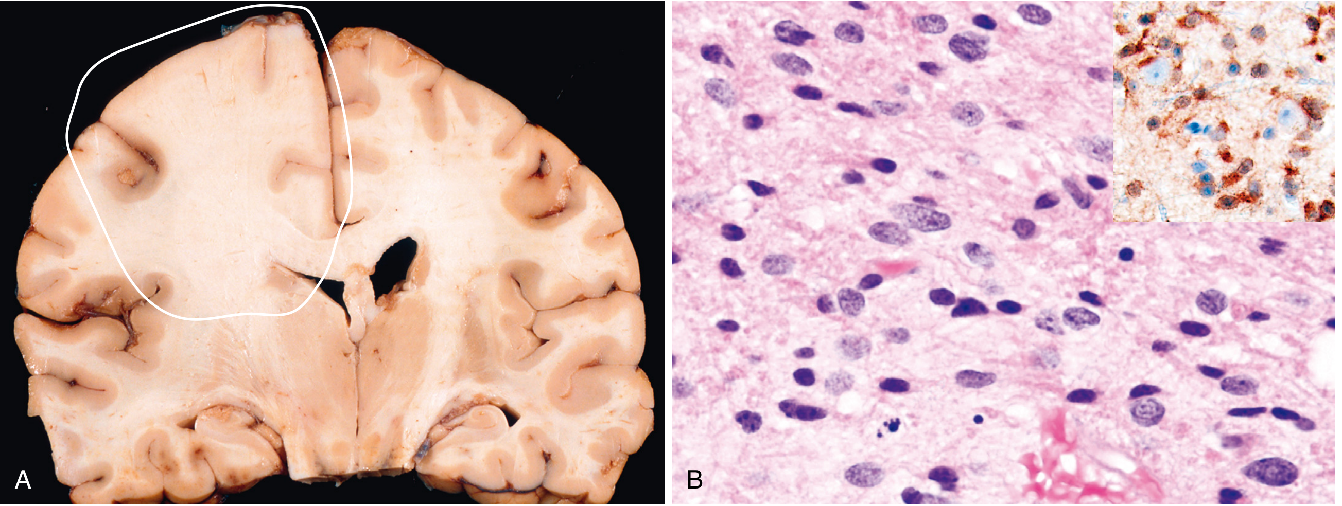
- Gross: poorly defined, gray, infiltrative masses that expand and distort brain without a discrete margin; cut surface firm or soft/gelatinous, sometimes with cystic degeneration; size ranges from a few cm to near-total hemispheric replacement
- Microscopic: hypercellular compared to normal white matter; enlarged, elongated/irregular hyperchromatic nuclei in a GFAP-positive fibrillary background; tumor cells infiltrate well beyond the visible margin (individual cells seen many cm away, including "perineuronal satellitosis" around cortical neurons)
- IDH1 p.R132H immunostain positive in up to 90% of cases; IDH sequencing reserved for immunonegative cases to detect less common mutations
- Nearly all cases show ATRX loss and p53 overexpression by IHC
- Grade 3: denser cellularity, more pleomorphism, mitoses appear
- Grade 4: cellular crowding, cytologic atypia, microvascular proliferation and/or necrosis (though typically less necrosis/hemorrhage than IDH-wildtype glioblastoma)
Prognosis: Median survival >10 years (grade 2), 5-10 years (grade 3), ~3 years (grade 4). Both texts stress that IDH-mutant astrocytomas have much better prognosis than glioblastoma even when they reach grade 4 histology.
2. Glioblastoma, IDH-Wild-Type
Epidemiology:
- Basic Pathology: ~50% of all primary malignant brain tumors in adults
- Cotran/Pathologic Basis: ~14% of all primary CNS tumors and over half of all CNS malignancies; occurs mostly in cerebral hemispheres of adults >55 years; arises de novo and does not evolve from a lower-grade astrocytoma (unlike the old "secondary glioblastoma" concept)
Grading: Always CNS WHO grade 4 by definition - no lower-grade precursor exists for this entity.
Pathogenesis (combined list of hallmark-acquiring alterations):
| Cancer hallmark | Genetic alteration |
|---|---|
| Evasion of senescence | TERT promoter mutation (or alternative telomere lengthening) |
| Escape from growth control | Homozygous CDKN2A deletion (loss of p16) |
| Growth factor signaling activation | EGFR or PDGFR gene amplification |
| Resistance to apoptosis | TP53 mutation |
| Altered chemosensitivity | MGMT promoter methylation (DNA repair enzyme downregulation -> better temozolomide response) |
Cotran specifically notes the diagnostic rule: an adult diffuse astrocytoma that is IDH-wildtype qualifies as glioblastoma if it shows at least one of: combined +7/-10 chromosomal copy-number change, TERT promoter mutation, or EGFR amplification - even without classic high-grade histology.
Morphology:
- Gross: strikingly heterogeneous - firm/white regions next to soft/yellow necrotic areas, cystic degeneration, and hemorrhage
- Microscopic: high cellularity, poorly differentiated pleomorphic cells, brisk mitoses, serpiginous "pseudopalisading" necrosis (tumor nuclei lined around anucleate necrotic zones), and microvascular proliferation
Clinical course: Short and aggressive; very poor prognosis regardless of treatment.
3. Oligodendroglioma, IDH-Mutant and 1p/19q-Codeleted
Epidemiology: 5-15% of gliomas; 4th-5th decades; frontal/temporal lobes; long antecedent history of seizures common.
Pathogenesis: Co-deletion of chromosomes 1p and 19q, occurring only in association with an IDH1/IDH2 mutation; most tumors also carry TERT promoter mutations.
Morphology:
- Grade 2: gelatinous, gray, infiltrative mass; cysts, hemorrhage, calcification (up to 90% of cases)
- Classic "fried egg" cells - round nuclei with finely granular chromatin and a clear cytoplasmic halo - set within a "chicken-wire" capillary network
- Grade 3: higher cellularity, nuclear anaplasia, more mitoses, microvascular proliferation and/or necrosis
Prognosis: Best among diffuse gliomas when corrected for grade - average survival 10-20 years (grade 2), 5-10 years (grade 3).
Combined Comparison Table (Table 28.5, Cotran - Pathologic Basis of Disease)
| Feature | Astrocytoma, IDH-mutant (Grade 2-4) | Oligodendroglioma, IDH-mutant + 1p/19q-codeleted (Grade 2-3) | Glioblastoma, IDH-wildtype (Grade 4) |
|---|---|---|---|
| IDH status | Mutant | Mutant | Wildtype |
| Other genetics | TP53-mut, ATRX-mut; CDKN2A homozygous deletion -> grade 4 | 1p/19q-codeleted | +7/-10, TERT promoter mutation, EGFR amplification |
| Typical morphology | Nuclear atypia; mitoses (grade 3); microvascular proliferation/necrosis (grade 4) | Round nuclei with clear halos ("fried egg"); mitoses, microvascular proliferation and/or necrosis in grade 3 | Nuclear atypia, mitoses, microvascular proliferation, necrosis |
| Prognosis | Better | Best | Worst |
4. Pilocytic Astrocytoma (WHO Grade 1) - circumscribed, non-diffuse glioma
Covered mainly in Basic Pathology: benign, well-circumscribed tumor of children and young adults, typically cerebellar, but also third ventricle, optic pathway, spinal cord, or cerebral hemispheres. Presents with mass effect, obstructive hydrocephalus, raised ICP; frequently cystic. Curable with complete resection given its circumscribed nature - unlike the diffusely infiltrating entities above.
Key synthesis point (why both editions matter for exam purposes)
- Basic Pathology gives the more streamlined, exam-friendly version (definitions, grading rules, survival numbers).
- Pathologic Basis of Disease (Cotran) adds mechanistic depth: the oncometabolite 2-hydroxyglutarate, precise epidemiologic proportions (14% of CNS tumors for GBM), the explicit de novo origin of glioblastoma (refuting the old low-grade-to-high-grade progression model for GBM), and the IHC correlate panel (IDH1 R132H, ATRX loss, p53 overexpression) used for diagnosis in practice.
Together, they form a complete answer for a "molecular basis, diagnostic markers, and pathology of gliomas" exam question: pathogenesis (driver mutations) -> diagnostic morphology/IHC -> grading rules -> prognosis, exactly as both Robbins editions structure it.
Search for recent research on emerging glioma biomarkers and therapies
Searching PubMed
glioma biomarker liquid biopsy
Searching PubMed
glioblastoma CAR T cell therapy
Searching PubMed
IDH mutant glioma vorasidenib
Searching PubMed
glioma tumor treating fields
Searching the Web
emerging glioma biomarkers and therapies 2026
Reading Web Page
https://www.mdanderson.org/newsroom/research-newsroom/biomar…
Searching PubMed
glioblastoma oncolytic virus biomarker
Searching PubMed
glioma DNA methylation classifier diagnosis
Quick answer
This is a shared conversation. Sign in to Orris to start your own chat.