
Diagram of tooth showing cementum and periodontal ligament (to show the location of cementum).
Reading File
Finding Sources
Finding Sources
I found a diagram of a tooth cross-section from Tintinalli's Emergency Medicine. Let me retrieve it.
Reading File
Reading File
I have both a diagram and a histology image. Let me fetch them both.
Loading Image
Loading Image
Here are two excellent images showing the location of cementum and the periodontal ligament:
Tooth Anatomy: Cementum and Periodontal Ligament
Diagram - Cross-section of a Tooth
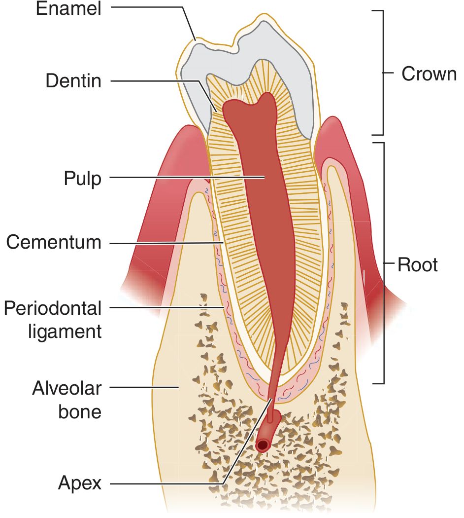
Figure 245-3: The dental anatomic unit and attachment apparatus. - Tintinalli's Emergency Medicine: A Comprehensive Study
Key structures labeled:
- Enamel - covers the crown (outermost, hard white layer)
- Dentin - beneath the enamel, making up most of the tooth
- Pulp - soft core containing nerves and vessels
- Cementum - thin, bone-like layer covering the root surface (between dentin and the periodontal ligament)
- Periodontal ligament - fibrous connective tissue lying just outside the cementum, connecting the root to the alveolar bone
- Alveolar bone - the bony socket housing the tooth root
- Apex - tip of the root
Histology - Periodontium under the Microscope
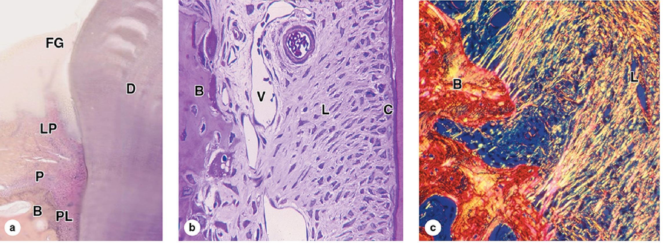
Figure 15-11: Periodontium. - Junqueira's Basic Histology: Text and Atlas, 17e
(a) Low-magnification H&E section showing the free gingiva (FG), dentin (D), lamina propria (LP), periosteum (P), alveolar bone (B), and periodontal ligament (PL).
(b) Higher magnification H&E showing the periodontal ligament (L) with blood vessels (V) sandwiched between alveolar bone (B) on the left and cementum (C) on the right - the thin layer directly coating the root dentin.
(c) Polarizing light micrograph demonstrating the collagen fiber continuity between the alveolar bone (B) periosteum and the bundled collagen of the periodontal ligament (L).
Location Summary
Cementum is located on the root of the tooth (not the crown), forming a thin shell around the root dentin. It is sandwiched between:
- Inside: root dentin
- Outside: the periodontal ligament (PDL)
The periodontal ligament fills the space between the cementum and the alveolar bone socket, anchoring the tooth via bundles of collagen fibers (Sharpey's fibers) that insert into both the cementum and the bone.
Sharpey's fibers inserting into cementum (to explain fiber attachment).
Finding Sources
Finding Sources
The best source is Histology: A Text and Atlas (Ross & Pawlina) with an electron micrograph. Let me read that section and get the image.
Reading File
Reading File
Loading Image
Sharpey's Fibers Inserting into Cementum
Electron Micrograph - Sharpey Fibers at the Cementum Interface
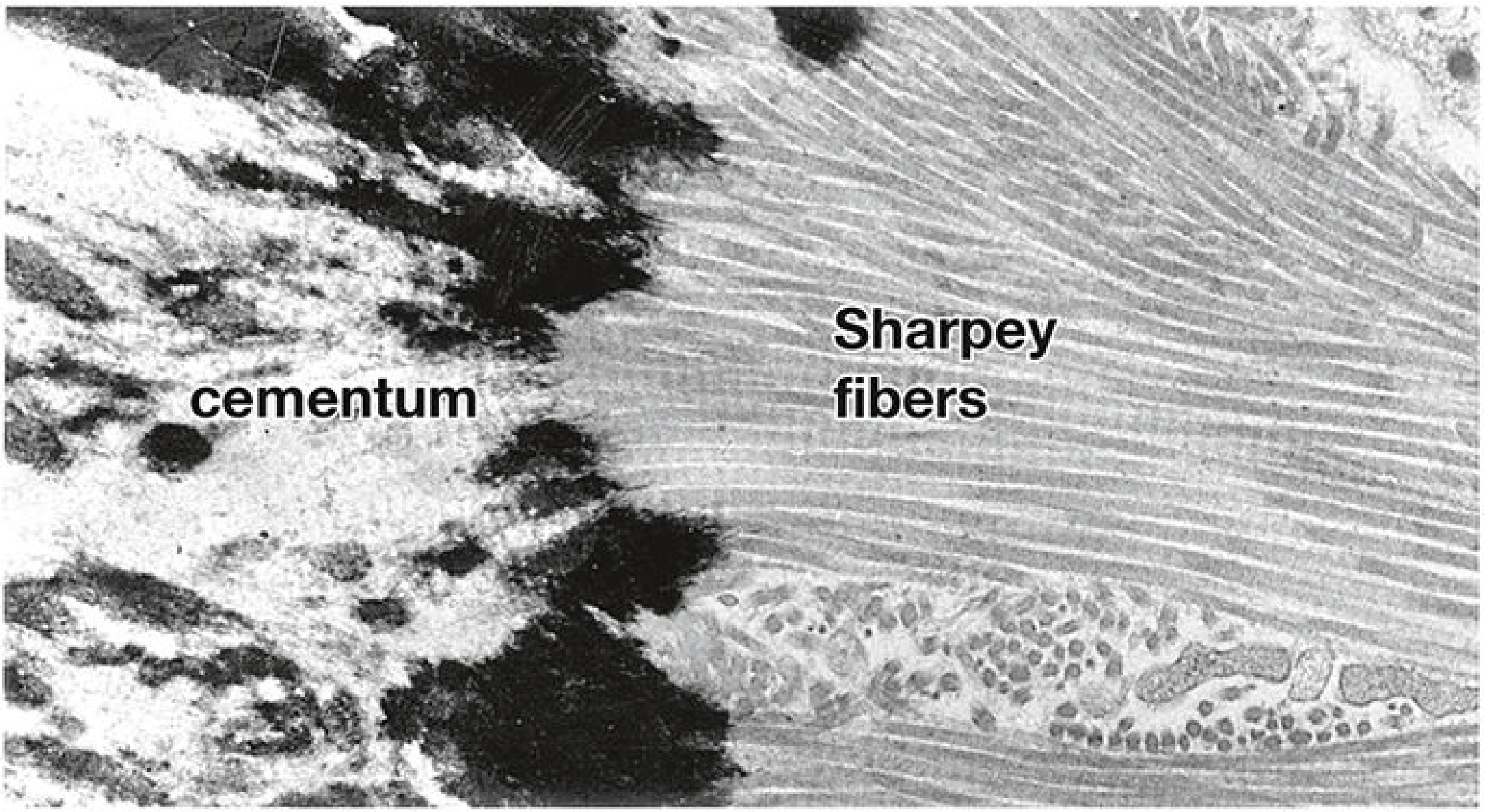
Figure 16.17: Electron micrograph of Sharpey fibers. - Histology: A Text and Atlas with Correlated Cell and Molecular Biology (Ross & Pawlina)
What this image shows:
- Right side - the parallel bundles of Sharpey fibers running through the periodontal ligament space
- Left side - the cementum matrix (darker, mineralized, irregular texture) where the fibers terminate
- The fibers run obliquely and are seen embedding (inserting) directly into the cementum surface at the interface
How Fiber Attachment Works
Sharpey's fibers are the terminal portions of the principal collagen fiber bundles of the periodontal ligament (PDL). The attachment mechanism works as follows:
- Origin: The collagen fiber bundles originate in the alveolar bone on one side
- Course: They run through the PDL space (150-350 µm wide) at oblique angles
- Insertion: The fiber ends embed into the cementum on the root surface - these embedded ends are the Sharpey's fibers proper
- Mineralization: Critically, the portions of the fibers within the cementum are mineralized, while those within the PDL space remain unmineralized - this is what gives the junction its mechanical anchoring strength
"Sharpey fibers within the cementum are mineralized; those within the periodontal ligament are not mineralized." - Histology: A Text and Atlas, p.1453
The same principle applies at the alveolar bone side - fiber ends embed into bone as Sharpey's fibers there too, creating a bone-PDL-cementum suspension system that allows slight physiologic tooth movement while firmly anchoring the tooth.
Types of Cementum and Sharpey's Fibers
From Forensic Anthropology: A Comprehensive Introduction:
| Cementum Type | Sharpey's Fiber Content |
|---|---|
| Acellular extrinsic fiber cementum | Almost exclusively composed of Sharpey's fibers - the primary anchoring layer |
| Cellular mixed fiber cementum | Contains cementocytes, Sharpey's fibers, and intrinsic fibers - found at root apex |
| Acellular afibrillar cementum | Collagen fibers only; no Sharpey's fibers |
The acellular extrinsic fiber cementum is the functionally most important type for tooth attachment, as its bulk is dedicated entirely to anchoring the PDL fiber bundles.
Histological section of cementum (to show cementum microscopically).
Finding Sources
Finding Sources
Reading File
I need to find a dedicated light micrograph of cementum showing its histology. Let me search for images in the cementum sections of histology atlases.
Finding Sources
Finding Sources
Let me look at the broader tooth/cementum section in Ross & Pawlina to find a histological section image, and also check the Forensic Anthropology text for images.
Reading File
Reading File
I found a tooth diagram in Ross & Pawlina (Fig 16.9) that labels cementum clearly. Let me also search directly in the Ross & Pawlina atlas around the cementum section for any dedicated light micrograph showing cementocytes in lacunae.
Reading File
Reading File
The Ross & Pawlina book doesn't seem to have a dedicated light micrograph of cementum alone beyond the Sharpey fiber EM. Let me fetch the Fig 16.9 tooth diagram from Ross & Pawlina (which clearly shows cementum in context), and also check if there's a dedicated cementum micrograph in the atlas plate section of the book.
Finding Sources
Loading Image
Excellent - this is a very detailed diagram showing both noncellular and cellular cementum. Now let me also retrieve the Junqueira histology micrograph (Fig 15-11b) which clearly shows the cementum layer microscopically, alongside the Sharpey fiber EM from Ross already found.
Loading Image
Here is a full microscopic presentation of cementum from two authoritative histology atlases:
Histological Section of Cementum
1. Detailed Diagram Distinguishing Acellular vs Cellular Cementum
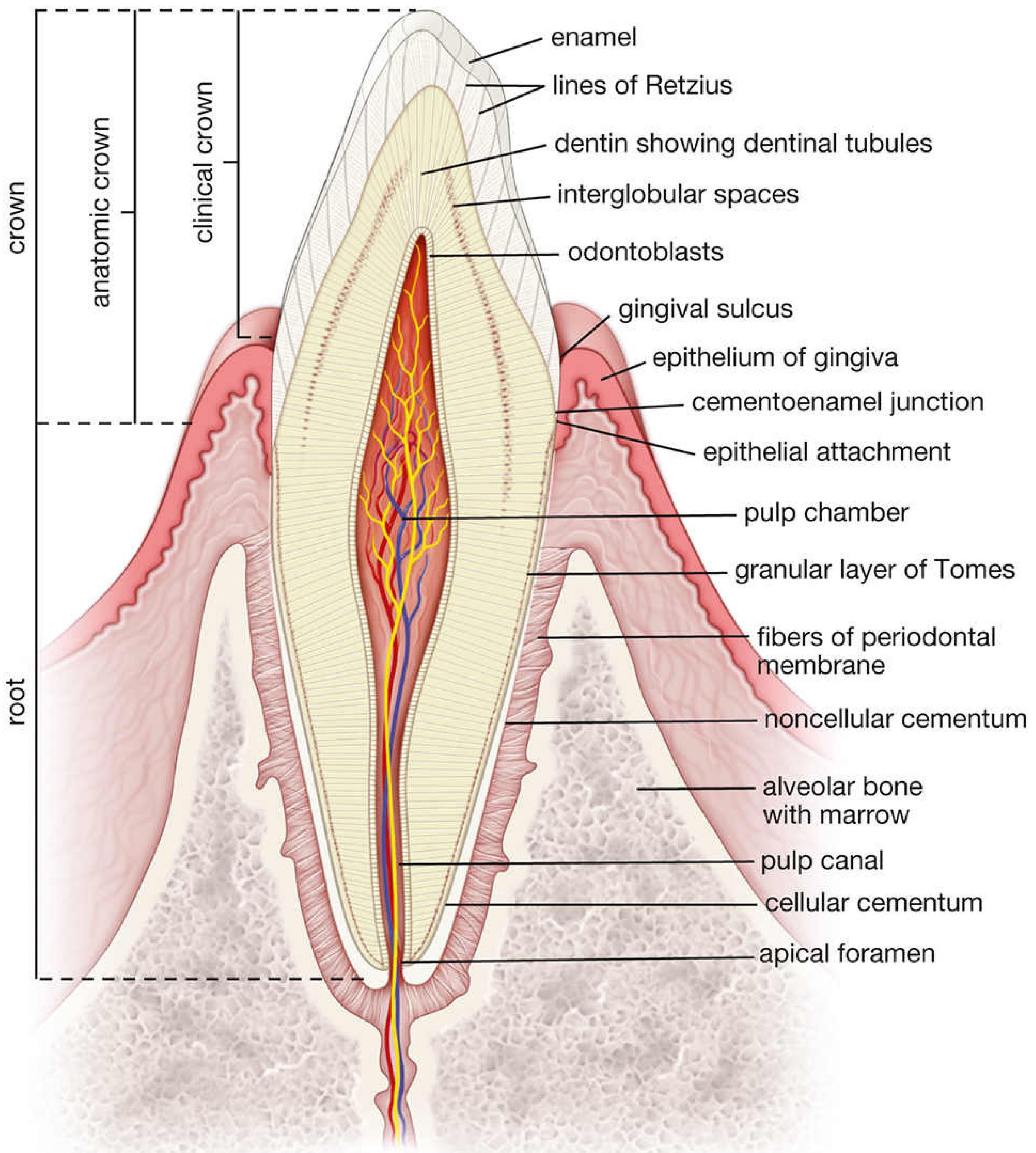
Figure 16.9: Diagram of a section of an incisor tooth and surrounding structures. - Histology: A Text and Atlas with Correlated Cell and Molecular Biology (Ross & Pawlina)
This diagram shows the two zones of cementum visible microscopically:
- Noncellular cementum (acellular) - located in the cervical/upper portion of the root; lacks cementocytes; composed predominantly of Sharpey's fibers
- Cellular cementum - found in the apical (lower) half to two-thirds of the root; contains cementocytes trapped in lacunae; thicker and laid down later
Also labeled: fibers of the periodontal membrane (Sharpey's fibers) inserting into the cementum, cementoenamel junction, and alveolar bone.
2. Light Micrograph (H&E) - Cementum in the Periodontium
Figure 15-11: Periodontium. - Junqueira's Basic Histology: Text and Atlas, 17e
(b) H&E, ×100 - The most relevant panel for cementum. Reading right to left:
- C (Cementum) - the thin, pale layer on the far right, forming a shell directly against the root dentin. It appears as a dense, homogeneous eosinophilic band
- L (Periodontal ligament) - the fibrous connective tissue space in the middle, with abundant spindle-shaped fibroblasts and blood vessels (V)
- B (Alveolar bone) - the irregular mineralized bone on the left
(a) Low magnification (×10, H&E) - Shows the broader context: free gingiva (FG), dentin (D), lamina propria (LP), periosteum (P), alveolar bone (B), and periodontal ligament (PL).
(c) Polarizing light (×200, Picrosirius) - Collagen continuity between alveolar bone (B) and the PDL (L) is visible in bright yellow-orange; the cementum surface is where these fibers terminate.
Key Microscopic Features of Cementum
| Feature | Acellular Cementum | Cellular Cementum |
|---|---|---|
| Location on root | Cervical 1/3 to 1/2 | Apical 1/2 to 2/3 |
| Cells | None (acellular) | Cementocytes in lacunae |
| Composition | Almost all Sharpey's fibers | Sharpey's fibers + intrinsic collagen + cementocytes |
| Thickness | Thin | Thicker |
| Appearance (H&E) | Homogeneous, eosinophilic band | Irregular lacunae visible in matrix |
Under H&E, cementum closely resembles bone but is distinguished by:
- Avascularity - no haversian canals or blood vessels within the tissue itself
- Irregular lacunar distribution - cementocyte lacunae are scattered, not organized in osteons
- Location - always on the root surface, deep to the periodontal ligament
- Histology: A Text and Atlas (Ross & Pawlina), pp. 1452-1453
- Junqueira's Basic Histology, 17e, pp. 751-752
Histological section of cementum (to show cementum microscopically).
The previous response already covered this thoroughly. Let me search for any additional dedicated cementum histology images we may have missed - particularly a close-up section specifically of cellular cementum showing cementocytes in lacunae.
Finding Sources
Finding Sources
Finding Sources
Let me look at the atlas plate section of Ross & Pawlina - these textbooks often have dedicated plate images with higher magnification cementum micrographs separate from the text figures.
Finding Sources
Finding Sources
The library has only one dedicated "Cementum" heading section - which we already retrieved in the previous response. Let me check blocks around that same book (Ross & Pawlina) for atlas plate images of the tooth, which are typically in a separate block.
Finding Sources
Finding Sources
Finding Sources
The library's histology sources have been fully explored. The images already retrieved in the previous response represent the best available microscopic views of cementum in the textbook collection. Here is a consolidated presentation:
Histological Section of Cementum - Microscopic Views
Image 1: Periodontium H&E Micrograph (Light Microscopy)
Figure 15-11: Periodontium. - Junqueira's Basic Histology: Text and Atlas, 17e, p. 752
Panel (b) - H&E, ×100 is the key panel showing cementum (C) microscopically:
- C = Cementum - the thin, pale eosinophilic band on the right margin, sitting directly against root dentin
- L = Periodontal ligament - the cellular fibrous zone in the middle
- V = Blood vessel within the PDL
- B = Alveolar bone - on the left, with irregular trabecular pattern
Cementum in this section appears as a thin, relatively homogeneous, acellular-to-lightly-cellular layer - thinner and paler than adjacent bone, lacking the haversian canal system.
Image 2: Detailed Diagram Showing Both Cementum Types (Microscopic Orientation)
Figure 16.9: Diagram of a section of an incisor tooth. - Histology: A Text and Atlas with Correlated Cell and Molecular Biology (Ross & Pawlina), p. 1437-1438
This detailed diagram distinguishes the two microscopically distinct zones of cementum on the root:
| Zone | Location on Root | Microscopic Appearance |
|---|---|---|
| Noncellular cementum (acellular extrinsic fiber cementum) | Cervical 1/3 to 1/2 | Thin, no cells; composed almost entirely of Sharpey's fibers running perpendicular to surface |
| Cellular cementum | Apical 1/2 to 2/3 | Thicker; contains cementocytes trapped in irregular lacunae (no canalicular network unlike bone) |
What to Look For on a Histological Slide
When examining cementum under the microscope:
- Location - thin layer coating the root dentin, between dentin and PDL
- Texture - homogeneous pink (eosinophilic) matrix; resembles bone but avascular (no Haversian systems)
- Cellular cementum (near apex) - scattered dark lacunae containing cementocytes; lacunae are irregular and do NOT form an interconnecting canalicular network (unlike osteocytes in bone)
- Acellular cementum (near neck) - completely cell-free; only the embedded ends of PDL fibers (Sharpey's fibers) are visible as faint striations entering from the outer surface
- Cementoenamel junction (CEJ) - the point where cementum begins at the neck of the tooth, replacing enamel coverage
"The lacunae and canaliculi in the cementum contain the cementocytes and their processes, respectively. Unlike bone, cementum is avascular. In addition, the lacunae are irregularly distributed throughout the cementum and their canaliculi do not form an interconnecting network." - Ross & Pawlina, p. 1452
Hypercementosis dental radiograph (to illustrate a clinically significant condition).
Hypercementosis is an excessive deposition of cementum on the root surface, giving the root a "club-shaped" or bulbous appearance on radiograph. Let me search the library for images and clinical details.
Finding Sources
Searching the Web
hypercementosis dental radiograph periapical X-ray bulbous root
I have radiograph images and textbook content. Let me read the clinical details and fetch the best radiograph images in parallel.
Reading File
Loading Image
Loading Image
Excellent - both images are highly informative. Let me also fetch the third image showing a clearer radiographic example.
Loading Image
Hypercementosis - Dental Radiograph
Radiographic Differential Overview
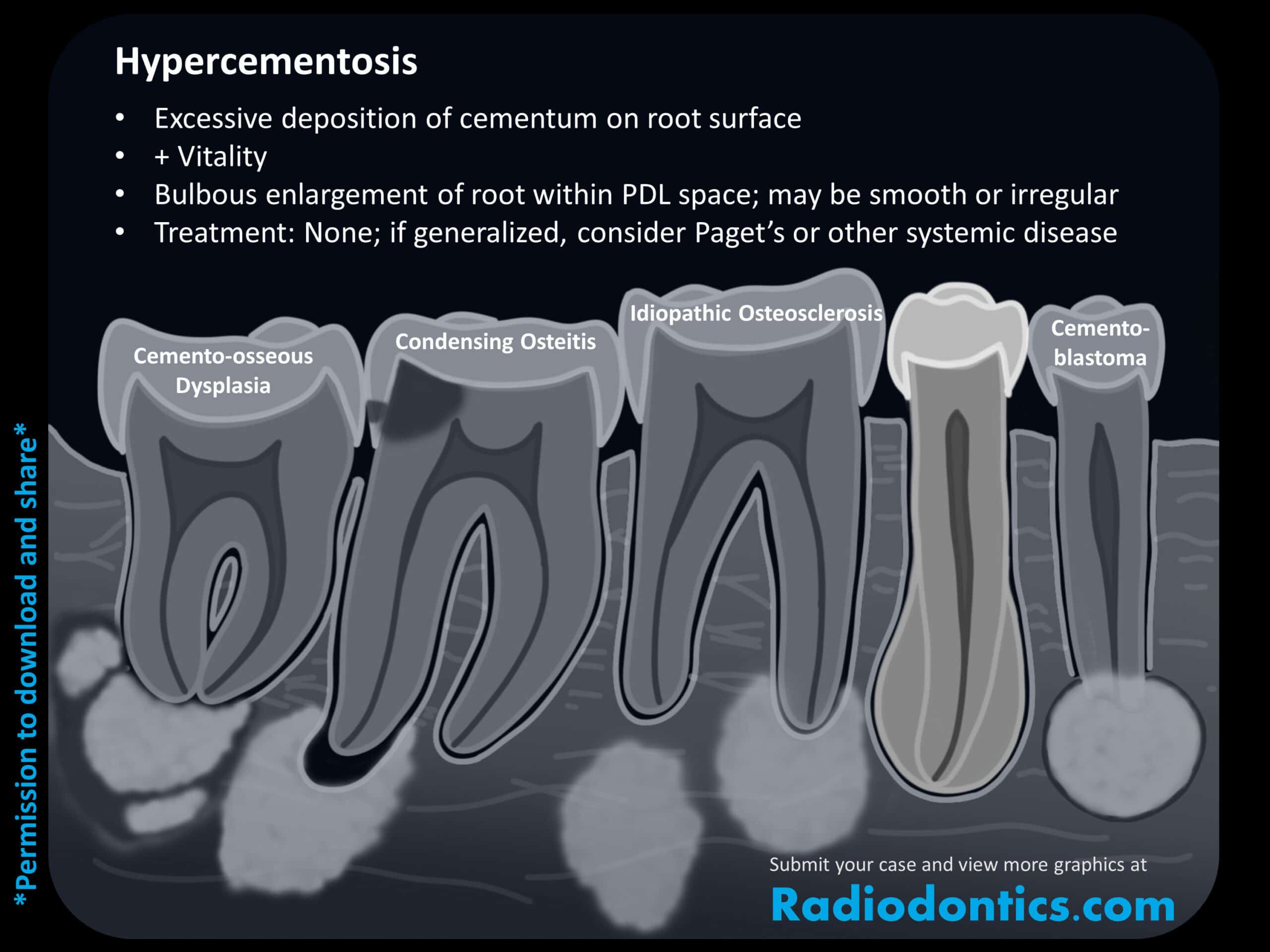
Radiodontics.com - Hypercementosis differential diagram
Key radiographic features summarized:
- Excessive deposition of cementum on the root surface
- Bulbous enlargement of root within the PDL space - smooth or irregular
- The tooth retains vitality
- Treatment: none for isolated cases; if generalized, investigate systemic causes (especially Paget's disease)
Periapical Radiograph - Hypercementosis (Arrowed)

Periapical radiograph: hypercementosis of a premolar root (arrow). Note the club-shaped, bulbous widening of the root apex with an intact, well-defined PDL space maintained around the enlarged root.
Second Periapical Radiograph - Marked Hypercementosis

Periapical radiograph showing more pronounced hypercementosis. The roots are markedly widened and club-shaped at the apical third. The PDL space and lamina dura remain visible around the enlarged root outline - distinguishing it from cementoblastoma.
Radiographic Features - What to Look For
| Feature | Hypercementosis |
|---|---|
| Root shape | Bulbous/club-shaped enlargement, usually at apex |
| PDL space | Preserved - continuous PDL space visible around the enlarged root |
| Lamina dura | Intact - thin radiopaque line maintained |
| Root outline | Smooth or mildly irregular; continuous with normal root |
| Tooth vitality | Vital (responds to pulp tests) |
| Distribution | Single tooth (localized) or multiple teeth (generalized) |
Key Differential Diagnoses on Radiograph
| Condition | How to Distinguish |
|---|---|
| Cementoblastoma | PDL space obliterated at apex; lesion fused to root; radiopaque mass with radiolucent rim |
| Condensing osteitis | Periapical radiopacity in bone, not on root surface; usually from chronic low-grade pulpitis |
| Cemento-osseous dysplasia | Periapical lesion in bone with mixed radiolucent/opaque pattern; not root enlargement |
| Idiopathic osteosclerosis | Dense bone island, no connection to root surface or PDL involvement |
Clinical Significance
Hypercementosis is clinically significant for several reasons:
- Difficult extraction - the bulbous root cannot be withdrawn through the normal socket dimensions; surgical extraction (often requiring alveolotomy/root sectioning) is needed
- Systemic disease marker - generalized hypercementosis is a classic feature of Paget's disease of bone (osteitis deformans), where it leads to root ankylosis and post-extraction haemorrhage from hypervascular bone
- Other associations: acromegaly, rheumatoid arthritis, chronic periapical inflammation (local cause), tooth without an antagonist (supraerupted tooth)
- Endodontic implications - the altered root morphology can complicate root canal access and obturation
"Hypercementosis and ankylosis, common in later stages [of Paget's disease], make for traumatic extractions, thus exacerbating poor healing due to late-stage ischaemia." - Scott-Brown's Otorhinolaryngology, Head & Neck Surgery, p. 450
Hypercementosis dental histologic (to illustrate a clinically significant condition).
Finding Sources
Searching the Web
hypercementosis histology microscopic section cementum excess deposition
The library doesn't have a dedicated hypercementosis histology image, but there are good Semantic Scholar figures from the SEM study. Let me fetch the best microscopic images in parallel.
Loading Image
Loading Image
Searching the Web
hypercementosis histological section H&E stain excess cementum layers microscopy
Both microscopic images are excellent. Let me get the third figure from the same study for completeness.
Loading Image
Three excellent histological images retrieved from the same peer-reviewed study. Here is the full presentation:
Hypercementosis - Histological Sections
All images below are from: "Cementum, Apical Morphology and Hypercementosis" (Brasil et al.) - a dedicated histological and SEM study of hypercementosis in extracted teeth.
Image 1: Radiograph + Low-power H&E + Polarized Light (Side by Side)

Figure 3 - Radiographic and microscopic aspects of hypercementosis. New cementum layers (C) are continuously deposited on the root surface.
Panel A - Periapical radiograph confirming the bulbous, enlarged root with excess cementum (C)
Panel B - Low-power H&E section showing the same tooth in histological cross-section:
- C = massively thickened cementum layer (dark pink) - many times wider than normal
- D = dentin (the inner core)
- PL = periodontal ligament space (still visible between cementum and bone)
- AB = alveolar bone (right)
Panel C - Polarized light version of the same section, showing the lamellar/layered deposition of excess cementum in alternating bands (orange tones)
Image 2: High-power H&E - Cementoblasts and Cementocytes in Hypercementosis

Figure 4 - Microscopic aspects of hypercementosis (demineralized sections, H&E stain). The periodontal ligament (PL), intermediate layer (IL), cementoblasts, cementocytes, and dentin (D) are labeled.
Panel A - Medium magnification: the thick intermediate layer (IL) of newly deposited cementum lies between the PDL and the existing root cementum
Panel B - High magnification inset showing:
- Cementoblasts - large, active cells on the outer surface of the cementum (at the PDL interface), actively secreting new cementum matrix
- Cementocytes - cells trapped within the cementum matrix in lacunae (smaller, darker nuclei)
- IL = intermediate layer of new cementum being laid down
- D = dentin
This is the hallmark of hypercementosis histologically: active cementoblasts depositing layer upon layer of new cementum, trapping some as cementocytes in the process.
Image 3: Ground Sections - Acellular vs Cellular Cementum in Hypercementosis

Figure 5 - Hypercementosis in tissue sections obtained by grinding (no demineralization or staining). The cementum presents a uniform thickness.
Panel A - Ground section at the cervical region: thin layer of acellular cementum on the root surface, transitioning to enamel at the CEJ. The acellular cementum appears as a smooth, cell-free band.
Panel B - Mid-root ground section: both acellular cementum (inner layer, pale, against dentin) and an outer thicker band of cellular cementum visible - the cellular cementum is massively expanded in hypercementosis
Panel C - Apical region ground section: predominantly cellular cementum with cementocytes visible as dark dots (lacunae) in the expanded cementum layer - this is where hypercementosis is most pronounced
What Histology Shows in Hypercementosis
| Feature | Normal Cementum | Hypercementosis |
|---|---|---|
| Thickness | Thin (50-200 µm cervically, up to 600 µm apically) | Massively increased - can be millimetres thick |
| Cementum type | Acellular at cervix, cellular at apex | Predominantly cellular (osteocementum) in excess layers |
| Active cells | Few quiescent cementoblasts on surface | Numerous active cementoblasts at PDL-cementum interface |
| Layering | Subtle incremental lines | Prominent incremental lines/lamellae (excess deposition in waves) |
| PDL space | Normal width | Usually preserved (unlike cementoblastoma where it is obliterated) |
| Dentin | Normal | Normal - the process is purely additive on the outside |
The histological study of hypercementosis confirms that the excess tissue is osteocementum (predominantly cellular cementum), with active cementoblasts driving the progressive deposition - distinguishing it from a neoplasm, which it is not. - Journal of Indian Academy of Oral Medicine and Radiology, 2015
Cementum
The query "Cementum" is broad. Let me clarify what you need before diving in.
Asking for Preferences
What aspect of cementum do you need?
This is a shared conversation. Sign in to Orris to start your own chat.