
Bones kf orbit with markings of every location and boundaries
Reading File
The orbit is formed by 7 bones. Let me search the medical library for detailed anatomy with images.
Searching sources...
Finding Sources
Finding Sources
Finding Sources
Finding Sources
Reading File
Reading File
Reading File
Loading Image
Here is a complete, well-sourced breakdown of the Bones of the Orbit with all walls, boundaries, and key markings.
Bones of the Orbit
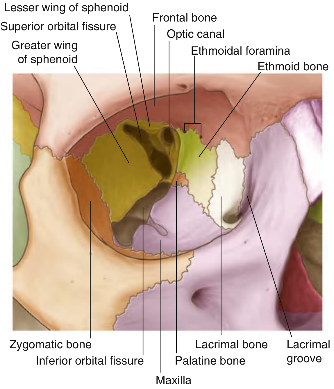
Fig. 8.79 - Bones of the Orbit (Gray's Anatomy for Students)
Overview
The bony orbit is a pyramid-shaped cavity with:
- Base (orbital rim) opening anteriorly onto the face
- Apex directed posteromedially (at the optic foramen)
- 4 walls: roof, floor, medial, lateral
7 bones form the orbit in total:
| Bone | Walls contributed to |
|---|---|
| Frontal bone | Roof (mainly), orbital rim (superiorly) |
| Sphenoid bone | Roof (posteriorly - lesser wing), Lateral wall (posteriorly - greater wing), Medial wall (small part) |
| Zygomatic bone | Lateral wall (anteriorly), Floor (laterally), Orbital rim (inferolateral) |
| Maxilla | Floor (mainly), Medial wall (anterior - frontal process), Orbital rim |
| Ethmoid bone | Medial wall (largest contributor - lamina papyracea) |
| Lacrimal bone | Medial wall (anterior part) |
| Palatine bone | Floor (small posterior contribution) |
Orbital Rim (Base)
The orbital rim is formed by:
- Superiorly: Frontal bone
- Medially: Frontal process of the maxilla
- Inferiorly: Zygomatic process of the maxilla + zygomatic bone
- Laterally: Zygomatic bone + frontal process of the zygoma + zygomatic process of the frontal bone
Wall-by-Wall Detail
Roof (Superior Wall)
- Main bone: Orbital part of the frontal bone
- Posterior completion: Lesser wing of the sphenoid bone
- Key markings:
- Trochlear fovea (anteromedially): attachment pit for the pulley (trochlea) of the superior oblique muscle
- Lacrimal fossa (anterolaterally): lodges the orbital part of the lacrimal gland
- May contain part of the frontal sinus anteriorly
Medial Wall
- Bones (anterior to posterior): Frontal process of maxilla → Lacrimal bone → Ethmoid bone (lamina papyracea) → small part of Sphenoid
- The ethmoid bone (lamina papyracea) is the largest contributor
- Key markings:
- Lacrimal groove: formed between the lacrimal bone (posterior lacrimal crest) and the frontal process of the maxilla (anterior lacrimal crest); contains the lacrimal sac
- Anterior ethmoidal foramen: at the frontoethmoidal suture; transmits the anterior ethmoidal nerve and vessels
- Posterior ethmoidal foramen: slightly posterior; transmits posterior ethmoidal nerve and vessels
Floor (Inferior Wall)
- Also forms the roof of the maxillary sinus
- Main bone: Orbital surface of the maxilla
- Small contributions: Zygomatic bone (anterolateral) and orbital process of the palatine bone (posteromedial corner)
- Key markings:
- Inferior orbital fissure: runs along the lateral boundary of the floor; bounded by greater wing of sphenoid, orbital surface of maxilla, and orbital process of palatine bone
- Transmits: infraorbital nerve (CN V2 branch), zygomatic nerve, branches of the sphenopalatine ganglion, infraorbital artery and vein, ophthalmic vein branch
Lateral Wall
- Anterior: Zygomatic bone
- Posterior: Greater wing of the sphenoid bone
- Key markings:
- Superior orbital fissure: between the greater wing (below) and lesser wing (above) of the sphenoid; transmits CN III, IV, V1, VI, superior ophthalmic vein, orbital branch of middle meningeal artery, recurrent lacrimal artery branch
- The lateral wall is the thickest and strongest wall
Key Foramina / Apertures at the Apex
| Opening | Location | Transmits |
|---|---|---|
| Optic canal | Formed by the lesser wing of sphenoid | CN II (optic nerve) + ophthalmic artery |
| Superior orbital fissure | Between lesser and greater wings of sphenoid | CN III, IV, V1, VI + ophthalmic veins |
| Inferior orbital fissure | Floor/lateral wall junction | CN V2 branches, infraorbital vessels |
| Anterior ethmoidal foramen | Medial wall (frontoethmoidal suture) | Anterior ethmoidal nerve and vessels |
| Posterior ethmoidal foramen | Medial wall (slightly posterior) | Posterior ethmoidal nerve and vessels |
Clinical Correlates
- Orbital blow-out fractures most commonly involve the floor (thinnest bone, roof of maxillary sinus) and medial wall (lamina papyracea of ethmoid). Floor fractures can trap the inferior oblique muscle causing vertical diplopia (upward gaze failure).
- Medial wall fractures allow air into the orbit from the ethmoidal sinuses - seen on X-ray as orbital emphysema; patients may feel orbital fullness when blowing the nose.
- Superior orbital fissure syndrome: compression of structures here causes ophthalmoplegia, ptosis, fixed dilated pupil (CN III, IV, VI involvement) - WITHOUT visual loss.
- Orbital apex syndrome: involves the optic canal too, so adds vision loss to the above findings.
Sources: Gray's Anatomy for Students, p. 1067; K.J. Lee's Essential Otolaryngology, p. 1132
Wheere is palatine bone
The palatine bone makes a very small contribution to the orbit - it sits at the posteromedial corner of the floor.
Searching sources...
Finding Sources
The textbooks confirm it clearly. Here is the precise answer:
Location of the Palatine Bone in the Orbit
The palatine bone contributes via its orbital process, and it sits in the posteromedial corner of the orbital floor - right at the junction where the maxilla, ethmoid, and sphenoid bones all meet.
It is the smallest contributor to the orbit - a tiny triangular wedge tucked deep at the back.
Exact Position
         MEDIAL WALL
              |
Ethmoid ------+-------- Sphenoid (lesser wing)
              |
         PALATINE  <-- sits HERE, at this 3-way junction
              |
       Maxilla (floor) ---- Zygoma
- Wall: Floor (inferior wall)
- Position on floor: Posterior + medial corner
- Neighbors: Maxilla (anteriorly), ethmoid (medially), sphenoid (posteriorly)
Also Contributes to the Inferior Orbital Fissure
The orbital process of the palatine bone forms part of the boundary of the inferior orbital fissure, along with:
- Greater wing of sphenoid
- Orbital surface of maxilla
Why It Matters Clinically
During orbital decompression surgery, the orbital process of the palatine bone is deliberately drilled away to improve infero-lateral access to the orbit (Cummings Otolaryngology).
In simple terms: look at the back of the orbital floor, in the innermost corner - that tiny bone completing the floor is the palatine bone's orbital process. It is barely visible on the labeled diagram but sits right where the floor meets the medial wall, deep near the apex.
Sources: Gray's Anatomy for Students; K.J. Lee's Essential Otolaryngology; Cummings Otolaryngology
This is a shared conversation. Sign in to Orris to start your own chat.