
Pathological calcification
Reading File
Pathological calcification is the abnormal deposition of calcium salts in tissues. It has two main forms: dystrophic (in dead/dying tissue, normal serum calcium) and metastatic (in normal tissue, due to hypercalcemia).
Searching sources...
Finding Sources
Finding Sources
Finding Sources
Reading File
Finding Sources
Finding Sources
Robbins Pathologic Basis of Disease has a dedicated section. Let me read it fully, along with the Basic Pathology entry.
Reading File
Reading File
Now let me get the rest of Robbins' section on metastatic calcification and fetch the key images.
Reading File
Loading Image
I now have comprehensive information from Robbins to write a detailed answer. Let me also fetch the psammoma body image.
Loading Image
That second image is a placeholder. I have all I need for a thorough response. Here is the complete answer:
Pathological Calcification
Pathological calcification is the abnormal tissue deposition of calcium salts (principally calcium phosphate/hydroxyapatite, along with smaller amounts of iron, magnesium, and other mineral salts) in locations where they do not normally occur. There are two distinct forms based on mechanism and serum calcium status.
1. Dystrophic Calcification
Definition: Calcium deposition occurring in dead or dying tissue despite normal serum calcium levels and no systemic disturbance of calcium metabolism.
Sites
- Areas of necrosis - coagulative, caseous, liquefactive, or enzymatic fat necrosis
- Atheromatous plaques of advanced atherosclerosis (nearly always present)
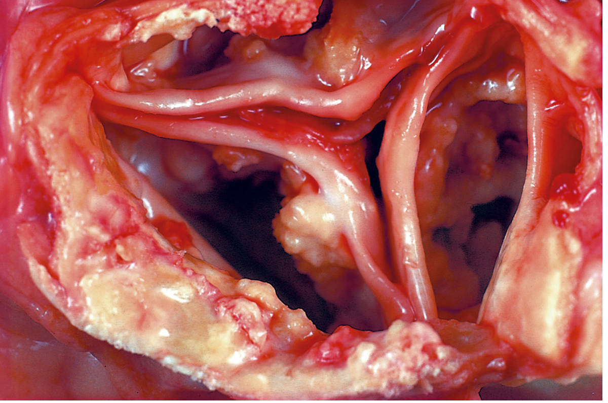
- Aging/damaged heart valves - causes calcific aortic stenosis and mitral annular calcification (Fig. 2.34 below)
- Tuberculous lymph nodes - may be entirely converted to stone
- Old infarcts, scarred tissue, thrombi
Pathogenesis
Initiated by two processes:
- Extracellular route: Membrane-bound vesicles (derived from injured/dying cells) concentrate calcium via affinity for membrane phospholipids. Phosphatases in the vesicle membrane generate phosphate ions, which combine with calcium to form crystals. These crystals are then propagated, accumulating larger deposits.
- Intracellular route: Dying cells lose the ability to regulate cytosolic calcium; calcium accumulates in mitochondria of dying cells, forming initial mineral deposits.
Morphology (H&E)
- Macroscopic: Fine white granules or clumps, gritty on sectioning
- Microscopic: Basophilic, amorphous granular/clumped deposits - can be intracellular or extracellular
- Over time, heterotopic bone may form at the site
- Single necrotic cells can become seed crystals encrusted with mineral, forming lamellated concentric rings called psammoma bodies (resemble grains of sand)
- Psammoma bodies are characteristic of: papillary thyroid carcinoma, papillary serous carcinoma of ovary/endometrium, meningioma, mesothelioma
- In asbestosis, calcium and iron salts gather on asbestos spicules forming asbestos bodies (beaded dumbbell shapes)
Serum calcium: Normal
Clinical significance
- May be an incidental marker of previous cell injury (e.g., healed tuberculosis)
- Can cause serious organ dysfunction:
- Calcific aortic stenosis - severely compromised valve motion
- Atherosclerosis - contributes to plaque rigidity and instability
2. Metastatic Calcification
Definition: Calcium deposition in otherwise normal tissues, almost always secondary to hypercalcemia.
Causes of Hypercalcemia (4 principal causes)
| Cause | Examples |
|---|---|
| 1. Excess PTH secretion | Primary hyperparathyroidism (parathyroid adenoma/hyperplasia); ectopic PTH-related protein (PTHrP) from malignant tumors |
| 2. Bone resorption/destruction | Multiple myeloma, leukemia, diffuse skeletal metastases; Paget disease; immobilization |
| 3. Vitamin D-related disorders | Hypervitaminosis D; sarcoidosis (macrophages convert vitamin D precursor to active form via 1-alpha hydroxylase) |
| 4. Renal failure | Phosphate retention → secondary hyperparathyroidism |
Additional causes: milk-alkali syndrome, thiazide diuretics, adrenal insufficiency.
Preferred Sites
Metastatic calcification can occur in any normal tissue, but preferentially affects tissues with an alkaline intracellular/interstitial pH (because alkalinity favors calcium phosphate precipitation):
- Gastric mucosa (secretes acid - interstitium is alkaline)
- Kidneys (renal tubules excrete acid - interstitium alkaline; leads to nephrocalcinosis)
- Lungs (CO2 excretion creates alkaline alveolar septa) - particularly alveolar septa, bronchiole walls, and pulmonary vessels
- Systemic arteries and pulmonary veins
Morphology
- Morphologically identical to dystrophic calcification: basophilic granular/amorphous or hydroxyapatite crystalline deposits
- Kidneys: nephrocalcinosis - visible on imaging, can lead to renal damage
- Lungs: patchy opacities on chest X-ray; HRCT is more sensitive; 99mTc-MDP bone scan shows increased uptake
Clinical Features
- Usually causes no clinical dysfunction
- Massive pulmonary involvement → respiratory compromise, restrictive deficits
- Massive renal involvement (nephrocalcinosis) → renal damage
- Clinical manifestations are most common in chronic renal failure patients on hemodialysis
Serum calcium: Elevated (hypercalcemia)
Comparison Table
| Feature | Dystrophic | Metastatic |
|---|---|---|
| Tissue | Dead/dying/abnormal | Normal |
| Serum calcium | Normal | Elevated (hypercalcemia) |
| Mechanism | Local tissue damage + vesicle nucleation | Systemic calcium dysregulation |
| Sites | Atheromas, valves, necrosis foci | Kidneys, lungs, gastric mucosa, vessels |
| Psammoma bodies | Yes (in necrotic foci) | No |
| Clinical significance | Valve disease, atherosclerosis | Nephrocalcinosis, pulmonary compromise |
Special Histological Structures
- Psammoma bodies: Concentrically lamellated calcified deposits. Occur in: papillary thyroid carcinoma, serous ovarian/endometrial carcinoma, meningioma, mesothelioma. Each layer represents a calcified necrotic cell acting as a nidus.
- Asbestos bodies (ferruginous bodies): Calcium + iron deposited on asbestos fibers → exotic beaded dumbbell shapes. Seen in asbestosis.
Sources: Robbins, Cotran & Kumar Pathologic Basis of Disease, p. 59-60, 75-76; Robbins & Kumar Basic Pathology, p. 37-38; Fishman's Pulmonary Diseases and Disorders, p. 1100
This is a shared conversation. Sign in to Orris to start your own chat.