
Gall Bladder histology
Reading File
This is a visual/histology topic. Let me search the library for gall bladder histology sections and images.
Finding Sources
Finding Sources
Finding Sources
Finding Sources
Excellent! Found it in the Histology textbook. Let me read those sections in parallel.
Reading File
Reading File
I have excellent content. Let me read more from the histology textbook for the complete cellular details, and fetch the histology images.
Reading File
Loading Image
Loading Image
I also saw a Rokitansky-Aschoff sinuses image. Let me fetch it too, along with the rest of the text.
Loading Image
Reading File
I now have comprehensive content from the Histology textbook (Ross & Pawlina) with all three histology images. Let me compile a complete, well-structured answer.
Gall Bladder Histology
The gallbladder is a blind pear-shaped pouch that concentrates and stores bile. It can remove ~90% of the water from incoming dilute bile, concentrating bile salts, cholesterol, and bilirubin up to 10-fold.
Wall Layers at a Glance
The gallbladder wall has 4 layers - and critically, it lacks a muscularis mucosa and submucosa (unlike the rest of the GI tract):
| Layer | Features |
|---|---|
| Mucosa | Epithelium + lamina propria; numerous deep folds when empty |
| Muscularis externa | Randomly oriented smooth muscle bundles |
| Perimuscular connective tissue | Dense CT with blood vessels, lymphatics, nerves |
| Adventitia / Serosa | Adventitia where attached to liver; serosa (mesothelium) on free surface |
Low-Power Photomicrograph (H&E, x175)
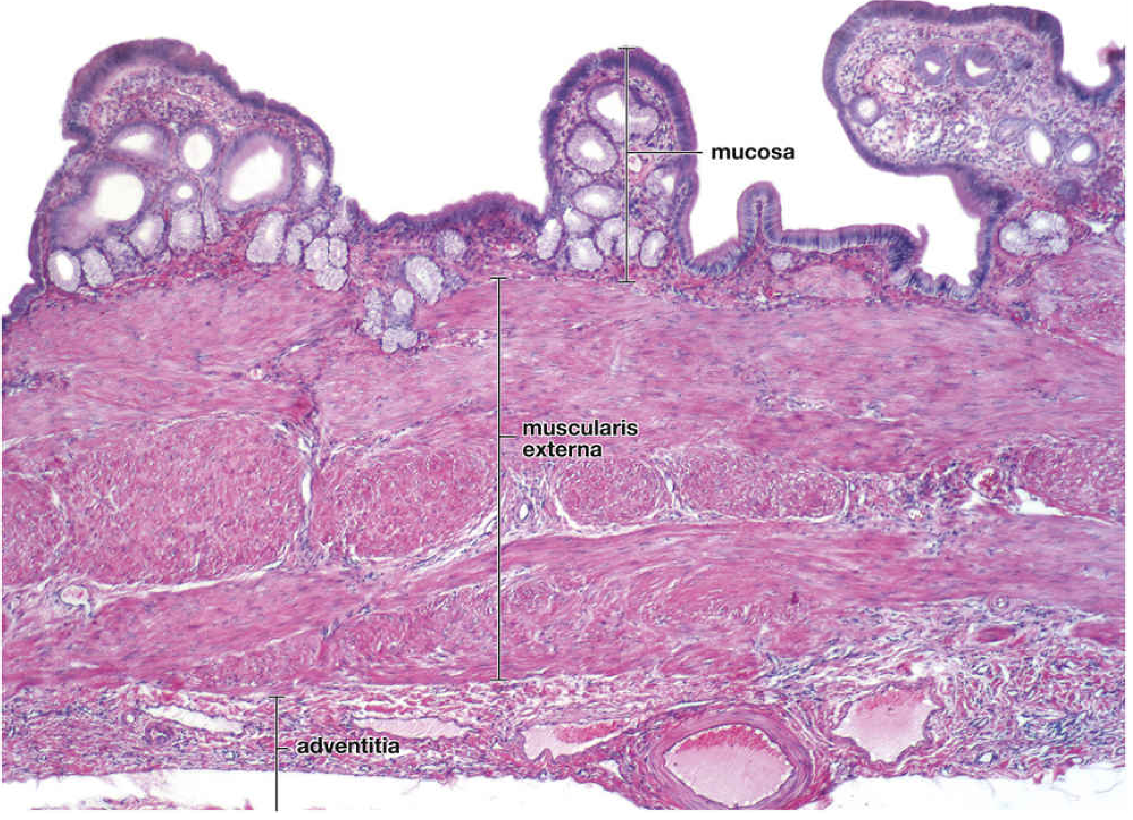
The wall shows prominent mucosal folds at the top, a thick muscularis externa in the middle, and the adventitia with large blood vessels at the bottom.
1. Mucosa
Epithelium
The mucosal surface is lined by simple columnar epithelium (tall cells also called cholangiocytes). These cells have:
- Short, not well-developed apical microvilli
- Apical junctional complexes (zonula occludens + zonula adherens) - these separate the lumen from the lateral intercellular space
- Numerous mitochondria concentrated in the apical and basal cytoplasm (needed for active transport)
- Complex lateral plications (interdigitations of lateral plasma membranes)
- Na+/K+-ATPase located in lateral plasma membranes (key for bile concentration)
- Secretory vesicles filled with glycoproteins in the apical cytoplasm
These cells closely resemble intestinal absorptive cells (enterocytes).
Lamina Propria
- Rich in fenestrated capillaries and small venules (for rapid absorption of water and electrolytes)
- No lymphatic vessels in this layer
- Highly cellular with large numbers of lymphocytes and plasma cells
- Mucin-secreting glands may be present (especially near the neck; more common in inflamed gallbladders)
- Enteroendocrine-like cells found within the mucosal glands
Electron Micrographs of Gallbladder Epithelium
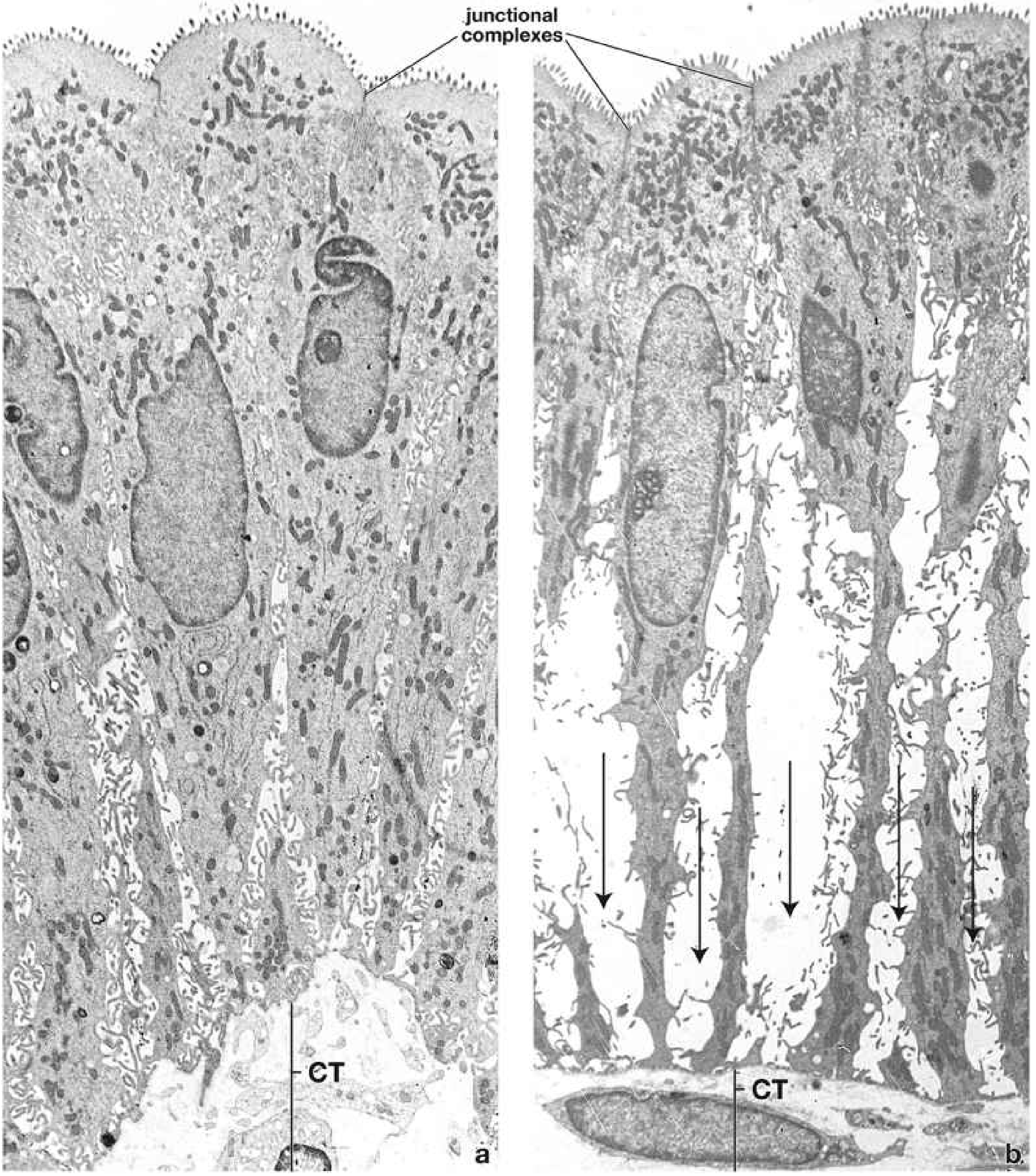
- Left (a): Resting state - tall columnar cells with apical microvilli, junctional complexes, mitochondria, and connective tissue (CT) below
- Right (b): Active fluid transport - intercellular spaces are greatly distended (arrows) as salt and water are pumped into them, then pass into the underlying CT and blood vessels
2. Muscularis Externa (No Muscularis Mucosa or Submucosa)
- Smooth muscle bundles are randomly oriented (no distinct inner circular/outer longitudinal layers as in intestine)
- Bundles intermixed with collagen and elastic fibers
- Contraction reduces bladder volume, expelling bile through the cystic duct
- Triggered by cholecystokinin (CCK) from duodenal enteroendocrine cells in response to fat
3. Perimuscular Connective Tissue / Adventitia / Serosa
- Thick layer of dense connective tissue external to the muscularis
- Contains large blood vessels, extensive lymphatic network, and autonomic nerves (parasympathetic neuron cell bodies are found in the wall of the cystic duct)
- Rich in elastic fibers and adipose tissue
- Adventitia where the gallbladder attaches to the liver surface
- Serosa (visceral peritoneum) - layer of mesothelium + thin loose connective tissue - covers the unattached (free) surface
4. Rokitansky-Aschoff Sinuses
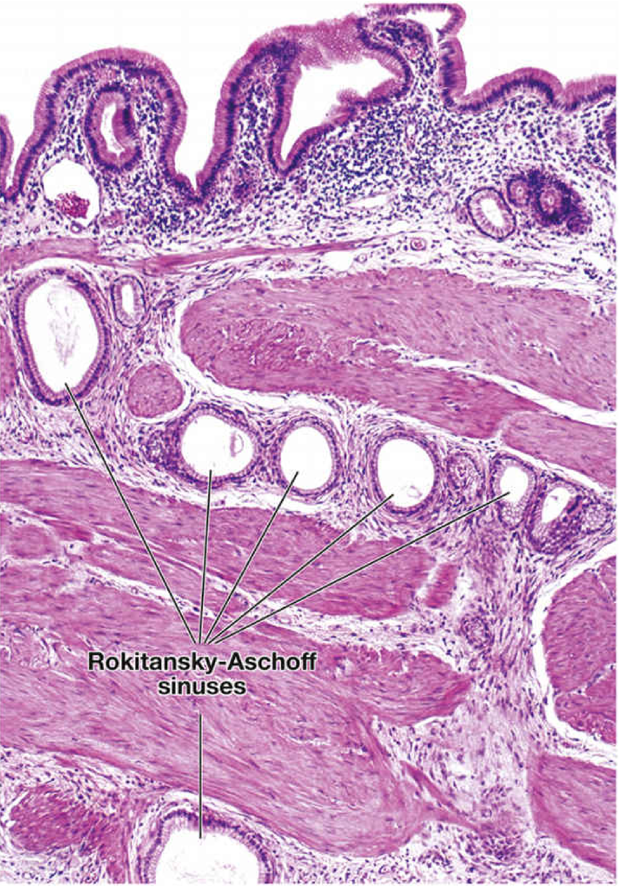
Rokitansky-Aschoff (R-A) sinuses are deep diverticula of the mucosa that extend through the muscularis externa into the perimuscular connective tissue. Key points:
- Result from epithelial hyperplasia and herniation through the muscularis
- Thought to presage pathological changes
- Bacteria may accumulate in these sinuses causing chronic inflammation - a risk factor for gallstone formation
- Prominent feature of adenomyomatosis of the gallbladder
5. Bile Concentration Mechanism
The gallbladder epithelium concentrates bile via coupled transport:
- Na+, Cl-, and HCO3- are actively transported (via Na+/K+-ATPase) into the lateral intercellular compartment
- This creates an osmotic gradient
- Water follows passively through aquaporin channels (AQP1 and AQP8) on apical and basolateral membranes
- Fluid passes from distended intercellular space across basal lamina → lamina propria → fenestrated capillaries
Key Distinguishing Features (Exam Points)
| Feature | Gallbladder | Rest of GI Tract |
|---|---|---|
| Muscularis mucosa | Absent | Present |
| Submucosa | Absent | Present |
| Smooth muscle orientation | Random | Layered (inner circular, outer longitudinal) |
| Epithelium | Simple columnar | Varies |
| Mucosal glands | At neck only | Throughout |
| Outer covering (liver side) | Adventitia | - |
| Outer covering (free surface) | Serosa | Serosa |
Sources: Histology: A Text and Atlas with Correlated Cell and Molecular Biology, pp. 1701-1707 | Sleisenger and Fordtran's Gastrointestinal and Liver Disease
This is a shared conversation. Sign in to Orris to start your own chat.