
Cartilages of nasal bone and their number
The nasal skeleton consists of bone and cartilage. The cartilaginous part includes: 1 septal cartilage, 2 upper lateral (lateral nasal) cartilages, 2 lower lateral (alar/greater alar) cartilages, and variable minor alar cartilages. That gives a typical count of 5 named cartilages (plus small accessory ones).
Searching sources...
Reading File
Finding Sources
Finding Sources
Reading File
Finding Sources
Reading File
Reading File
Finding Sources
Loading Image
Reading File
Reading File
Reading File
Loading Image
I now have comprehensive information from the textbooks along with excellent anatomical diagrams. Here is the full answer:
Cartilages of the Nose - Complete Overview
The external nose is supported by a cartilaginous framework below the nasal bones. There are 5 named cartilages (some paired, giving a total of 9 individual cartilaginous pieces including accessory cartilages), plus variable small accessory ones.
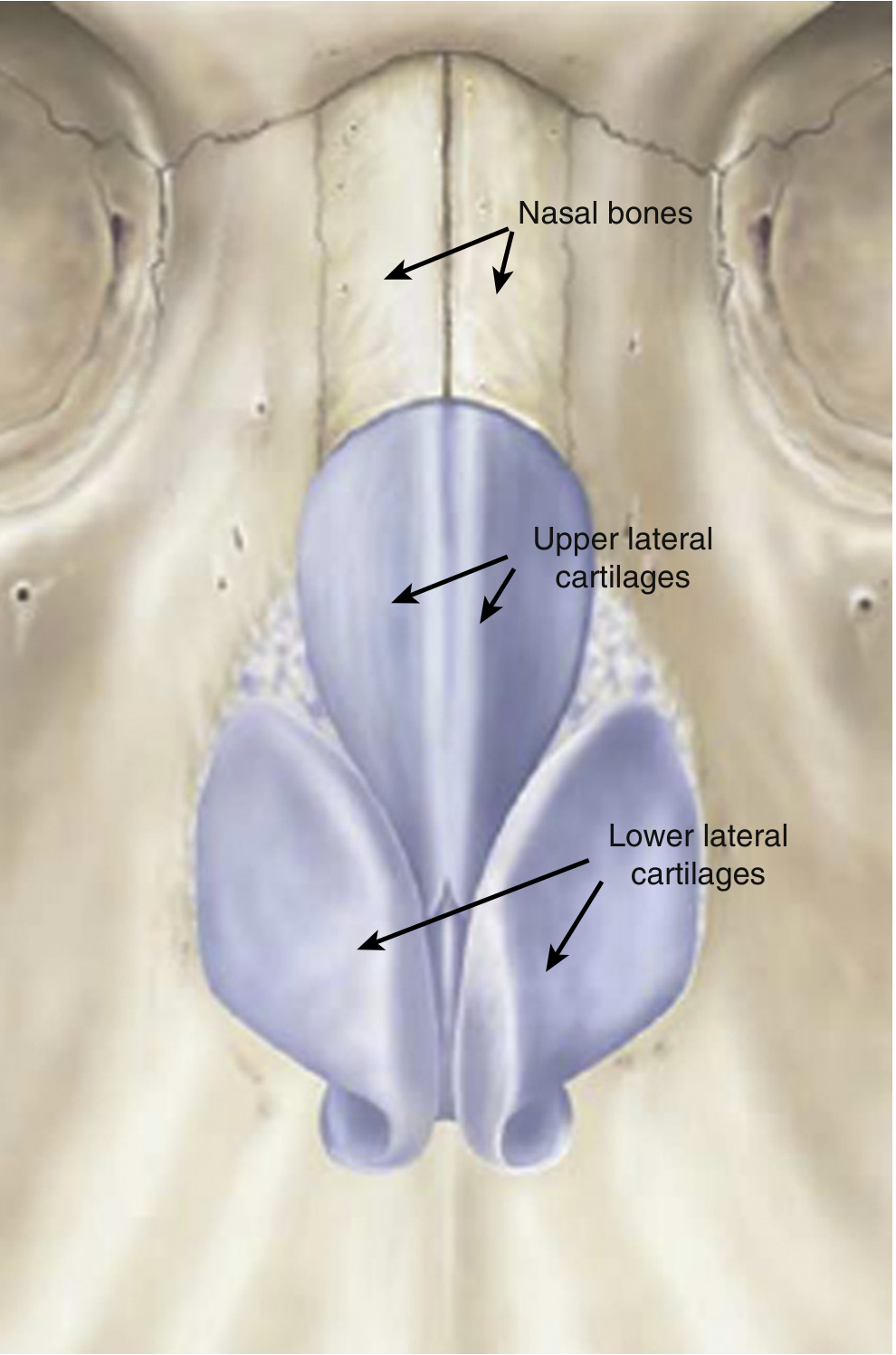
Summary Table
| Cartilage | Number | Location / Region |
|---|---|---|
| Septal cartilage (quadrilateral/quadrangular) | 1 | Midline - forms anterior nasal septum |
| Upper lateral cartilages (ULC) | 2 (paired) | Middle third of nose |
| Lower lateral cartilages (LLC / alar cartilages) | 2 (paired) | Lower third / nasal tip |
| Sesamoid (minor alar) cartilages | Variable (2-4) | Lateral to LLC |
| Vomeronasal cartilages | 2 (paired) | Along inferior edge of septal cartilage |
Total named cartilages = 5 types; typical individual pieces = 9 (excluding variable accessory ones)
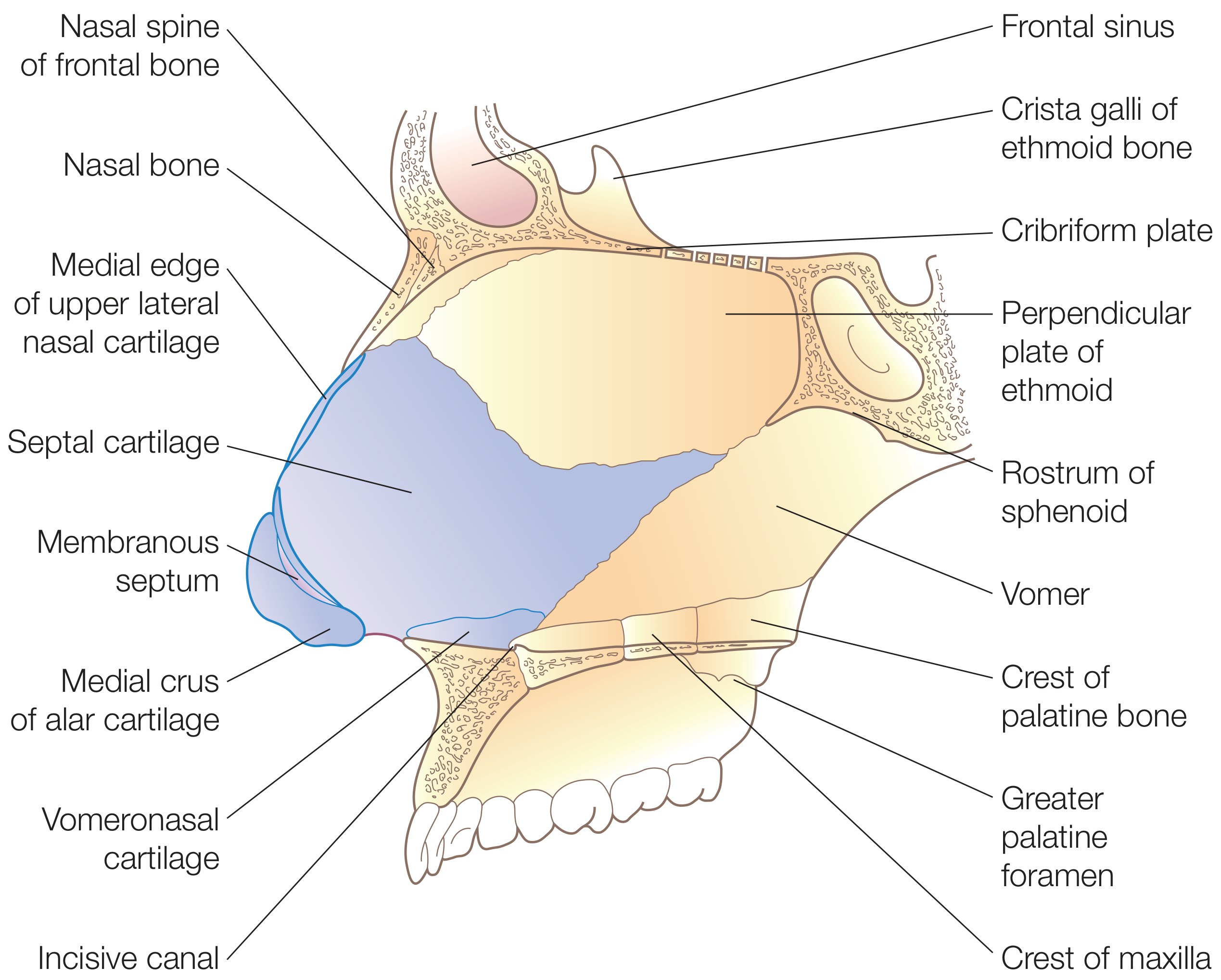
1. Septal Cartilage (Quadrilateral Cartilage) - 1
- Also called the quadrangular cartilage
- Forms the cartilaginous anterior nasal septum
- Bound to the nasal bones superiorly, perpendicular plate of ethmoid posterosuperiorly, and vomer posteroinferiorly
- Continuous with both upper lateral cartilages at the nasal bridge ("keystone area")
- A posterior projection called the sphenoidal process (septal tail) extends between the vomer and the perpendicular plate
(Scott-Brown's Otorhinolaryngology, p. 1014; Cummings Otolaryngology)
2. Upper Lateral Cartilages (ULC) - 2 (paired)
- Also called the triangular cartilages or lateral nasal cartilages
- Support the middle third of the nose
- Their cephalic edge slides under the nasal bones at the keystone area
- Their medial edges are fused with the dorsal septal cartilage (they are actually continuous with the septal cartilage superiorly and only become separate structures inferiorly)
- The angle between the ULC and septum forms the internal nasal valve (most important airway resistance point in the nose)
(Murray & Nadel's Respiratory Medicine, p. 1608; Scott-Brown's, p. 1222)
3. Lower Lateral Cartilages (LLC / Greater Alar Cartilages) - 2 (paired)
- Also called the alar cartilages or greater alar cartilages
- Define the lower third and tip of the nose
- Each has three crura:
- Medial crus - forms the columella, attaches to caudal septal cartilage
- Middle crus (intermediate crus) - forms the dome/nasal tip
- Lateral crus - extends laterally to form the alar rim
- Contrary to common belief, the lower lateral cartilages do not extend to the very margins of the nostril rim
(Scott-Brown's Otorhinolaryngology, p. 1222; Cummings Otolaryngology)
4. Sesamoid (Minor Alar / Accessory) Cartilages - Variable (2-4)
- Small fibrofatty cartilages situated lateral to the lower lateral cartilages
- Part of the "sesamoid complex" that bridges the lateral crus to the piriform aperture
- They are a minor tip support mechanism - they extend support of the lateral crura to the piriform aperture
(K.J. Lee's Essential Otolaryngology)
5. Vomeronasal Cartilages (Cartilages of Jacobson) - 2 (paired)
- Narrow strips of cartilage located between the inferior edge of the septal cartilage and the vomer
- Embryological remnants of the vomeronasal organs (Jacobson's organs), which regress before birth
- Often found in association with a septal spur
- Can be used as graft material when septal/auricular cartilage is absent or exhausted
(Cummings Otolaryngology, p. 564)
Anatomical Diagram of the Nasal Septum
Key Structural Relationships (for exam purposes)
- Upper third of nose = nasal bones
- Middle third = upper lateral cartilages
- Lower third = lower lateral cartilages
- The internal nasal valve = angle between ULC and dorsal septum (primary airway resistance zone)
- The external nasal valve = formed by lower lateral cartilage + septum + inferior turbinate
The 3 major tip support mechanisms are: (1) shape/strength of the lower lateral cartilages, (2) attachment of medial crural footplates to caudal septum, and (3) attachment of ULC to LLC (cephalic border).
- Cummings Otolaryngology Head and Neck Surgery
- Scott-Brown's Otorhinolaryngology Head & Neck Surgery
- Murray & Nadel's Textbook of Respiratory Medicine
- K.J. Lee's Essential Otolaryngology
This is a shared conversation. Sign in to Orris to start your own chat.