I now have all the content needed for a complete, exam-focused overview. Let me compile the full answer.
Inflammation - Complete Medical Student Overview
Source: Robbins & Kumar Basic Pathology (Robbins Pathology)
Definition
Inflammation is a protective response of vascularized tissues to harmful stimuli (microbes, necrotic cells, foreign substances). Its goal is to eliminate the cause of injury, clear debris, and initiate tissue repair. It is classified as acute or chronic.
ACUTE INFLAMMATION
The Three Core Components
- Vasodilation - dilation of small vessels (responsible for redness and warmth)
- Increased vascular permeability - allows protein-rich fluid to escape into tissues
- Leukocyte emigration - movement of leukocytes (mainly neutrophils) from blood into tissue
All reactions occur mainly in postcapillary venules and are triggered by inflammatory mediators.
Cardinal Signs (Celsus + Virchow)
| Sign | Latin | Mechanism |
|---|
| Redness (rubor) | Rubor | Vasodilation |
| Warmth (calor) | Calor | Vasodilation |
| Swelling (tumor) | Tumor | Exudation of fluid |
| Pain (dolor) | Dolor | Prostaglandins, bradykinin |
| Loss of function | Functio laesa | Combined |
Exudate vs Transudate
- Exudate: high protein, cellular debris, caused by increased vascular permeability (inflammation)
- Transudate: low protein, mainly albumin, caused by osmotic/hydrostatic imbalance (not inflammation)
- Pus (purulent exudate): rich in neutrophils and dead cell debris
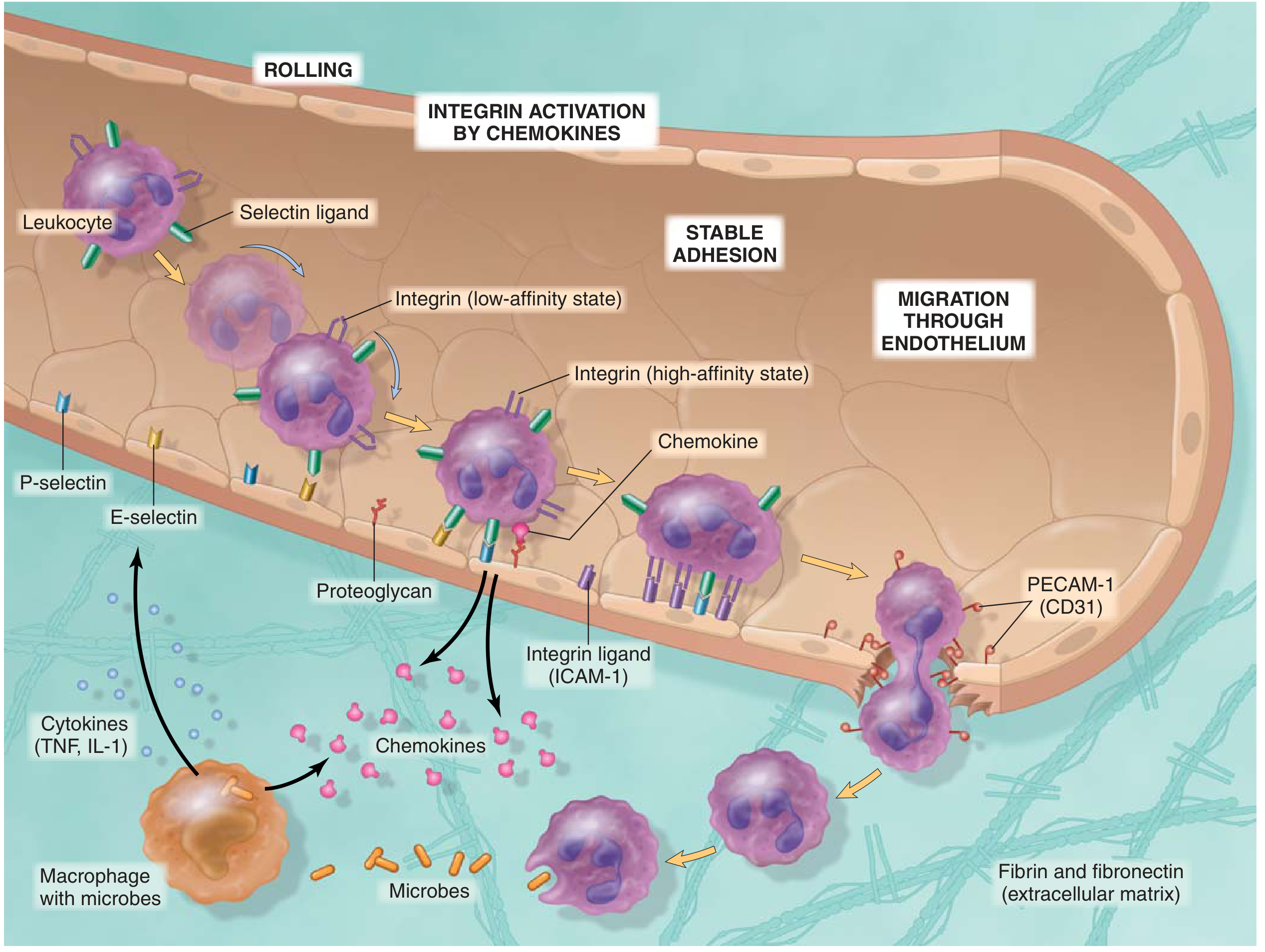
LEUKOCYTE RECRUITMENT (Multi-Step Process)
| Step | Process | Molecules |
|---|
| 1 | Margination (slowing, peripheral displacement) | Stasis |
| 2 | Rolling | Selectins (P-, E-selectin on endothelium; L-selectin on leukocytes) |
| 3 | Firm adhesion | Integrins (activated by chemokines) + ICAM-1 on endothelium |
| 4 | Transmigration (diapedesis) | PECAM-1 (CD31) |
| 5 | Migration to site of injury | Chemokines, C5a, LTB4 |
Key memory aid: "Rolling, Sticking, Crawling, and Migrating" - Selectins → Integrins → PECAM-1
INFLAMMATORY MEDIATORS (High-Yield)
Cell-Derived Mediators
| Mediator | Source | Key Actions |
|---|
| Histamine | Mast cells, basophils, platelets | Vasodilation, increased permeability |
| Prostaglandins (PGE2, PGI2) | Mast cells, macrophages, endothelium | Vasodilation, edema, fever, pain |
| Leukotrienes (LTC4, LTD4, LTE4) | Mast cells, leukocytes | Increased permeability; bronchoconstriction (key in asthma) |
| Leukotriene B4 (LTB4) | Leukocytes | Chemotaxis |
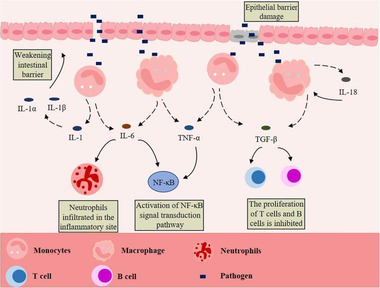
| TNF, IL-1 | Macrophages | Fever, endothelial activation, leukocyte recruitment |
| Chemokines | Leukocytes, macrophages | Directional migration of leukocytes |
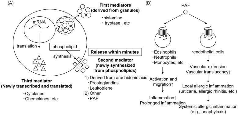
| PAF (Platelet-Activating Factor) | Mast cells, endothelium | Vasodilation, increased permeability, bronchoconstriction |
| ROS (reactive oxygen species) | Leukocytes | Tissue damage |
| NO (nitric oxide) | Endothelium, macrophages | Vasodilation |
Plasma-Derived Mediators
| System | Key Products | Role |
|---|
| Complement | C3a, C5a | Increased permeability, chemotaxis, opsonization; C5b-9 = membrane attack complex |
| Kinin system | Bradykinin | Vasodilation, pain, increased permeability |
| Coagulation | Thrombin, fibrin | Vessel sealing, fibrinous exudate |
Summary Table (Reactions → Principal Mediators)
(From Robbins Basic Pathology)
| Reaction | Principal Mediators |
|---|
| Vasodilation | Histamine, NO |
| Increased permeability | Histamine, C3a/C5a, LTC4/D4/E4 |
| Chemotaxis/leukocyte recruitment | TNF, IL-1, chemokines, C5a, LTB4 |
| Fever | IL-1, TNF, prostaglandins |
| Pain | Prostaglandins, bradykinin, neuropeptides |
| Tissue damage | Lysosomal enzymes, ROS |
ARACHIDONIC ACID PATHWAY (Key for Pharmacology)
Membrane phospholipids
        ↓ Phospholipase A2 (blocked by CORTICOSTEROIDS)
Arachidonic Acid
    ↙                    ↘
COX-1, COX-2              Lipoxygenase
(blocked by NSAIDs/        (blocked by 5-LOX inhibitors)
 Aspirin)                        ↓
    ↓                    Leukotrienes (LTB4, LTC4, D4, E4)
Prostaglandins (PGE2,
PGI2, TXA2, PGD2)
- TXA2 (platelets): vasoconstriction, platelet aggregation
- PGI2/Prostacyclin (endothelium): vasodilation, inhibits platelet aggregation
- PGE2, PGD2: vasodilation, edema, fever
MORPHOLOGIC PATTERNS OF ACUTE INFLAMMATION
| Pattern | Features | Example |
|---|
| Serous | Watery, low-cell fluid in body cavities or tissue | Skin blister (burn), pleural effusion |
| Fibrinous | Fibrin deposition due to large permeability increase | Fibrinous pericarditis ("bread and butter" pericarditis) |
| Suppurative (purulent) | Pus-forming; abundant neutrophils | Abscess, bacterial pneumonia |
| Ulcerative | Necrotic epithelium, underlying acute/chronic inflammation | Peptic ulcer |
OUTCOMES OF ACUTE INFLAMMATION
- Complete resolution - removal of debris by macrophages, resorption of edema via lymphatics (ideal outcome)
- Healing by connective tissue / fibrosis (scarring) - when tissue cannot regenerate or fibrin cannot be cleared
- Progression to chronic inflammation - when injurious agent persists
CHRONIC INFLAMMATION
Chronic inflammation = prolonged (weeks-months) response in which inflammation, tissue injury, and repair coexist.
Key Features vs Acute
| Feature | Acute | Chronic |
|---|
| Duration | Minutes to days | Weeks to months |
| Dominant cells | Neutrophils | Macrophages, lymphocytes, plasma cells |
| Main mediators | Histamine, eicosanoids | Cytokines (IL-1, TNF, IFN-γ) |
| Vascular changes | Prominent | Less prominent |
| Tissue injury | Mild | Often progressive |
| Fibrosis | Absent | Common |
Causes of Chronic Inflammation
- Persistent infections - mycobacteria (TB), fungi, parasites
- Hypersensitivity/autoimmune diseases - rheumatoid arthritis, lupus, Crohn disease, asthma
- Prolonged exposure to toxic agents - silica (silicosis), endogenous lipids (atherosclerosis)
Key Cells in Chronic Inflammation
- Macrophages - dominant cell; activated by IFN-γ; secrete cytokines, ROS, proteases; central to tissue damage AND repair
- Lymphocytes - T cells (activate macrophages), B cells/plasma cells (antibodies)
- Eosinophils - parasite infections, allergic reactions
- Mast cells - immediate hypersensitivity
GRANULOMATOUS INFLAMMATION
A special pattern of chronic inflammation with high diagnostic significance.
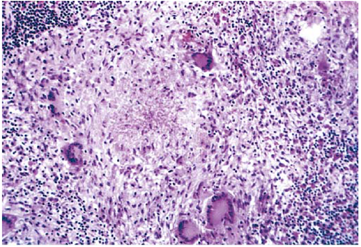
Definition: Collections of activated macrophages (epithelioid cells), often with T lymphocytes, sometimes with central caseous necrosis.
Histologic Components
- Epithelioid macrophages - large, pink, granular cytoplasm; indistinct cell borders
- Langhans giant cells - multinucleated (40-50 µm), formed by fusion of macrophages
- Collar of lymphocytes surrounding the core
- Fibroblasts and connective tissue at the rim (older granulomas)
Types of Granulomas
| Type | Central necrosis | Examples |
|---|
| Caseating | Yes (cheesy appearance) | Tuberculosis, histoplasmosis |
| Noncaseating | No | Sarcoidosis, Crohn disease, leprosy, foreign body reactions |
Key Diseases with Granulomatous Inflammation
| Disease | Cause | Notes |
|---|
| Tuberculosis | M. tuberculosis | Caseating; acid-fast bacilli; Langhans giant cells |
| Leprosy | M. leprae | Acid-fast bacilli in macrophages; noncaseating |
| Sarcoidosis | Unknown | Noncaseating; "naked granulomas" |
| Crohn disease | Immune reaction to intestinal bacteria | Noncaseating in bowel wall |
| Syphilis | Treponema pallidum | Gumma; plasma cell infiltrate |
| Cat-scratch disease | Bartonella henselae | Stellate granuloma with neutrophils |
| Foreign body | Talc, sutures, silica | Material identifiable by polarized light |
SYSTEMIC EFFECTS OF INFLAMMATION (Acute-Phase Response)
Fever: IL-1, TNF → induce COX-2 in hypothalamus → PGE2 → raises thermostat set-point
Acute-Phase Proteins (liver-derived, stimulated by IL-1, TNF, IL-6):
- CRP (C-reactive protein) - opsonin, fixes complement; marker of cardiovascular risk
- Fibrinogen - causes rouleaux formation → elevated ESR
- Serum amyloid A (SAA) - prolonged elevation → secondary amyloidosis
Leukocytosis: Most bacterial infections → neutrophilia; viral infections → lymphocytosis; allergies/parasites → eosinophilia; typhoid, rickettsiae → leukopenia
Anemia of chronic disease: IL-6 → hepcidin → reduces iron availability → normocytic/normochromic anemia
Sepsis (extreme systemic response): massive TNF, IL-1, IL-6 → DIC, hypoglycemia, cardiovascular failure, multiorgan failure
HIGH-YIELD EXAM SUMMARY
| Concept | Key Fact |
|---|
| First leukocyte in acute inflammation | Neutrophil (first 6-24 hr) |
| Predominant cell in chronic inflammation | Macrophage |
| Mediator of fever | IL-1, TNF → PGE2 |
| Mediator of pain | Prostaglandins, bradykinin |
| Mediator of vasodilation | Histamine, NO |
| Rolling = mediated by | Selectins |
| Firm adhesion = mediated by | Integrins + ICAM-1 |
| Transmigration = mediated by | PECAM-1 (CD31) |
| Asthma mediators | LTC4, LTD4, LTE4 (cysteinyl leukotrienes) |
| NSAIDs block | COX-1 and COX-2 |
| Corticosteroids block | Phospholipase A2 (entire AA pathway) |
| Caseating granuloma | TB (also fungal) |
| Noncaseating granuloma | Sarcoidosis, Crohn disease, leprosy |
| ESR elevated because of | Fibrinogen → rouleaux |
| CRP clinical use | Marker of inflammation and cardiovascular risk |
| Anemia of chronic disease mechanism | Hepcidin ↑ → iron sequestration |
Robbins & Kumar Basic Pathology - Chapter 2 (Inflammation and Repair)