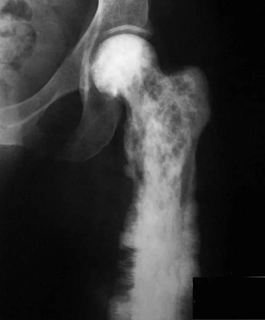
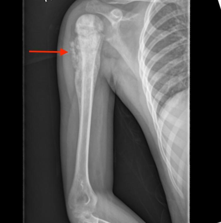
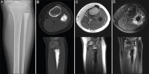
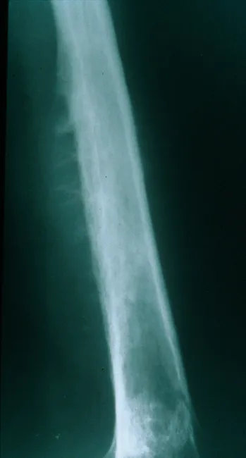
Here is the complete bone tumors pathology reference, compiled from Robbins & Kumar Basic Pathology and supplementary textbooks.
Bone Tumours - Complete Pathology
(Source: Robbins & Kumar Basic Pathology; Rheumatology 2-Vol Set; Miller's Review of Orthopaedics; Campbell's Operative Orthopaedics)
SECTION 1: CLASSIFICATION OF PRIMARY BONE TUMOURS
Bone tumors are classified primarily by the tissue of origin (histogenetic type) and behavior (benign vs. malignant). The WHO recognizes >60 subtypes.
| Category | Behavior | Tumor Type | Common Location | Age (yr) | Key Morphology |
|---|
| Cartilage-forming | Benign | Osteochondroma | Metaphysis of long bones | 10-30 | Bony excrescence with cartilage cap |
| Benign | Chondroma (Enchondroma) | Small bones of hands & feet | 30-50 | Intramedullary hyaline cartilage nodule |
| Malignant | Chondrosarcoma | Pelvis, shoulder, ribs | 40-60 | Infiltrates marrow; chondrocytes with atypia |
| Bone-forming | Benign | Osteoid osteoma | Metaphysis of long bones | 10-20 | Small cortical nidus; interlacing woven bone trabeculae |
| Benign | Osteoblastoma | Vertebral column (posterior) | 10-20 | Like osteoid osteoma but >2 cm |
| Malignant | Osteosarcoma | Metaphysis: distal femur, proximal tibia | 10-20 | Malignant cells producing woven bone/osteoid |
| Unknown origin | Benign | Giant cell tumor (GCT) | Epiphysis of long bones | 20-40 | Osteoclast-type giant cells + mononuclear stromal cells |
| Benign | Aneurysmal bone cyst | Femur, tibia, vertebra | 10-20 | Multiloculated blood-filled spaces |
| Malignant | Ewing sarcoma | Diaphysis of long bones | 10-20 | Sheets of small round blue cells; t(11;22) |
Memory tip for tumor locations: OsteoSarcoma = Metaphysis | Giant Cell Tumor = Epiphysis | Ewing = Diaphysis
SECTION 2: BONE-FORMING TUMOURS
2A. OSTEOID OSTEOMA
| Feature | Details |
|---|
| Incidence | Most common benign bone-forming tumor in young patients |
| Age/Sex | 10-20 yr; M > F |
| Location | Cortex of metaphysis of long bones (femur, tibia); posterior vertebral elements |
| Size | < 2 cm (key distinguishing feature from osteoblastoma) |
| Symptoms | Night pain, relieved by aspirin/NSAIDs (prostaglandin-mediated) |
Radiology: Round lucent nidus (<1.5 cm) surrounded by dense reactive sclerosis
Histology:
- Well-circumscribed round-oval mass
- Delicate interlacing trabeculae of woven bone rimmed by a single layer of osteoblasts
- Stroma: loose connective tissue with dilated, congested capillaries
Treatment: Radiofrequency ablation (RFA) or conservative NSAIDs
2B. OSTEOBLASTOMA
| Feature | Details |
|---|
| Size | > 2 cm (larger than osteoid osteoma) |
| Location | Posterior vertebral elements (laminae, pedicles) |
| Pain | Unresponsive to aspirin |
| Histology | Similar to osteoid osteoma; no marked reactive bony sclerosis around it |
Treatment: Curettage or en bloc excision
2C. OSTEOSARCOMA (Osteogenic Sarcoma)
Definition
Malignant tumor producing osteoid matrix or mineralized bone. The most common primary malignant bone tumor (excluding hematopoietic tumors).
Epidemiology
| Feature | Details |
|---|
| Age | Bimodal: ~75% <20 yr (adolescent); second peak in older adults (secondary type) |
| Sex | M > F (1.6:1) |
| Site | Metaphysis; ~50% near the knee (distal femur or proximal tibia) |
| Incidence | 1-1.5 per million |
Pathogenesis
Peak incidence at adolescent growth spurt - rapid proliferation near growth plate predisposes to mutations.
| Gene | Role |
|---|
| RB | Mutated in 70% of sporadic cases; germline mutation = 1000-fold increased risk |
| TP53 | Germline mutation in Li-Fraumeni syndrome; common in sporadic tumors |
| MDM2 / CDK4 | Overexpressed in low-grade osteosarcomas (inhibit p53 and RB) |
| CDKN2A | Inactivated (encodes p16 and p14 tumor suppressors) |
| MYC | Amplified in up to 50%; associated with poor prognosis |
No specific chromosomal translocation (unlike Ewing sarcoma). Instead: complex karyotypes with numerous chromosomal aberrations.
Secondary Osteosarcoma
Occurs in older adults in association with:
- Paget disease of bone
- Bone infarcts
- Prior radiation to bone
Gross Pathology
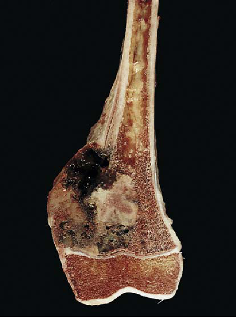
- Bulky, gritty, tan-white tumor with areas of hemorrhage
- Destroys cortex and invades soft tissue
- Extensive intramedullary spread
- Confined to metaphyseal side of growth plate (rarely crosses the epiphyseal plate)
Radiology
| Sign | Description |
|---|
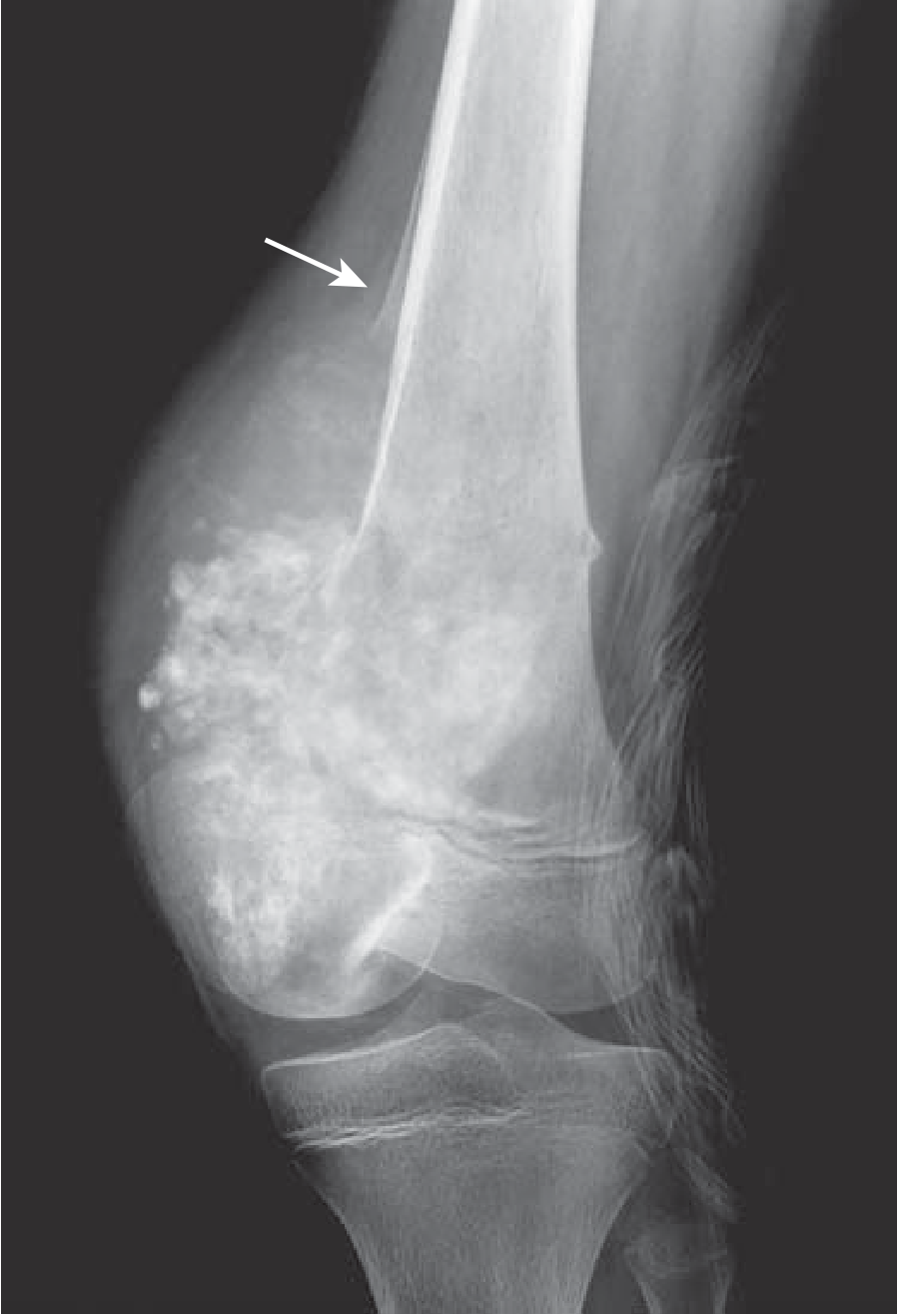
| Codman triangle | Periosteum lifted by tumor → triangular wedge of reactive subperiosteal bone. Indicates aggressive tumor; not pathognomonic |
| Sunburst pattern | Perpendicular periosteal spicules radiating from bone surface |
| Mixed lytic/sclerotic | Combined bone destruction + new bone formation |
| Soft tissue mass | Extension beyond cortex |
Histology (Microscopy)
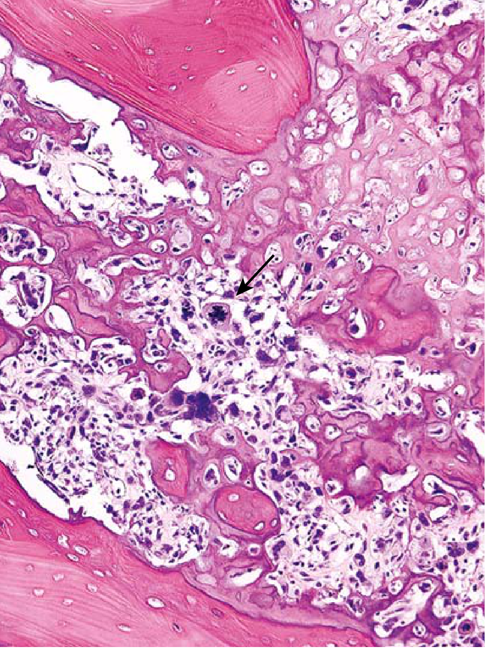
Diagnostic hallmark: Malignant tumor cells producing unmineralized osteoid or mineralized bone
Microscopic features:
- Cellular pleomorphism - large hyperchromatic nuclei
- Bizarre tumor giant cells
- Abundant mitoses including abnormal (bipolar) forms
- Fine lacelike osteoid produced by malignant cells
- Extensive necrosis
- Intravascular invasion
- Bridges preexisting lamellar bone
Clinical Features & Prognosis
| Feature | Details |
|---|
| Presentation | Painful, progressively enlarging mass; may present as pathologic fracture |
| Metastasis | All assumed to have occult metastases at diagnosis. Hematogenous spread to lungs (most common), then bone, brain |
| Treatment | Neoadjuvant chemo → Surgery (limb-sparing) → Adjuvant chemo |
| 5-year survival | ~70% without overt metastases at diagnosis |
| With metastases | Significantly worse prognosis |
SECTION 3: CARTILAGE-FORMING TUMOURS
3A. OSTEOCHONDROMA (Exostosis)
| Feature | Details |
|---|
| Definition | Benign cartilage-capped tumor arising from the bone surface |
| Type | Sessile or pedunculated (with bony stalk); 85% solitary |
| Age/Sex | Late adolescence/early adulthood; M:F = 3:1 |
| Location | Metaphysis near growth plate; especially around the knee |
| Origin | Bones of endochondral origin only |
| Genetics | Loss-of-function mutations in EXT1 or EXT2 genes (encode heparan sulfate enzymes) → disrupts hedgehog signaling → abnormal chondrocyte differentiation |
| Syndrome | Multiple hereditary exostoses (familial; bones bowed and shortened) |
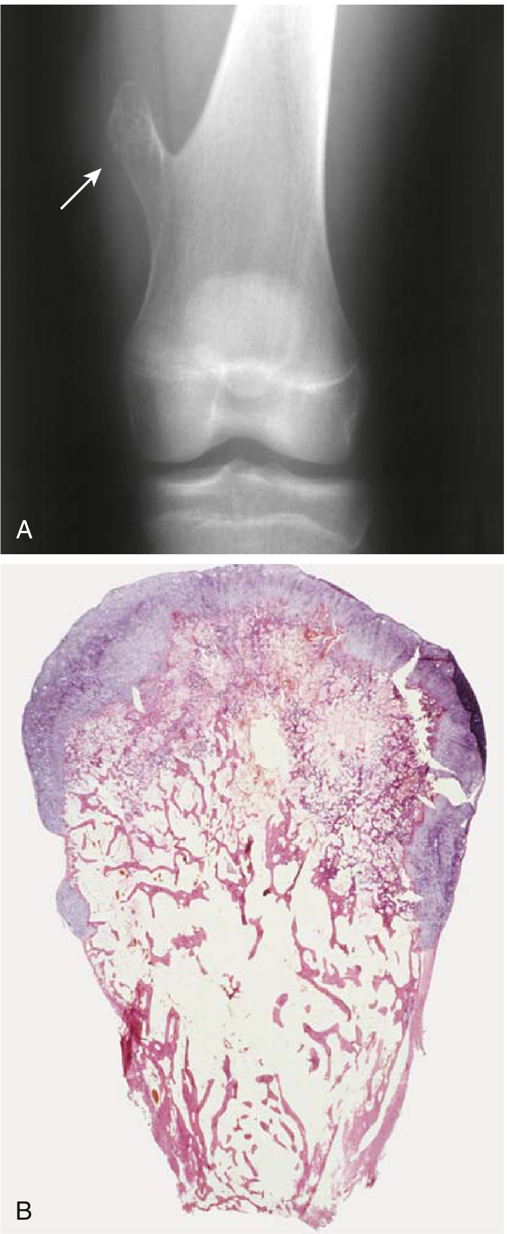
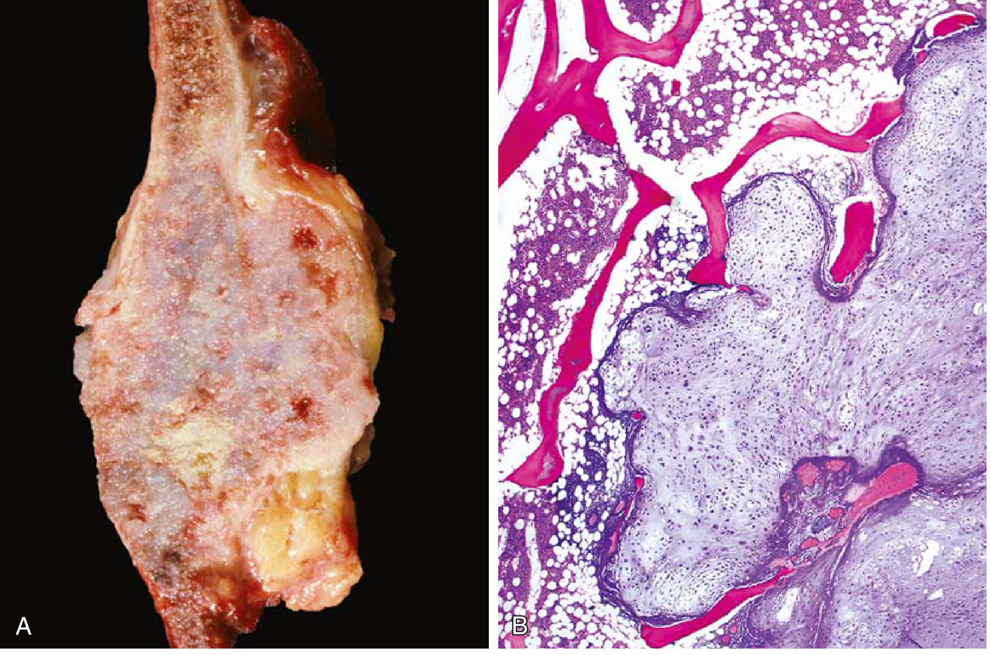
Gross: Bony excrescence with hyaline cartilage cap covered by perichondrium
Histology:
- Cap = hyaline cartilage recapitulating growth plate
- Undergoes endochondral ossification inward
- Medullary cavity of osteochondroma is continuous with host bone
Clinical Features:
- Slow-growing; stops at growth plate closure
- Can cause pain if nerve impingement or stalk fracture
- Sarcomatous transformation rare in solitary lesions; more common in multiple hereditary exostoses
- Treatment: simple excision when symptomatic
3B. CHONDROMA (ENCHONDROMA)
| Feature | Details |
|---|
| Definition | Benign tumor of hyaline cartilage within the medullary cavity (enchondroma) or on bone surface (juxtacortical) |
| Age | 20-50 yr; equal sex distribution |
| Location | Most common site: small bones of hands and feet (proximal/middle phalanges); most common bone tumor of the hand |
| Genetics | IDH1 / IDH2 mutations → 2-hydroxyglutarate ("oncometabolite") → altered DNA methylation |
| Syndromes | Ollier disease (multiple enchondromas); Maffucci syndrome (enchondromas + hemangiomas, higher malignant potential) |
Radiology: Circumscribed lucency with central irregular calcifications, sclerotic rim, intact cortex
Histology:
- Benign-appearing chondrocytes in well-circumscribed nodules of hyaline cartilage
- Peripheral nodules may undergo endochondral ossification
- Center may calcify and become infarcted
- Syndromic enchondromas: more cellular + more atypia
Clinical: Usually asymptomatic; may cause pathologic fracture. Solitary lesions rarely undergo sarcomatous change.
3C. CHONDROSARCOMA
| Feature | Details |
|---|
| Definition | Malignant tumor producing cartilage |
| Incidence | 2nd most common malignant matrix-producing bone tumor (after osteosarcoma) |
| Age/Sex | 40s or older; M:F = 2:1 |
| Location | Axial skeleton - pelvis, shoulder, ribs (contrast with osteosarcoma which favors extremities) |
| Secondary type | 15% arise from preexisting enchondroma or osteochondroma |
| Genetics | EXT1/EXT2 mutations (in osteochondroma-related); IDH1/IDH2 mutations; CDKN2A silencing |
Subtypes: Conventional (90%), dedifferentiated, clear cell, mesenchymal
Gross:
- Bulky nodules of glistening gray-white, translucent cartilage with gelatinous myxoid areas
- Focal calcifications; central necrosis creating cystic spaces
- Extracortical extension common
Histology:
- Cartilage infiltrates marrow space and entraps normal bony trabeculae
- Graded 1-3 based on cellularity, atypia, mitoses (Grade 1 = low grade; Grade 3 = high grade with bizarre giant cells)
- Grade 1: low cellularity, plump vesicular nuclei, small nucleoli
- Grade 3: high cellularity, extreme pleomorphism, frequent mitoses
X-ray: "Flocculent" (popcorn/ring-and-arc) calcifications within cartilage matrix; cortical destruction; soft tissue mass
Clinical/Treatment:
- Painful, progressively enlarging mass
- Most are grade 1 (low grade, good prognosis)
- Treatment: Wide surgical resection only (chemo and radiotherapy largely ineffective - key exam point!)
- 10-year survival: Grade 1 = ~83%; Grade 2 = ~53%; Grade 3 = ~22%
SECTION 4: TUMOURS OF UNKNOWN ORIGIN
4A. GIANT CELL TUMOUR (GCT) OF BONE
| Feature | Details |
|---|
| Behavior | Benign but locally aggressive |
| Age | 3rd-5th decade (20-40 yr); more common in young women |
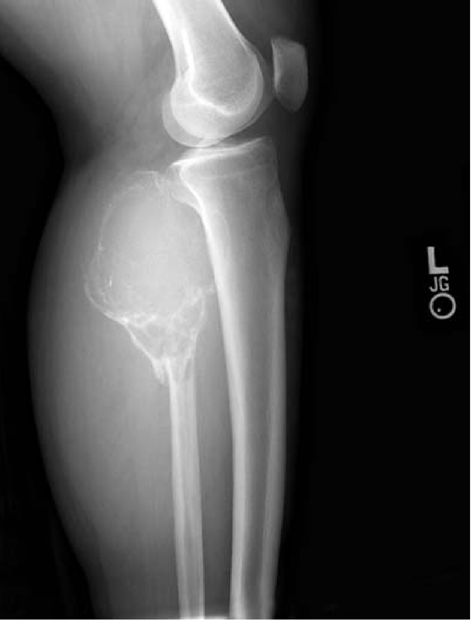
| Location | Epiphysis of long bones - most commonly distal femur and proximal tibia (around the knee); also distal radius |
| Key distinction | Epiphyseal location sets it apart from most other bone tumors |
Pathogenesis
- Neoplastic cells: primitive osteoblast precursors that express high levels of RANKL
- RANKL drives proliferation and differentiation of osteoclast precursors
- Multinucleate giant cells: are the non-neoplastic osteoclasts (express RANK receptor)
- Absence of normal osteoblast-osteoclast feedback → localized highly destructive bone resorption
Gross & Histology
Gross: Red-brown mass; destroys overlying cortex → bulging soft tissue mass bounded by thin shell of reactive bone; frequently undergoes cystic degeneration
Histology:
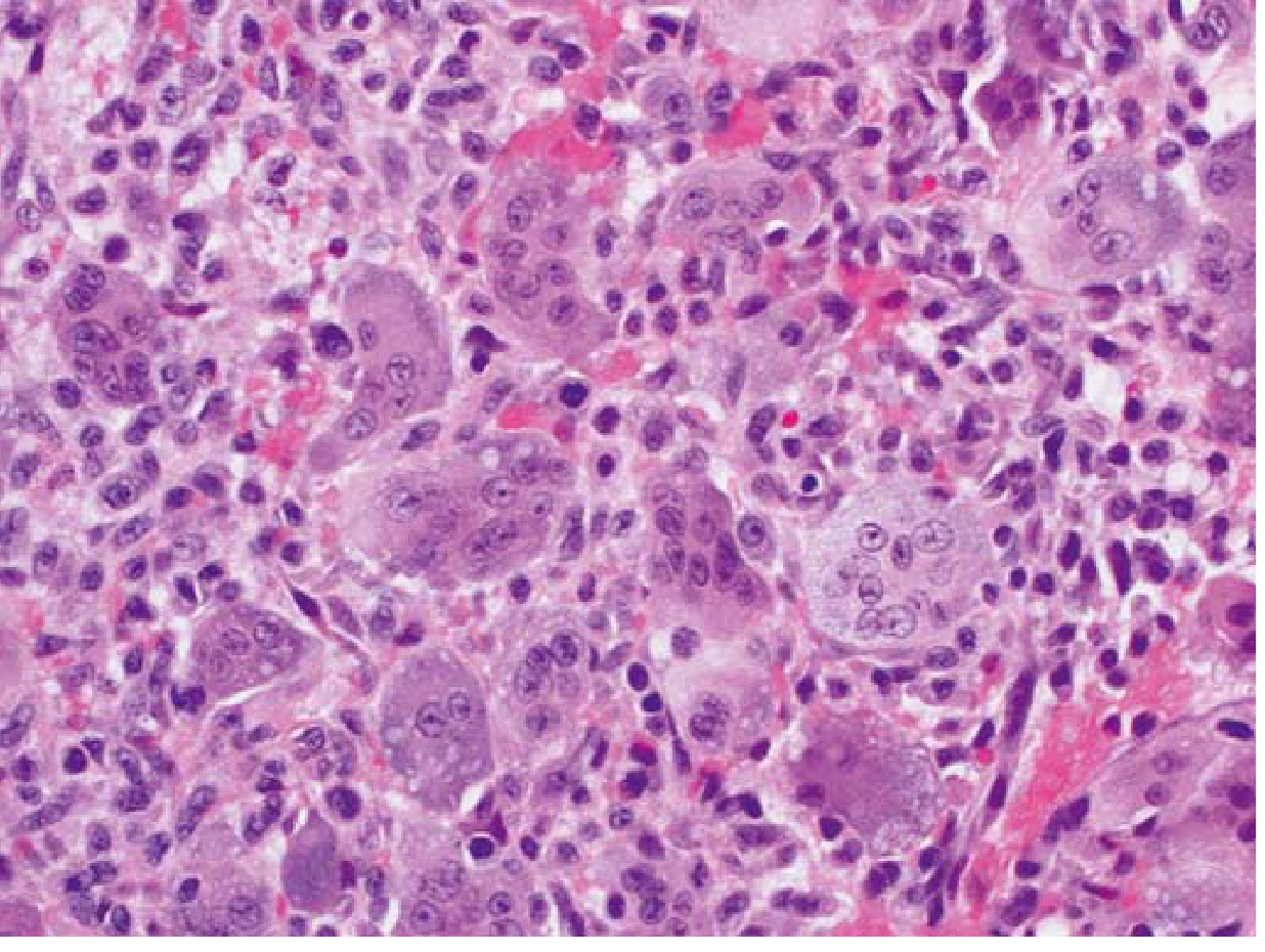
- Numerous multinucleate osteoclast-type giant cells (up to 100+ nuclei)
- Background of uniform oval mononuclear neoplastic stromal cells
- Giant cells are reactive (non-neoplastic); stromal cells are the true neoplastic component
Radiology
Predominantly lytic and expansile with cortical destruction; "soap-bubble" appearance; may show pathologic fracture
Clinical Features & Treatment
| Feature | Details |
|---|
| Symptoms | Joint pain (near epiphysis), arthritis-like symptoms, pathologic fracture |
| Treatment | Curettage (primary); 40-60% recur locally |
| Alternative | Denosumab (anti-RANKL antibody) - inhibits osteoclast maturation; used when surgery would be deforming |
| Metastasis | ~4% develop lung metastases; most cured by excision of metastases |
4B. EWING SARCOMA
| Feature | Details |
|---|
| Definition | Malignant tumor of small round cells |
| Incidence | ~10% of primary malignant bone tumors; 2nd most common bone sarcoma in children |
| Age/Sex | 80% <20 yr; slight male predominance |
| Location | Diaphysis of long bones (femur most common); 20% extraskeletal |
Pathogenesis
- >90% have balanced translocation t(11;22)(q24;q12) - EWSR1 + FLI1 genes
- Produces chimeric EWS/FLI1 fusion protein - binds chromatin, dysregulates transcription → uncontrolled growth
- Cell of origin: mesenchymal stem cells or primitive neuroectodermal cells (uncertain)
Gross & Histology
Gross:
- Arises in medullary cavity → invades cortex, periosteum, and soft tissue
- Periosteal reaction: layered reactive bone = "onion-skin" appearance on X-ray
- Soft, tan-white tumor; areas of hemorrhage and necrosis
Histology:
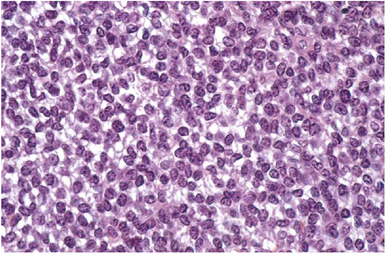
- Sheets of uniform small round cells - slightly larger and more cohesive than lymphocytes
- Scant cytoplasm, clear (rich in glycogen; PAS positive)
- Homer-Wright rosettes (circular cell groupings with central fibrillary core) may be present
- Tumor cells do NOT produce bone or cartilage
- One of the small round blue cell tumors (SRBCT) of childhood
Radiology
- Permeative, destructive diaphyseal lesion
- Onion-skin periosteal reaction (layered periosteal new bone)
- Soft tissue mass
Clinical Features & Treatment
| Feature | Details |
|---|
| Symptoms | Painful, enlarging mass; site is tender, warm, swollen (may mimic osteomyelitis/infection) |
| Fever/ESR | May be elevated - simulates infection |
| Spread | Hematogenous; lungs and other bones |
| Treatment | Multiagent chemotherapy + radiation ± surgery (chemo is highly effective; often curative) |
| 5-year survival | ~65-75% with localized disease |
4C. ANEURYSMAL BONE CYST (ABC)
| Feature | Details |
|---|
| Definition | Benign lesion characterized by multiloculated blood-filled spaces |
| Age | Adolescence (all ages affected) |
| Location | Femur, tibia, vertebral posterior elements |
| Behavior | Benign but locally aggressive |
| Genetics | Rearrangements of chromosome 17p13 → USP6 gene fusion (commonly with COL1A1) → USP6 overexpression → NF-κB activation → matrix metalloprotease upregulation → cystic bone resorption |
| Association | 20-40% are secondary to a preexisting lesion (most commonly GCT) |
Radiology:
- Eccentric, expansile, lytic metaphyseal lesion with well-defined margins
- MRI: fluid-fluid levels (pathognomonic - blood sediment by density)
Gross: Multiple blood-filled cystic spaces separated by thin tan-white septa
Histology:
- Cystic spaces lack endothelial lining (not true aneurysm)
- Septa contain: plump uniform fibroblasts + multinucleate osteoclast-like giant cells + reactive woven bone
Treatment: Curettage or excision; 10-50% recur
SECTION 5: NON-NEOPLASTIC LESIONS SIMULATING TUMOURS
Fibrous Cortical Defect / Nonossifying Fibroma (NOF)
| Feature | Details |
|---|
| Prevalence | Fibrous cortical defects in up to 50% of children >2 yr |
| Location | Eccentric metaphysis of distal femur / proximal tibia (often bilateral) |
| Size | <0.5 cm = fibrous cortical defect; 5-6 cm = nonossifying fibroma |
| Behavior | Sharply demarcated radiolucent mass with thin rim of sclerosis; rarely needs biopsy |
| Outcome | Most regress spontaneously |
SECTION 6: COMPARATIVE SUMMARY TABLE
| Feature | Osteosarcoma | Chondrosarcoma | Ewing Sarcoma | GCT | Osteochondroma |
|---|
| Age | 10-20 yr | 40-60 yr | 10-20 yr | 20-40 yr | 10-30 yr |
| Location | Metaphysis | Pelvis/axial | Diaphysis | Epiphysis | Metaphysis |
| Genetics | RB, TP53 mutations; complex karyotype | IDH1/2, EXT1/2 | t(11;22) EWS-FLI1 | RANKL overexpression | EXT1/EXT2 |
| X-ray | Mixed lytic/sclerotic; Codman triangle; sunburst | Flocculent calcifications; soft tissue mass | Onion-skin periosteal reaction | Lytic/expansile; soap-bubble | Bony excrescence with cap |
| Histology | Malignant cells + osteoid production | Malignant chondrocytes in cartilage matrix | Small round blue cells; no bone/cartilage | Giant cells + mononuclear stromal cells | Cartilage cap + endochondral ossification |
| Chemo response | Good (70% survival) | Poor/resistant | Excellent | N/A | N/A |
| Treatment | Neoadjuvant chemo + surgery + adjuvant chemo | Wide resection (no chemo) | Chemo + radiation ± surgery | Curettage ± denosumab | Excision |
SECTION 7: APPROACH TO A BONE TUMOUR (CLINICAL PEARL)
Patient >50 yr with destructive bone lesion → Think metastasis first!
Most common primary sites to metastasize to bone: Breast, Lung, Thyroid, Kidney (renal), Prostate
Mnemonic: "BLT with a Kosher Pickle"
Biopsy principles:
- Benign, asymptomatic lesions (fibrous dysplasia, simple bone cyst, enchondroma, osteochondroma) → observation, no biopsy needed
- Night pain + any suspicious lesion → mandatory biopsy + expert pathologic opinion
- All malignant lesions: biopsy before treatment planning
Source: Robbins & Kumar Basic Pathology (Table 19.1, pp. 781-787), Rheumatology 2-Vol Set 2022 (p. 3062), Miller's Review of Orthopaedics 9th Ed.