
F Difference between malignant and being
The difference between malignant and benign (I think you mean "benign") tumors/conditions is a core concept in pathology. Here's a quick answer while I pull from the medical library:
Benign = non-cancerous, localized, slow-growing, does not invade or spread.
Malignant = cancerous, invasive, capable of metastasizing (spreading to distant sites), often life-threatening.
Searching sources...
Reading File
Finding Sources
Finding Sources
Reading File
Finding Sources
Robbins has a comparison table. Let me read it.
Reading File
I have excellent content from Robbins Pathology. Here is the full, detailed answer:
Benign vs. Malignant Tumors
(You likely meant "benign" - a common autocorrect error!)
These two terms describe the fundamental biological behavior of neoplasms (abnormal new growths). The four key distinguishing characteristics, directly from Robbins Pathologic Basis of Disease (Table 7.2):
Comparison Table
| Characteristic | Benign | Malignant |
|---|---|---|
| Differentiation / Anaplasia | Well differentiated; structure often typical of tissue of origin | Some lack of differentiation (anaplasia); structure often deranged |
| Rate of Growth | Usually progressive and slow; may come to a standstill or regress; mitotic figures rare and normal | Erratic - may be slow to rapid; mitotic figures may be numerous and abnormal |
| Local Invasion | Usually cohesive, expansile, well-demarcated masses that do NOT invade or infiltrate surrounding normal tissues | Locally invasive, infiltrating surrounding tissue; sometimes misleadingly cohesive and expansile |
| Metastasis | Absent | Frequent; more likely with large, undifferentiated primary tumors |
Key Concepts Explained
1. Differentiation
- Benign: Tumor cells closely resemble the normal parent cells. A benign tumor of fat (lipoma) looks and behaves like normal fat cells.
- Malignant: Cells may be poorly differentiated or anaplastic - they lose resemblance to their tissue of origin, have abnormal nuclei (large, hyperchromatic), and may show bizarre mitotic figures.
2. Rate of Growth
- Benign: Slow, predictable growth. The tumor may even stop growing or shrink spontaneously.
- Malignant: Unpredictable - can be slow or rapid. Rapid growth often means increased mitotic activity and may indicate aggressive behavior.
3. Local Invasion
- Benign: Grows by expansion, pushing neighboring tissue aside. Usually has a well-defined capsule or border - this makes surgical removal easy and complete.
- Malignant: Grows by infiltrating and destroying surrounding tissue (local invasion). This finger-like invasion into adjacent structures makes complete surgical excision difficult and is a hallmark of cancer.
4. Metastasis
- Benign: Cannot metastasize. This is actually the single most reliable distinguishing feature between benign and malignant tumors.
- Malignant: Can spread to distant sites via:
- Lymphatics (most common in carcinomas)
- Blood vessels / Hematogenous (most common in sarcomas) - commonly to liver and lungs
- Seeding of body cavities (e.g., ovarian cancer seeding the peritoneum)
Quick Memory Aid
| Feature | Benign | Malignant |
|---|---|---|
| Capsule | Often present | Usually absent |
| Borders | Well-defined | Irregular, ill-defined |
| Mitoses | Rare, normal | Frequent, abnormal |
| Necrosis | Rare | Common |
| Recurrence after removal | Rare | Common |
| Effect on patient | Usually not life-threatening | Can be fatal |
Important Note - Exceptions Exist
- Some benign tumors can be dangerous depending on location (e.g., a benign brain tumor can be fatal by compressing vital structures).
- Some malignant tumors grow slowly (e.g., some thyroid cancers), so "slow growth" alone does not guarantee benignity.
- Metastasis is the only absolute proof of malignancy - if a tumor has spread to a distant site, it is definitively malignant.
"The distinguishing features of benign and malignant tumors are summarized [by these four characteristics]... the most reliable indicator of malignancy is the presence of metastasis."
- Robbins, Cotran & Kumar - Pathologic Basis of Disease
Pleomorphism
Pleomorphism refers to variability in the size and shape of cells and their nuclei - it's a hallmark feature of malignant/anaplastic tumors. Normal tissues have uniform cells; pleomorphic cells look bizarre and irregular.
Searching sources...
Finding Sources
Finding Sources
Reading File
Reading File
Now let me grab the Robbins section specifically on anaplasia/pleomorphism and check for any images:
Finding Sources
Finding Sources
Reading File
Reading File
Excellent - rich content from Robbins. Let me fetch the key histology image of pleomorphism:
Loading Image
Loading Image
Here is a thorough breakdown of pleomorphism, straight from Robbins Pathology:
Pleomorphism
Definition
Pleomorphism (from Greek: pleion = more, morphe = form) refers to variation in the size and shape of cells and their nuclei within the same tumor. It is one of the hallmark features of anaplasia - the loss of differentiation seen in malignant tumors.
"Pleomorphism refers to variation in cell size and shape. Thus, cells within the same tumor are not uniform, but range from small cells with an undifferentiated appearance to tumor giant cells many times larger than their neighbors."
- Robbins, Cotran & Kumar - Pathologic Basis of Disease
What You See Under the Microscope
Pleomorphism is part of a cluster of anaplastic features. Here is what the pathologist looks for:
1. Cellular Pleomorphism
- Cells vary widely in size (some tiny, some enormous)
- Cells vary widely in shape (round, spindle, irregular, bizarre)
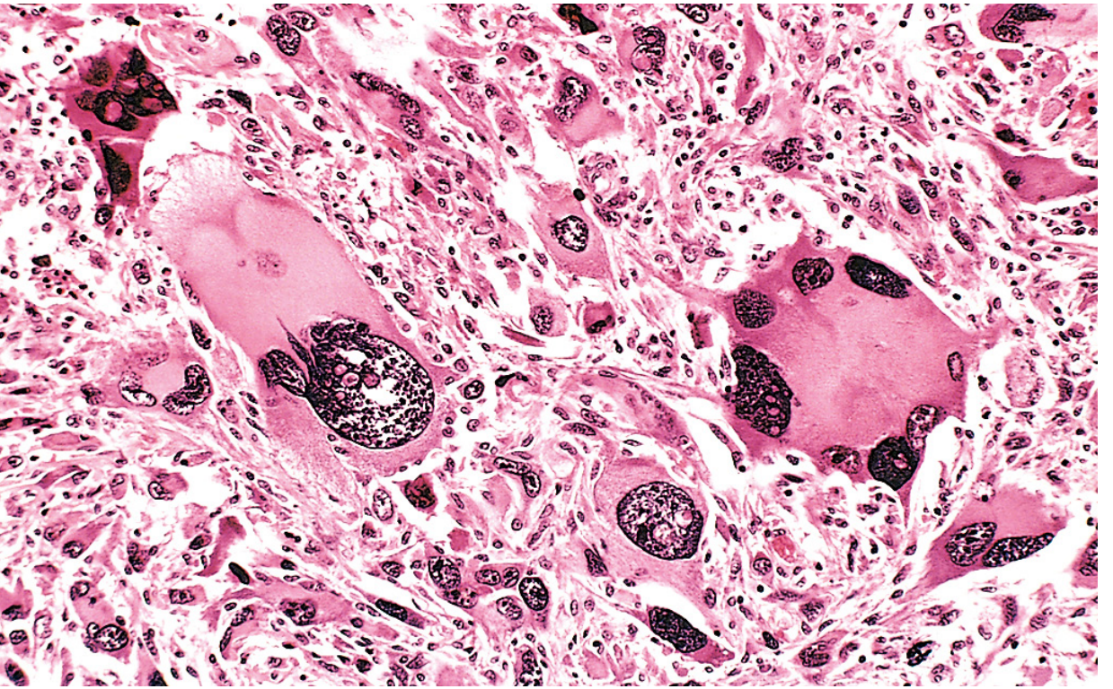
- Tumor giant cells - individual cells many times larger than their neighbors, containing one huge polymorphic nucleus OR two or more large hyperchromatic nuclei
2. Nuclear Pleomorphism
- Nuclei vary in size and shape within the same tumor
- High nuclear-to-cytoplasmic (N:C) ratio - may approach 1:1 instead of normal 1:4 to 1:6
- Nuclear shape is variable and often irregular
- Chromatin is coarsely clumped and distributed along the nuclear membrane
- Hyperchromasia - nuclei stain darker than normal (dark purple on H&E)
- Prominent, enlarged nucleoli (macronucleoli)
3. Abnormal Mitoses
- Numerous mitotic figures
- Atypical/bizarre mitoses - tripolar spindles, star-shaped, ring forms (these are the most important indicator of malignancy)
4. Loss of Polarity
- Cells lose their normal orientation relative to each other and to basement membranes
- Sheets of tumor cells grow in a completely disorganized fashion
Histology Images from Robbins
Fig. 7.6 - Anaplastic tumor showing cellular and nuclear variation in size and shape. The arrow points to a tripolar spindle - an abnormal mitosis:
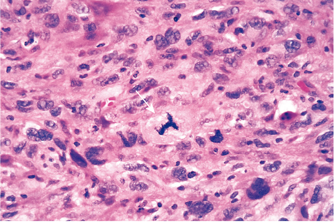
Fig. 7.8 - Rhabdomyosarcoma (pleomorphic tumor of skeletal muscle) showing marked cellular and nuclear pleomorphism, hyperchromatic nuclei, and tumor giant cells:
Pleomorphism vs. Anaplasia - How They Relate
| Term | Meaning |
|---|---|
| Anaplasia | Lack of differentiation; the overall loss of resemblance to normal parent tissue |
| Pleomorphism | One specific feature of anaplasia - variation in cell/nuclear size and shape |
| Dysplasia | Disordered growth; a precursor/premalignant state - dysplastic cells also exhibit pleomorphism |
Pleomorphism is a component of anaplasia, not a synonym. An anaplastic tumor shows pleomorphism PLUS abnormal mitoses, loss of polarity, hyperchromasia, and other features.
Important Distinction - Tumor Giant Cells
Malignant tumor giant cells (seen in pleomorphic tumors) must be distinguished from:
- Langhans giant cells - seen in granulomatous inflammation (TB, sarcoidosis) - derived from macrophages, contain many small normal-appearing nuclei arranged at the periphery in a horseshoe pattern
- Foreign body giant cells - also macrophage-derived, nuclei scattered throughout
Clinical Significance
- High pleomorphism = poorly differentiated = higher grade = more aggressive tumor
- Pleomorphism is used by pathologists to grade tumors (e.g., Grade 1 = low pleomorphism; Grade 3 = high pleomorphism)
- Seen prominently in: rhabdomyosarcoma, pleomorphic undifferentiated sarcoma (formerly MFH), anaplastic carcinomas, glioblastoma, melanoma
Bailey & Love's Short Practice of Surgery also notes that none of these features is diagnostic of malignancy in isolation - the full histological picture must be interpreted together. - Bailey and Love's Short Practice of Surgery, 28th Ed.
This is a shared conversation. Sign in to Orris to start your own chat.