
Orthostatic hypotension
"orthostatic hypotension"[MeSH Terms] AND management
orthostatic hypotension blood pressure postural change diagram
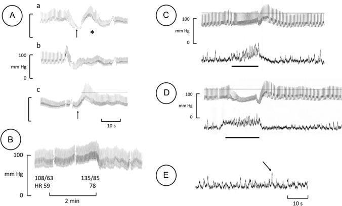
This physiological tracing panel illustrates cardiovascular and autonomic nervous system responses in a patient with orthostatic hypotension. Panel A shows continuous blood pressure (BP) recordings during postural transitions from lying to standing, highlighting rapid hypotension leading to presyncope and the compensatory effect of leg crossing (arrow in 'c'). Panel B displays a qualitatively normal hypertensive and tachycardic response to sustained handgrip. Panels C and D provide integrated microneurography recordings of muscle sympathetic nerve activity (MSNA, lower traces) alongside BP (upper traces). In Panel C (apnea test), a marked increase in MSNA precedes a rise in BP, with subsequent sympathetic inhibition during the post-apnea BP rebound. Panel D (Valsalva maneuver) demonstrates a similar pattern: initial sympathetic activation during the strain phase followed by profound inhibition during the phase IV BP overshoot. Panel E illustrates skin sympathetic nerve activity (SSNA) at rest, showing a characteristic arousal response (arrow). These findings help evaluate baroreceptor-governed sympathetic outflow and peripheral autonomic integrity in neurogenic orthostatic disorders.
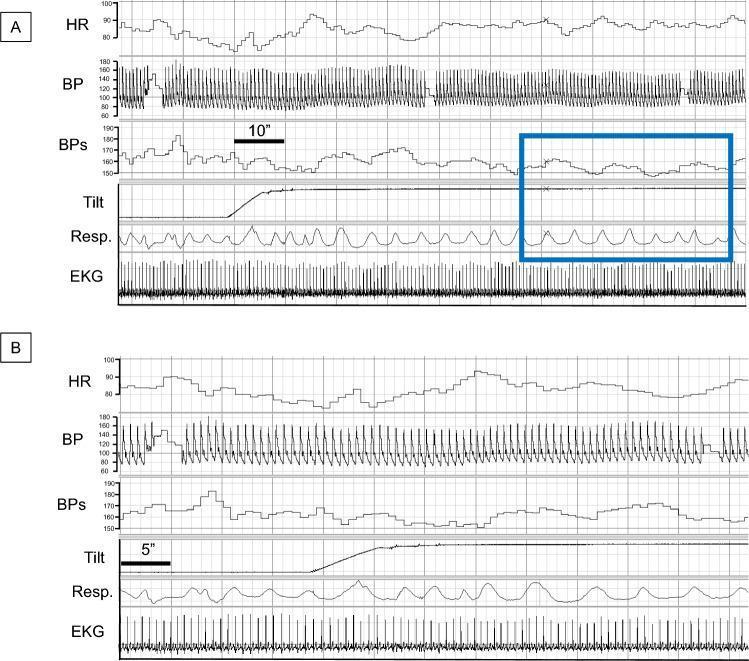
This diagnostic composite shows physiological recordings from a healthy volunteer during head-up tilt testing, presented in two panels (A and B) with different time scales. Each panel displays synchronized waveforms for Heart Rate (HR), Blood Pressure (BP), Systolic Blood Pressure (BPs), Tilt angle, Respiration (Resp.), and Electrocardiogram (EKG). The primary educational focus is the identification of Mayer waves—low-frequency oscillations in blood pressure—and their relationship to respiration and orthostatic stress. Panel A features a blue rectangle highlighting that the BPs oscillations (Mayer waves) occur at a significantly lower frequency than the respiratory cycles, indicating they are not respiratory-driven. Following the upward tilt, the BPs maintains stability without the precipitous drop characteristic of orthostatic hypotension. The EKG and HR tracings provide context for cardiac autonomic response during the postural change. This visualization is used in cardiology and neurology to distinguish between normal autonomic blood pressure regulation and neurogenic orthostatic hypotension.
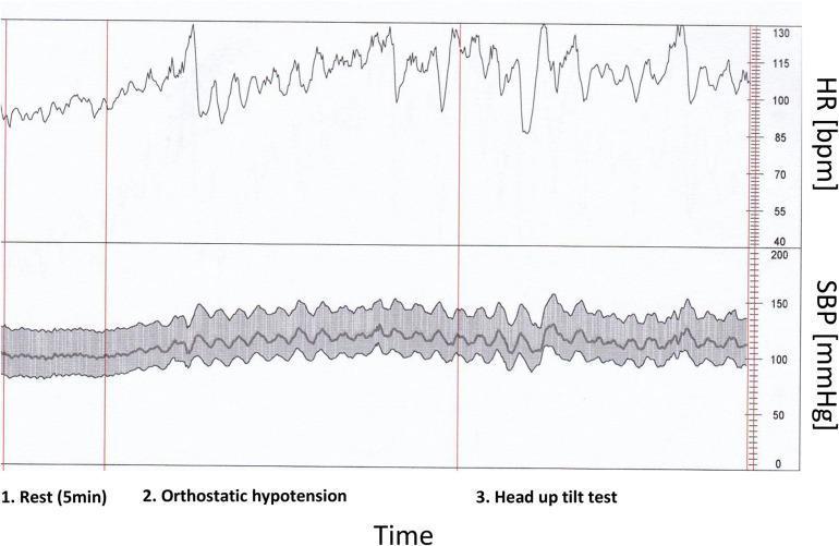
This diagnostic line graph displays a real-time beat-to-beat analysis of heart rate (HR) and systolic blood pressure (SBP) in a patient with COVID-19 and low baroreflex sensitivity. The data is divided into three clinical phases: 1. Rest (5 min), 2. Orthostatic hypotension (active standing), and 3. Head up tilt test. The upper panel shows the HR in beats per minute (bpm), showing an upward trend and increased variability from a baseline of ~95 bpm at rest to over 115 bpm during postural challenges. The lower panel displays SBP in mmHg, oscillating between 100 and 150 mmHg. A central line indicates mean SBP, while a surrounding shaded area represents the beat-to-beat variability or standard deviation. The visual demonstrates impaired autonomic nervous system (ANS) regulation, characterized by significant fluctuations in hemodynamic parameters during orthostatic stress. This educational tool illustrates the cardiovascular response to autonomic provocation in the context of post-viral dysautonomia.
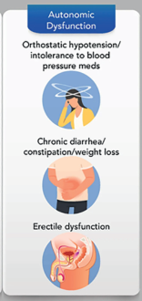
Summary : This illustration summarizes key symptoms of autonomic dysfunction, highlighting orthostatic hypotension/intolerance to blood pressure medications, chronic gastrointestinal issues, and erectile dysfunction. illustration: # Title & Main Text : • Title: "Autonomic Dysfunction" (blue header). • Symptom 1: "Orthostatic hypotension/intolerance to blood pressure meds". • Symptom 2: "Chronic diarrhea/constipation/weight loss". • Symptom 3: "Erectile dysfunction". # Visual Elements : • Symptom 1: Cartoon of a person holding their head, surrounded by swirling lines indicating dizziness. • Symptom 2: Illustration of a person holding their abdomen, suggesting gastrointestinal discomfort. • Symptom 3: Diagram of male pelvic anatomy, highlighting erectile dysfunction. # Layout : • Vertical arrangement: Title at top, followed by three symptom sections, each with corresponding illustration and text. • Each symptom is paired with a circular icon-style illustration. # Analysis : • The figure visually organizes and emphasizes three major symptom domains of autonomic dysfunction: cardiovascular (orthostatic hypotension), gastrointestinal (chronic diarrhea/constipation/weight loss), and genitourinary (erectile dysfunction). • The use of distinct icons and clear text aids rapid recognition of symptom clusters associated with autonomic dysfunction.
Orthostatic Hypotension (OH)
Definition
- In hypertensive patients: drop of ≥30/15 mmHg SBP/DBP
- Initial OH: >40 mmHg SBP drop within 15-30 seconds of standing (transient; more common in young adults)
- Delayed OH: BP drops after 3-5 min (or up to 10+ min); often reflects autonomic dysfunction or medications
- Postprandial OH: BP drop within 2 hours of a meal, especially high-carbohydrate meals or with alcohol
Epidemiology
| Age Group | Prevalence |
|---|---|
| <50 years | <5% |
| >70 years | up to 20% |
Pathophysiology
- Baroreceptor activation
- Increased sympathetic outflow → arteriolar and venous constriction
- Mild heart rate increase
- Net result: SBP maintained, DBP rises slightly
Neurogenic vs. Non-Neurogenic OH
| Feature | Non-Neurogenic OH | Neurogenic OH (NOH) |
|---|---|---|
| HR increase on standing | >15 bpm (intact reflex) | <15 bpm (blunted) |
| ΔHR/ΔSBP ratio | >0.5 beats/min/mmHg | <0.5 beats/min/mmHg |
| Mechanism | Volume depletion, drugs, cardiac | Impaired NE release, autonomic failure |
| Prognosis | Better | Worse (44% mortality at 30 months; >60% at 10 years) |
Etiology / Causes
Primary Autonomic Disorders (Neurogenic)
- Parkinson's disease and Parkinsonism
- Multiple system atrophy (MSA)
- Pure autonomic failure (Bradbury-Eggleston syndrome)
- Lewy body dementia
- Autoimmune autonomic ganglionopathy
Secondary / Systemic Causes
- Metabolic: Diabetes mellitus (autonomic neuropathy), vitamin B12 deficiency, amyloidosis
- Cardiac: Heart failure, chronotropic insufficiency
- Renal: Chronic kidney disease
- Infectious: HIV, Chagas disease
- Other neuropathies: Paraneoplastic, hereditary amyloid, Fabry disease
Non-Neurogenic Causes
- Volume depletion: hemorrhage, dehydration, adrenal insufficiency, prolonged bed rest
- Medications (see below)
- Deconditioning / prolonged bed rest
- Pregnancy
Drug-Induced OH (Common Culprits)
- Diuretics
- Antihypertensives (especially alpha-blockers, beta-blockers, vasodilators)
- Tricyclic antidepressants (TCAs)
- Antipsychotics (especially clozapine, chlorpromazine, olanzapine - via alpha-1 blockade)
- Dopamine agonists (levodopa, pramipexole)
- PDE-5 inhibitors (sildenafil, tadalafil)
- Alcohol
Clinical Presentation
Classic Symptoms (on standing)
- Lightheadedness / presyncope (most common)
- Dizziness (wobbly, unstable - rarely true vertigo)
- Fatigue and generalized weakness
- Palpitations, flushing, or pallor
Atypical / Indirect Symptoms
- "Coat-hanger" syndrome: neck and shoulder pain from hypoperfusion of cervical paraspinal and trapezius muscles
- Orthostatic dyspnea (platypnea): from lung apex hypoperfusion
- Orthostatic angina: from myocardial hypoperfusion
- Orthostatic headache: may mimic low-CSF-pressure headache
- Cognitive impairment
Triggers
Functional Classification
| Class | Features |
|---|---|
| I | Asymptomatic; may have syncope/falls |
| II | Symptoms weekly/monthly; mild-moderate limitation |
| III | Frequent severe symptoms; markedly limit daily activity |
| IV | Daily severe symptoms; significant disability |
Diagnosis
- Measure BP and HR lying and after 1 and 3 minutes of standing
- Confirm with head-up tilt test (HUT) at 60° when standing is not possible
- Calculate ΔHR/ΔSBP ratio to distinguish neurogenic from non-neurogenic
- Rule out volume depletion (clinical exam, renal function, urine sodium)
- Consider autonomic function tests (Valsalva maneuver, QSART, thermoregulatory sweat test) in suspected NOH
- Review all medications
Management
Step 1 - Remove Aggravating Factors
- Reduce or discontinue offending drugs (diuretics, antihypertensives, TCAs, antipsychotics, dopamine agonists)
- Treat iron deficiency, B12 deficiency, and dehydration
Step 2 - Non-Pharmacological Measures
- Rise slowly; sit on the edge of bed before standing
- Avoid Valsalva maneuvers (use urinal or sit to urinate)
- Avoid hot/humid environments
- Small, frequent, low-carbohydrate meals
- Alcohol only in the evening (at bedtime)
- Fluid intake: 2-3 L/day
- Salt supplementation: 1-2 teaspoons/day (or salt tablets 0.5-1.0 g); aim for increased Na intake
- Leg crossing, toe standing, stooping, squatting, buttock clenching
- High-waist compression stockings (≥15-20 mmHg)
- Abdominal binder (reduces splanchnic pooling)
- Bolus water drinking (500 mL/16 oz rapidly): raises BP within 5-10 min, peaks at 30 min, via portal osmoreceptor reflex - useful as an acute rescue measure
Step 3 - Pharmacological Treatment
| Drug | Dose | Mechanism | Key Notes |
|---|---|---|---|
| Fludrocortisone | 0.1-0.2 mg/day (up to 0.4 mg/day) | Mineralocorticoid → expands plasma volume, mild α-agonist effect | First-line; monitor for supine HTN, hypokalemia, pedal edema, headache; check bone density long-term |
| Midodrine | Start 2.5 mg TID → up to 15 mg TID (most respond to 7.5-10 mg TID) | Selective α1-adrenoceptor agonist → arterial and venous constriction | Onset 30-60 min, duration 4 h; last dose before 6 PM; side effects: scalp tingling, goosebumps, urinary urgency |
| Droxidopa | 100 mg TID → 400-600 mg TID | NE precursor → converted to norepinephrine in sympathetic terminals and non-neuronal tissues | FDA-approved for NOH; improves symptoms and reduces falls in Parkinson's; supine HTN risk |
| Pyridostigmine | Start 30 mg TID → 60 mg TID | Cholinesterase inhibitor → enhances ganglionic transmission | Does not promote supine HTN (advantage); benefits patients with slow GI motility; side effects: cramps, diarrhea |
| Atomoxetine | 10-18 mg/morning | NE reuptake inhibitor (SNRI-like) | Adjuvant therapy; monitor for CNS stimulation (anxiety, tremor) |
- Acarbose (50-100 mg before meals): alpha-glucosidase inhibitor for postprandial hypotension
- Octreotide (0.2-0.4 mcg/kg SC): splanchnic vasoconstriction for postprandial hypotension
- Erythropoietin: in patients with anemia of chronic disease
Managing the Neurogenic OH + Supine Hypertension Paradox
- Daytime: OH management as above
- Avoid supine position during the day
- Nighttime head-of-bed elevation to 30-45° (reverse Trendelenburg) - lowers nocturnal BP, reduces pressure diuresis and natriuresis, and thereby improves daytime OH
- High-glycemic carbohydrate snacks or wine at bedtime to temporarily reduce supine HTN
- Short-acting antihypertensives at bedtime if needed: captopril 25 mg, losartan 50 mg, nifedipine 1-2 mg, amlodipine 2.5-5 mg, or a nitroglycerin patch (0.1 mg) - use with caution given fall risk on nocturnal rising
Drug-Induced OH: Psychiatric Medications (Special Focus)
- Titrate doses slowly; consider temporary dose reduction if OH develops
- Use modified-release preparations to reduce peak plasma levels
- Smaller and more frequent dosing
- Pharmacological: midodrine or fludrocortisone when antipsychotic cannot be changed (e.g., clozapine) and other strategies have failed
Key Clinical Pearls
- OH is often overlooked - most patients are asymptomatic or have atypical complaints; always check orthostatic vitals in elderly, Parkinson's patients, and those on multiple medications
- Goal of treatment is symptom relief, not a target BP number
- Neurogenic OH carries a markedly worse prognosis than non-neurogenic - identify it early via HR response
- Supine hypertension coexists in ~50% of NOH patients - managing one can worsen the other; head-of-bed elevation is the cornerstone for this scenario
- POTS (postural orthostatic tachycardia syndrome) is distinct: HR increases ≥30 bpm without sustained OH; a different management approach is required