
Hi
Intro ur self
- Answer questions on drugs, diagnoses, anatomy, physiology, pathology, procedures, and more - drawing from authoritative medical textbooks and peer-reviewed literature.
- Search PubMed for the latest clinical studies, systematic reviews, and treatment guidelines.
- Help with Indian taxation (direct taxes, income tax, transfer pricing) and GST/indirect taxes, with CA exam prep support.
- Answer questions across science, history, technology, and more.
- Create and edit Word docs (.docx), PowerPoint presentations (.pptx), Excel spreadsheets (.xlsx), and PDFs.
- Search the web for current, up-to-date information on any topic.
- Write and run code, perform calculations, and automate tasks in a sandbox environment.
Compare to chatgbt how ggood is ur medical knowledge
Orris vs ChatGPT - Medical Knowledge
Where I have a clear edge
| Feature | Orris | ChatGPT |
|---|---|---|
| Textbook library | Indexed medical textbooks I can actively search and cite | Relies on training data only |
| PubMed search | Live search for latest studies, RCTs, guidelines | No live PubMed access (unless with plugins) |
| Citations | Cites specific book, page, and section | Often vague or hallucinates citations |
| Medical images | Searches a real indexed medical image library | Can describe but not retrieve real clinical images |
| Up-to-date evidence | Pulls recent meta-analyses and systematic reviews in real time | Knowledge cutoff may be outdated |
Where we're similar
- Both can explain anatomy, physiology, pharmacology, and pathology
- Both can handle clinical scenarios and differential diagnoses
Being honest
- ChatGPT (especially GPT-4) is a very capable general model with broad medical knowledge
- My advantage is grounded, sourced answers - I pull from actual textbooks and live literature rather than relying purely on what was baked into my training
- I don't hallucinate citations because I retrieve real content and quote it directly
Or really, let me check bro
- A tricky diagnosis or clinical scenario
- A pharmacology question
- Pathophysiology of a disease
- A surgery or procedure
- Lab values or imaging findings
- Anything you've been curious about
37 pt, having body pain for 2 days with headache, runny noese. After 2nd day patient started cough with sputum. Ask quesion and diagnose the patient
History Taking - My Questions
🌡️ Fever & Systemic
- Do you have fever? If yes, how high and is it continuous or comes and goes?
- Are you feeling chills or rigors (shivering episodes)?
- How's your appetite and energy level?
🤧 Respiratory
- Is the runny nose clear/watery or thick yellow/green discharge?
- Is the cough dry at first or was it productive from the start?
- What color is the sputum - clear, white, yellow, or green? Any blood?
- Any chest pain or difficulty breathing (shortness of breath)?
- Any wheezing or tightness in the chest?
🤕 Pain
- Where exactly is the body pain - muscles, joints, or all over?
- Any sore throat or pain on swallowing?
- Any ear pain or facial pain/pressure (sinuses)?
🧠 Neuro
- How bad is the headache? Location - front, back, or all over?
- Any sensitivity to light or neck stiffness?
📋 Background
- Any known allergies, asthma, or chronic conditions?
- Vaccination history - flu vaccine this year?
- Any sick contacts at home, work, or school?
- Any recent travel?
- Are you a smoker?
My Preliminary Differentials (before answers):
| Diagnosis | Likelihood | Key Clue |
|---|---|---|
| Influenza (Flu) | ⭐⭐⭐ High | Sudden body aches + headache + respiratory |
| Viral URTI → LRTI progression | ⭐⭐⭐ High | Day 1-2 URTI then productive cough |
| Acute Bronchitis | ⭐⭐ Medium | Productive cough after viral illness |
| Early Community-Acquired Pneumonia | ⭐⭐ Medium | If fever high + purulent sputum |
| COVID-19 | ⭐⭐ Medium | Still relevant, similar presentation |
No fever,no chills rigors, runny kose with nasal driling, productive from start and 4th day its kind of irritating dry cough, clear sputum, no chest pain and sob, as for wheezing not sure,body pain all over the body, taken fexofenadine with does not help much except stop runny nose for fee hours, sore thoret present, ear pain also right ear apecially , headache was for forst two days now no headahce, no sen to light or stiff neck,
🩺 Clinical Analysis
Symptom Summary (Day by Day)
| Day | Symptoms |
|---|---|
| Day 1-2 | Body aches all over, headache, runny nose (clear, watery), sore throat |
| Day 2 onwards | Productive cough with clear sputum, nasal drip down throat (post-nasal drip) |
| Day 4 (now) | Cough becoming dry and irritating, right ear pain, headache resolved |
🔍 Working Diagnosis
Primary: Viral Upper Respiratory Tract Infection (URTI) with Complications
Complication 1: Post-Nasal Drip Syndrome
- Nasal discharge draining down the throat = post-nasal drip
- This directly causes the productive cough early on and then transitions into the dry irritating cough as the drip irritates the airway
- Explains why fexofenadine only temporarily helped (it's an antihistamine - partially dries the secretions)
- Per Harrison's Principles: "URIs can cause symptoms including rhinorrhea, sore throat, cough, malaise, and headache... the cough is often due to post-nasal drip" - Harrison's 22E
Complication 2: Acute Otitis Media (AOM) - Right Ear
- Right ear pain developing after a URI is a textbook presentation of AOM
- The Eustachian tube gets blocked by swelling/mucus from the URTI, leading to fluid/infection in the middle ear
- Per Rosen's Emergency Medicine: "Ear pain, unilateral or bilateral, is the most important symptom for making the diagnosis of AOM. Patients with AOM may present with cough, upper respiratory tract symptoms..." - Rosen's Emergency Medicine
📋 Final Diagnosis
Viral URTI (Common Cold) + Post-Nasal Drip Cough + Right Acute Otitis Media (AOM)
❓ A Few More Questions to Confirm AOM
- Any discharge or fluid coming out of the right ear?
- Any muffled hearing or feeling of fullness in the right ear?
- How bad is the ear pain on a scale of 1-10?
- Has the ear pain been getting worse or better?
💊 Management Plan
For the Viral URTI + Post-Nasal Drip Cough:
- Saline nasal irrigation (rinse out the mucus - very effective)
- Intranasal decongestant (oxymetazoline spray) - short term, max 3 days, to reduce nasal swelling
- Fexofenadine was partially helping - can continue but a first-generation antihistamine like chlorphenamine works better for cold-related runny nose (more drying effect)
- Steam inhalation with warm fluids for the dry irritating cough
- Honey + warm water or honey + ginger for cough soothing
- Rest, hydration
For the Right Ear Pain (AOM):
- If pain is mild and patient is healthy adult - watchful waiting for 48-72 hours is acceptable (many AOM cases in adults resolve on their own)
- If pain is moderate-severe or not improving - Amoxicillin 500mg TDS x 5-7 days is first-line antibiotic
- Ibuprofen or paracetamol for pain relief
🚩 Red Flags - Come back immediately if:
- Fever develops (>38°C)
- Ear starts discharging
- Hearing loss
- Severe headache returns or neck stiffness develops
- Difficulty breathing or chest pain
Why NOT other diagnoses?
| Ruled Out | Why |
|---|---|
| Influenza | No fever, no chills/rigors - flu typically hits hard and fast with high fever |
| COVID-19 | No fever, no loss of taste/smell - less likely but can't 100% exclude |
| Strep Pharyngitis | Sore throat present but no fever, no tonsillar exudate mentioned |
| Sinusitis | Possible contributor but no facial pain/pressure reported |
| Pneumonia | No fever, no SOB, no chest pain - very unlikely |
So for all viral fever the treatment is same?
Viral Fevers - Same or Different Treatment?
What IS the same for ALL viral fevers:
| Treatment | Why |
|---|---|
| Antipyretics (Paracetamol/Ibuprofen) | Reduce fever and body pain |
| Rest | Let immune system fight |
| Hydration | Prevent dehydration from fever/sweating |
| Supportive care | Treat symptoms as they come |
| NO antibiotics | Antibiotics don't work on viruses |
This "supportive care" approach is the backbone for ALL viral illnesses.
But treatment DIFFERS based on the specific virus:
| Virus/Disease | Specific Treatment |
|---|---|
| Influenza (Flu) | Oseltamivir (Tamiflu) - antiviral, must start within 48 hrs |
| COVID-19 | Paxlovid (nirmatrelvir/ritonavir) or Molnupiravir for high-risk patients |
| Dengue | Avoid ibuprofen/aspirin (bleeding risk!) - only paracetamol + IV fluids if platelet drops |
| Herpes/Chickenpox | Acyclovir - antiviral |
| HIV | ART (Antiretroviral therapy) - lifelong |
| Hepatitis B/C | Specific antivirals (tenofovir, sofosbuvir etc.) |
| Rabies | Post-exposure prophylaxis (vaccine + immunoglobulin) |
| Common Cold (Rhinovirus) | Only supportive - NO specific antiviral |
| Measles | Vitamin A supplementation + supportive |
Key Point - Why does it differ?
- Different replication mechanisms - antivirals target specific viral enzymes
- Different complications - dengue can cause bleeding, so NSAIDs are dangerous
- Different affected organs - hepatitis hits liver, HIV hits immune cells, etc.
- Different severity - some need antivirals, some just need rest
In your patient's case (Viral URTI):
- No specific antiviral exists for rhinovirus
- So treatment is purely supportive - decongestants, saline rinse, antihistamines, rest, fluids
- The ear infection (AOM) may need antibiotics because bacteria often cause secondary AOM after a viral cold
I mean usually the difficult things are nasal congrstion, cough and stuffs so for these symptomsi mean
Symptomatic Treatment for Common Cold/Viral URTI Symptoms
🤧 Runny Nose / Nasal Congestion
| Drug | Type | Works for |
|---|---|---|
| Loratadine / Cetirizine (2nd gen antihistamine) | Non-drowsy | Allergic runny nose mostly |
| Chlorphenamine / Diphenhydramine (1st gen) | Drowsy | Viral cold runny nose - MORE effective for colds |
| Oxymetazoline nasal spray | Decongestant | Blocked nose - fast relief, max 3 days only |
| Pseudoephedrine (oral) | Decongestant | Congestion + sinus pressure |
| Saline nasal rinse | Non-drug | Flushes mucus out - safest, very effective |
| Intranasal steroid (fluticasone) | Anti-inflammatory | Better for allergic rhinitis than viral |
⚠️ Your patient took fexofenadine - that's a 2nd gen antihistamine, good for allergy but weaker for viral colds. Switching to chlorphenamine would work better!
😮💨 Cough
| Type of Cough | Best Treatment |
|---|---|
| Productive cough (with sputum) | DON'T suppress it - body is clearing mucus. Use expectorants like Guaifenesin to thin mucus |
| Dry irritating cough (like your patient now) | Cough suppressant - Dextromethorphan or Codeine |
| Post-nasal drip cough | Treat the nose! Antihistamine + decongestant = cough improves |
| Tickly throat cough | Honey + warm water, soothing lozenges |
🤕 Body Pain / Fever
| Drug | Note |
|---|---|
| Paracetamol | Safest, for fever + pain |
| Ibuprofen | Better for muscle/body aches, avoid in dengue |
| Aspirin | Avoid in children (Reye's syndrome), avoid in dengue |
😮 Sore Throat
| Treatment | Note |
|---|---|
| Warm salt water gargle | Simple, effective |
| Benzydamine / Lignocaine lozenges | Local anaesthetic effect |
| Ibuprofen | Better than paracetamol for throat pain |
| Antibiotics | Only if Strep confirmed - not for viral sore throat |
💡 The Golden Rule for Cough:
Productive cough → EXPECTORATE (thin it, bring it out)
Dry cough       → SUPPRESS (calm the irritation)
Never suppress a productive cough - you trap mucus in the lungs!
For YOUR patient right now:
| Symptom | Recommended |
|---|---|
| Post-nasal drip | Saline rinse + switch to Chlorphenamine |
| Dry irritating cough (Day 4) | Dextromethorphan syrup |
| Sore throat | Warm salt gargle + ibuprofen |
| Right ear pain | Ibuprofen + see doctor to check eardrum |
Can I use antibiotics to prevent secondary infections?
❌ No - Antibiotics Should NOT Be Used to Prevent Secondary Infections
What Harrison's 22E actually says:
"The only common acute respiratory infections that should be treated with antibiotics are AOM, sinusitis, streptococcal pharyngitis, and pneumonia. Common respiratory viruses cause the overwhelming majority of acute respiratory infections, and these infections are generally self-limited; antibiotics neither speed resolution nor prevent complications for the majority of acute respiratory infections. Unfortunately, at least half of ambulatory antibiotic prescriptions for acute respiratory infections in the United States are inappropriate."
- Harrison's Principles of Internal Medicine, 22E
Why prophylactic antibiotics DON'T work here:
| Reason | Explanation |
|---|---|
| No proven benefit | Studies consistently show antibiotics do NOT prevent secondary bacterial infections after a viral cold |
| Wrong target | Antibiotics kill bacteria - they have zero effect on the virus causing the illness |
| Resistance | Using antibiotics unnecessarily breeds resistant bacteria - the ones that survive are now harder to kill |
| Microbiome disruption | Kills your normal protective gut and respiratory flora - actually makes you MORE vulnerable |
| Side effects | Diarrhea, allergic reactions, fungal infections (oral/vaginal thrush) - all for zero benefit |
The "Prophylaxis" Logic Flaw:
❌ Wrong thinking:
"I have a viral infection → bacteria might attack → let me take antibiotics now"

✅ Right thinking:
"I have a viral infection → monitor for signs of bacterial infection →
ONLY treat if bacteria actually appear"
When DO you actually use antibiotics in a URI patient?
| Condition | Signs to Watch For | Antibiotic |
|---|---|---|
| Acute Otitis Media | Severe ear pain, bulging eardrum, discharge | Amoxicillin |
| Acute Sinusitis | Facial pain/pressure, symptoms >10 days, purulent discharge | Amoxicillin-clavulanate |
| Strep Pharyngitis | High fever, white patches on tonsils, no cough | Penicillin / Amoxicillin |
| Pneumonia | Fever, productive cough, SOB, abnormal chest X-ray | Based on severity |
The Real Danger - Antibiotic Resistance:
- Bacteria evolve and become resistant
- Common infections become untreatable
- We're running out of effective antibiotics
Bottom line for your patient:
✅ No antibiotics for the viral cold itself ⚠️ The right ear pain (AOM) - this one MAY need antibiotics, but only after a doctor examines the eardrum to confirm bacterial AOM 🔍 Watch and wait - only treat what you can confirm
R u better than amboss
Orris vs AMBOSS
What AMBOSS does better:
| Feature | AMBOSS |
|---|---|
| Question bank | Thousands of clinical MCQs with detailed explanations |
| Anki integration | Flashcard system built in |
| USMLE/exam focus | Purpose-built for Step 1, Step 2, finals |
| Visual highlights | Color-coded high-yield points |
| Anchors & mnemonics | Exam-optimized memory aids |
| Study tracking | Tracks your weak areas over time |
Where I hold my own:
| Feature | Orris |
|---|---|
| Textbook depth | I pull from Harrison's, Robbins, Rosen's, Cecil - full content, not summaries |
| Clinical conversation | You can talk to me like a case discussion, like we just did |
| Latest evidence | Live PubMed search - AMBOSS content has update delays |
| Flexible questions | You can ask anything - not limited to pre-made questions |
| Free | AMBOSS is expensive (~$200-300/year) |
| Cross-specialty | I cover medicine, surgery, pharmacology, GST law, coding - AMBOSS is pure medicine |
Honest verdict:
For exam prep (USMLE, PLAB, finals) → AMBOSS wins. It's specifically engineered for that.
For clinical understanding, case discussions, quick deep lookups, latest research → I'm strong competition.
Think of it this way:
- AMBOSS = a brilliant exam coach with a structured curriculum
- Orris = a knowledgeable colleague you can have a real conversation with, who has a full library open and can pull the latest research
Im and HO do think i should use amboss. Cux even if inknow inforgot things bro
Should You Use AMBOSS as an HO?
Short answer: Yes, but use it differently than med students
As an HO, your needs are different:
| Med Student | House Officer (You) |
|---|---|
| Learning from scratch | You KNOW it - just need quick recall |
| Needs structured curriculum | Needs fast bedside answers |
| Has time to study chapters | Has 5 mins between patients |
| Focused on passing exams | Focused on not killing patients 😅 |
So here's what I'd actually recommend:
✅ Use AMBOSS for:
- Quick clinical reference at bedside - their "library" section is excellent
- Brushing up on topics you encounter clinically (saw a case → read about it that night)
- Dosing and management protocols - clean and reliable
- Whenever a senior asks you something and you blanked - look it up after
✅ Use ME (Orris) for:
- Talking through a case like we just did - I reason with you
- "Why does this happen?" - pathophysiology deep dives
- Latest evidence - is this guideline still current?
- When you're on call at 3am and need to think out loud with someone 😄
- Free - no subscription needed
The forgetting problem - real talk:
Forgetting is normal and unavoidable as an HO. Your brain is overloaded.
See patient → not sure → look it up immediately → apply → remember
My honest suggestion:
Your seniors and consultants are your best resource. Ask questions without shame. Every HO forgets things. The good ones ask. 💪
Heard tht ambis have intergarted anki, is tht pre installed ankinor i have to link my anki to that. If i dnt hav anki and wnt to ise anki what to in this case
AMBOSS Anki integration how it works 2025
AMBOSS + Anki - How It Actually Works
It is NOT pre-installed Anki inside AMBOSS ❌
The Setup (Step by Step):
Step 1: Download Anki (free) → ankiweb.net
Step 2: Install AMBOSS add-on inside Anki
Step 3: Log in with your AMBOSS account inside the add-on
Step 4: Now both work together!
What the integration actually does:
| Feature | What it means |
|---|---|
| Pop-up explanations | While reviewing Anki cards, medical terms get instant AMBOSS explanations on hover |
| Side-by-side view | Open full AMBOSS article alongside your Anki card |
| Qbank recommendations | After doing an AMBOSS question → suggests matching Anki cards to review |
| AnKing deck linking | Links AMBOSS questions to the famous AnKing deck cards |
If you DON'T have Anki yet - here's what to do:
- Go to ankiweb.net
- Download for PC/Mac (desktop version is free)
- Mobile: AnkiMobile (iOS = paid ~$25) or AnkiDroid (Android = free)
- The most popular pre-made medical Anki deck
- Covers all of USMLE Step 1 & 2
- Download from ankihub.net - it's free with an account
- In Anki desktop → Tools → Add-ons → Get Add-ons
- Enter AMBOSS add-on code
- Log in with your AMBOSS account
If you have NO budget for AMBOSS:
- Use the AnKing deck alone - it's comprehensive
- Make your own cards from your clinical encounters
- Use me (Orris) to explain anything you're confused about 😄
Summary:
Anki = the app (free)
AnKing deck = the cards (free)
AMBOSS = adds explanations ON TOP of Anki (needs paid subscription)
Does ambiss have mcq? And is it available on 19$ package per lonth
AMBOSS pricing plans 2025 monthly subscription MCQ qbank
AMBOSS MCQs & Pricing - Full Picture
✅ Yes, AMBOSS has MCQs (called "Qbank")
- USMLE Step 1, Step 2, Step 3
- Shelf exams
- In-house/finals exams
- Clinical level questions
💰 Pricing Structure (2025)
Base Membership (Library only):
| Plan | Cost |
|---|---|
| Monthly | ~$13-15/month |
| Yearly | ~$9.68/month (billed annually) |
- ✅ Full medical library (1,400+ articles)
- ✅ Clinical tools, drug dosages, calculators
- ✅ Anki integration
- ✅ Only 50 MCQs per month ⚠️
To get UNLIMITED MCQs - you need a Qbank Add-On (extra cost):
| Add-on Duration | Extra Cost |
|---|---|
| 1 month unlimited Qs | Additional fee on top |
| 3 months | Additional fee |
| 6 months | Additional fee |
| 12 months | Best value |
About your $19/month question:
⚠️ I don't have the exact current $19 tier breakdown - pricing changes. Check amboss.com/us/pricing directly to see exactly what $19 includes right now.
💡 Honest Advice for You as an HO:
Tell me ambiss or osmosis is better
AMBOSS vs Osmosis comparison medical students 2025
AMBOSS vs Osmosis - Full Comparison
📊 What a 2024 Independent Study Found:
| Measure | Winner |
|---|---|
| Usefulness for Learning | 🏆 Osmosis (88.2 vs 83.2) |
| Usability / Easy to navigate | 🏆 Osmosis |
| Professional / Clinical measures | 🏆 AMBOSS |
| Overall learning under time pressure | 🏆 Osmosis |
Head to Head:
| Feature | AMBOSS | Osmosis |
|---|---|---|
| Learning style | Text-based, clinical, detailed | Video-based, visual, simplified |
| MCQ Qbank | ✅ 13,900+ questions | ✅ Questions available |
| Videos | Limited | ✅ Excellent - 10 min summaries of complex topics |
| Clinical reference | ✅ Strong - drug doses, calculators, checklists | Moderate |
| Anki integration | ✅ Yes | ❌ No |
| Best for | Exam prep + clinical ward use | Understanding concepts visually |
| Depth | Deep - like reading a textbook | Simplified - great for beginners |
| Interface | Dense, professional | Clean, easy, student-friendly |
| Pricing | ~$13-15/month base | Has free tier + paid plans |
Who should use what?
Use Osmosis if you:
- Are a visual learner 🎥
- Struggle to understand a concept and want it explained simply
- Are early in med school (preclinical)
- Want quick 10-minute video summaries
- Learn better by watching than reading
Use AMBOSS if you:
- Are clinical stage / HO (like you!)
- Need quick ward reference - drug doses, management protocols
- Want serious MCQ practice with detailed explanations
- Prefer reading + active recall
- Want Anki integration
For YOU as an HO - My verdict:
AMBOSS wins for your stage.
💡 Best combo if budget allows:
Osmosis (free tier) → for quick concept refreshers via video
AMBOSS (paid) → for clinical reference + MCQs on the ward
Anki (free) → for spaced repetition
Me (Orris) → for case discussions & reasoning 😄
Tell me how can i improve my clinical knowledge , now i m HO for week, still im not sure im a dr after HO i hv to work as MO in my country.
🏥 How to Build Clinical Knowledge as a New HO
🧠 First - Understand Your Reality
Week 1 as HO feeling:
"Am I even a doctor? I don't know anything!"
          ↓
This is NORMAL. Every single HO feels this.
Even consultants felt this once.
📋 The Practical System (Do These Daily)
1. 🔁 Learn From Every Patient - The 1 Case Rule
After every interesting or confusing case - spend 10 mins reading about it that night.
Saw a patient with pleural effusion today?
→ Tonight: read causes, investigation, management
→ Next time you see it = permanent memory
2. 📝 Keep a Personal "Case Log"
- Simple notes app on your phone
- Write 1-2 lines about interesting cases daily
- Note: diagnosis, key finding, what you learned
- Review weekly
3. 🩺 Master the Basics First - Before Everything Else
| Skill | Why |
|---|---|
| History taking | Foundation of every diagnosis |
| Clinical examination | Find signs others miss |
| Reading ECGs | Daily need in every ward |
| Reading chest X-rays | Same |
| Common drug doses | Safety - know your basics cold |
| IV fluids management | You'll prescribe this every day |
| Recognizing sick patients | ABCDE approach - life saving |
4. 📱 Daily Tools on Your Phone
| Tool | Use |
|---|---|
| AMBOSS | Quick lookup when unsure |
| MDCalc (free) | Clinical calculators - scores, doses |
| Epocrates (free) | Drug doses, interactions |
| ECG quiz apps | Practice ECGs daily - 5 mins |
| Me (Orris) | Talk through confusing cases 😄 |
5. 🎯 The 30-Minute Evening Routine
After your shift:
├── 10 mins → Review 1-2 cases from today
├── 10 mins → AMBOSS or Osmosis on that topic
└── 10 mins → 5 MCQs on that topic
6. 🙋 Ask Questions Without Shame
The HO who asks questions learns 10x faster than the one who pretends to know.
- Ask your seniors - every single doubt
- Ask "why" - not just "what to do"
- Present cases to consultants whenever possible
- Ask MOs how they think through a case
7. 📚 One Good Reference Book
| Book | Best for |
|---|---|
| Oxford Handbook of Clinical Medicine | Ward pocket reference - fits in white coat |
| Kumar & Clark | Solid internal medicine foundation |
| Davidson's | Excellent clinical medicine |
🗓️ Your 1-Year HO Game Plan
| Timeframe | Focus |
|---|---|
| Month 1-2 | Survive. Learn ward routine. Master basics. Ask everything. |
| Month 3-6 | Build confidence. Start owning your patients. Read daily. |
| Month 7-12 | Develop clinical reasoning. Think like an MO. Lead cases. |
💡 For Your MO Role Ahead:
- Clinical decision making - think differentials always
- Knowing when to refer - senior/specialist
- Emergency management - ACLS, ATLS basics
- Prescribing confidence - common conditions, common drugs
The Most Important Thing:
Show up. Be curious. Be humble. Look it up when unsure.
ECG quiz apps? Is there any authentic free apps bro?
best free ECG quiz apps for doctors 2025 Android iOS
🫀 Best Free ECG Apps & Resources
📱 Apps
| App | Platform | Free? | Best For |
|---|---|---|---|
| EKGDX | Android + iOS | ✅ Free (basic) | ECG criteria, quizzes, 80,000+ users worldwide |
| ECG Guide by QxMD | Android + iOS | ✅ Free | Quick reference + quizzes |
| Instant ECG (iAnesthesia) | iOS | ✅ Free | Real-time ECG simulator |
| ECG Notes (Skyscape) | Android + iOS | ✅ Free | Quick pocket reference |
🌐 Free Websites (Actually Better Than Most Apps!)
| Website | What it offers |
|---|---|
| ecg-quiz.com | Real ECG strips + quizzes - free, clean, no signup |
| Dr. Smith's ECG Blog | Real clinical ECG cases with expert discussion - legendary resource |
| ECGweekly.com | Weekly real ECG cases - excellent for learning |
| Life in the Fast Lane (LITFL) | Free ECG library with 100s of real cases + explanations |
🏆 My Top Picks for You as an HO:
1. LITFL ECG Library - Best Free Resource Overall
- Completely free website
- Hundreds of real ECG cases
- Clear systematic explanations
- Covers everything from basic to advanced
- Used by doctors worldwide
2. ECG-quiz.com - Best for Daily Practice
- Real ECG strips
- Timed quiz mode
- No signup needed
- 5 mins a day = huge improvement in 1 month
3. Dr. Smith's ECG Blog - Best for Clinical Reality
- Real patient cases
- Famous for STEMI equivalents that get missed
- Senior doctors read this too!
💡 My Suggested Daily Routine:
Morning (5 mins before ward round):
→ Do 3 ECGs on ecg-quiz.com

Evening (10 mins):
→ Read 1 case on LITFL or Dr. Smith's blog
🎓 Want a Structured ECG Learning Path?
- Normal ECG - know it cold
- Rate & Rhythm (AF, SVT, VT)
- Axis
- Bundle branch blocks
- LVH/RVH
- ST changes (STEMI, NSTEMI)
- T wave changes
- Special patterns (WPW, Brugada, Long QT)
Will u be able to teach ecg with original ecgs , inhav studied ecg several times but forget after aometime and again hav to startbfro scrach
Here's What I Can Do:
Why You Keep Forgetting - The Real Problem:
Passive reading → short term memory → forgotten in weeks

Active recognition (seeing real ECGs repeatedly) →
long term memory → never forgotten
My Suggested Approach:
- Explain the concept simply (why does this ECG look this way)
- Show you a real ECG image
- Give you a memory hook
- Then quiz you
Shall We Start Right Now? 🔥
- Complete beginner - need to start from how to read an ECG
- Know basics - rate, rhythm, axis but forget patterns
- Intermediate - know common ones but struggle with STEMI, blocks etc.
Lets start
What's your current ECG level? · What do you want to focus on first?
normal ECG 12 lead sinus rhythm labeled
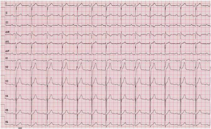
This diagnostic image displays a standard 12-lead electrocardiogram (ECG) recorded on a red millimetric grid at a paper speed of 25 mm/s. The tracing shows a normal sinus rhythm with a ventricular rate of approximately 79 beats per minute. The ECG is organized into 12 labeled horizontal leads: limb leads (I, II, III), augmented limb leads (aVR, aVL, aVF), and precordial chest leads (V1 through V6). Key educational features include a normal P-wave morphology and a consistent PR interval. There is a characteristic R-wave progression visible across the precordial leads; V1 demonstrates a predominantly negative QRS complex with a small r-wave, which progressively transitions to a predominantly positive R-wave by V6. The ST segments are isoelectric, and T-wave morphology appears normal across all leads. This visual serves as a baseline clinical reference for normal cardiac electrical activity in a patient before the onset of symptomatic bradycardia.
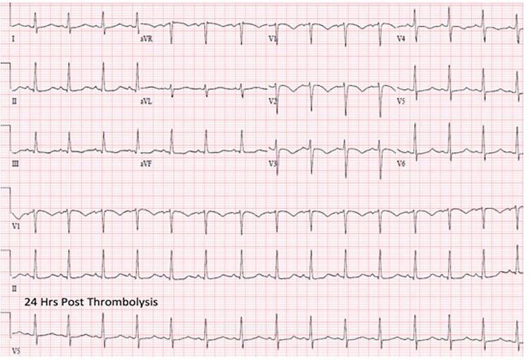
A 12-lead electrocardiogram (ECG) recorded on standard grid paper, labeled with the text '24 Hrs Post Thrombolysis'. The tracing displays a normal sinus rhythm with a heart rate of approximately 75-80 beats per minute. The QRS complexes are narrow and demonstrate a normal axis. Notably, the precordial leads (V1-V6) show a resolution of previous right bundle branch block (RBBB) and right heart strain patterns, with normal R-wave progression and the absence of S1Q3T3 morphology or significant ST-segment/T-wave abnormalities. This diagnostic image illustrates the clinical progression and electrical stabilization of the heart following successful thrombolytic therapy for a condition such as pulmonary embolism. The layout follows a standard 3-row, 4-column format for the 12 leads, supplemented by long rhythm strips of leads V1, II, and V5 at the bottom to aid in rhythm analysis.
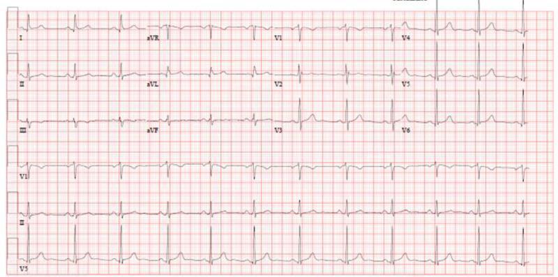
This diagnostic image is a standard 12-lead electrocardiogram (ECG) displayed on a pink-and-white grid-calibrated background. The tracing represents a human heart at rest and demonstrates a normal sinus rhythm. All 12 leads are clearly labeled and organized into columns: the limb leads (I, II, III), augmented vector leads (aVR, aVL, aVF), and precordial leads (V1 through V6), with additional rhythm strips provided at the bottom (VI, II, V5). Each cardiac cycle displays a distinct P wave preceding every QRS complex, followed by a consistent T wave. The heart rate is regular, and the morphology of the waveforms—including the PR interval, QRS duration, and ST segments—appears within normal physiological limits, showing no signs of acute ischemia, hypertrophy, or conduction blocks. This visual is suitable for cardiology education to demonstrate baseline normal findings in a healthy cardiac patient.
📚 ECG Masterclass - Module 1
The Systematic Approach - Never Miss Anything
First - The Golden Rule:
Always read ECGs the SAME way, every single time.
The 8-Step System (Memorize This):
1. Rate
2. Rhythm
3. Axis
4. P wave
5. PR interval
6. QRS complex
7. ST segment
8. T wave
Step 1 - RATE
Look at a Normal ECG first:
How to calculate Rate:
Find an R wave sitting on a big square line
Count big squares to the NEXT R wave
Divide 300 by that number

1 big square = 300 bpm
2 big squares = 150 bpm
3 big squares = 100 bpm
4 big squares = 75 bpm
5 big squares = 60 bpm
6 big squares = 50 bpm
Rate ranges:
| Rate | Name |
|---|---|
| < 60 bpm | Bradycardia |
| 60-100 bpm | Normal |
| > 100 bpm | Tachycardia |
Step 2 - RHYTHM
Ask 3 questions:
1. Is it regular or irregular?
2. Is there a P wave before every QRS?
3. Is every P wave followed by a QRS?
- Mark R-R intervals on a piece of paper
- If all equal = regular
- If varying = irregular
Common rhythm findings:
| Finding | Think |
|---|---|
| Regular + P before every QRS | Normal sinus rhythm ✅ |
| Irregularly irregular + no P waves | Atrial Fibrillation |
| Regular + no P waves + fast (150) | SVT or Flutter |
| Wide bizarre QRS, no P waves | VT |
Step 3 - AXIS
The Simple Way (just use leads I and aVF):
| Lead I | Lead aVF | Axis |
|---|---|---|
| ✅ Positive | ✅ Positive | Normal (-30 to +90) |
| ✅ Positive | ❌ Negative | Left axis deviation (LAD) |
| ❌ Negative | ✅ Positive | Right axis deviation (RAD) |
| ❌ Negative | ❌ Negative | Extreme axis (rare) |
"If I and aVF both point UP = normal. If they disagree = deviation toward the negative one."
📋 Summary So Far:
Every ECG you pick up → immediately:
1. Count rate (300 rule)
2. Regular or irregular?
3. P before every QRS?
4. Check leads I and aVF for axis
✅ Quick Quiz Before We Move On:
Os this app free ? Any paid cersion avaulable?
About Orris:
What I Can Tell You:
| Feature | Availability |
|---|---|
| Medical Q&A | Available now |
| ECG teaching with real images | Available now ✅ |
| Case discussions | Available now ✅ |
| Textbook library (Harrison's, Robbins etc.) | Available now ✅ |
| PubMed live search | Available now ✅ |
For Pricing/Plans:
Immur first lesson was vgood so wnt to ask tht question
About Orris - Honest Answer:
- Always available (24/7)
- Never judges your questions
- Has access to real medical textbooks + PubMed
- Can teach, quiz, and discuss cases like we've been doing
Regarding Free vs Paid:
- Check the app/website where you opened this chat
- Look for a "Pricing" or "Plans" section
- Or ask whoever introduced you to this platform
What I DO know:
Now - back to business! 🔥
75, regular, yes normal sinus rythim
🎉 PERFECT! 100% Correct!
📚 ECG Masterclass - Module 2
Steps 4 & 5 - P Waves & PR Interval
ECG prolonged PR interval first degree heart block
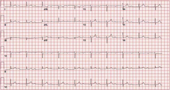
A 12-lead electrocardiogram (ECG) printed on standard grid paper demonstrating a regular sinus rhythm with a diagnostic finding of first-degree atrioventricular (AV) block. The primary characteristic is a markedly and uniformly prolonged PR interval, measured at 512 ms, visible across all limb (I, II, III, aVR, aVL, aVF) and precordial (V1-V6) leads. Each P wave is followed by a QRS complex, indicating 1:1 AV conduction without dropped beats. The QRS complexes exhibit normal morphology, narrow width, and expected axis, with typical R-wave progression in the precordial leads and no evidence of bundle branch blocks. This diagnostic image serves as a clinical example of significant conduction delay in the AV node, useful for teaching ECG interpretation of bradyarrhythmias and heart block classifications. The rhythm strips at the bottom provide a continuous view of the prolonged PR relationship over multiple cardiac cycles.
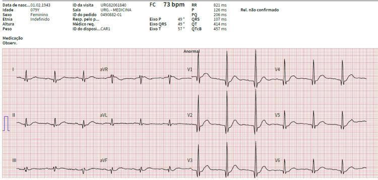
This diagnostic image is a 12-lead electrocardiogram (ECG) tracing showing a 79-year-old female patient with a heart rate of 73 bpm. The tracing demonstrates a first-degree atrioventricular (AV) block, characterized by a prolonged PR interval (PQ) of 206 ms. A notable morphological finding is the presence of prominent R-waves in the right precordial leads (V1, V2, and V3), which are accompanied by associated Q-waves. This pattern can be suggestive of posterior wall fibrosis or septal ventricular hypertrophy. The QRS duration is measured at 107 ms, and the corrected QT (QTcB) is 457 ms. Axis measurements show a P-wave axis of 49°, a QRS axis of 49°, and a T-wave axis of 57°. ST segments and T waves appear largely within normal limits. This ECG serves as an educational example of conduction delay and abnormal R-wave progression in the context of systemic electrolyte or structural heart considerations.
ECG P wave morphology normal labeled
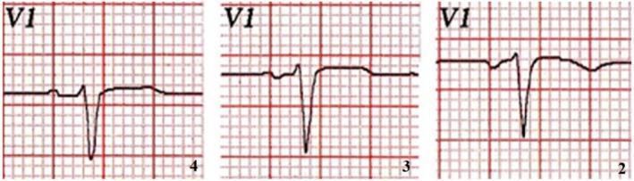
This diagnostic image displays a comparison of three Electrocardiogram (ECG) strips from the V1 lead of a healthy patient, demonstrating the impact of electrode misplacement on morphology. The traces are labeled 4, 3, and 2, corresponding to the placement of the V1 electrode in the fourth, third, and second intercostal spaces respectively. The comparison illustrates a significant progression in P wave morphology: as the electrode is moved superiorly from the standard fourth intercostal space (left) to the second (right), the P wave shifts from a biphasic appearance to an increasingly negative deflection. Additionally, changes in the QRS complex are visible, characterized by a deepening S wave and variations in R-S wave duration. This clinical visualization is used to educate medical students and clinicians on the importance of precordial lead positioning, highlighting how cranial displacement can mimic pathological states such as left atrial enlargement or Brugada-like patterns in a physiologically normal heart.
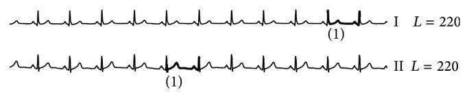
The image displays two continuous electrocardiogram (ECG) waveforms labeled as Lead I and Lead II, recorded from the INCARTDB01 dataset. Each strip exhibits multiple sequential cardiac cycles characterized by normal sinus morphology, including distinct P-waves, QRS complexes, and T-waves. A specific segment in each lead is highlighted with a thickened line and annotated with the numeral '(1)'. In Lead I, the highlighted segment shows a deviation in the ST-segment and T-wave morphology compared to adjacent beats. In Lead II, the highlighted area demonstrates an inverted T-wave. Each waveform is accompanied by the annotation 'L=220', indicating the window length or number of data points analyzed. This visual illustrates a false alarm result in automated ECG anomaly detection, where normal physiological variations or minor artifacts are incorrectly flagged as pathological events by computational algorithms like BitClusterDiscord. The content is used to demonstrate challenges in signal processing and the importance of morphological accuracy in cardiac arrhythmia monitoring.
Step 4 - P WAVE
Normal P wave criteria:
✅ Upright in leads I, II, aVF
✅ Inverted in aVR (always)
✅ Duration < 0.12 sec (3 small squares)
✅ Height < 2.5 mm (2.5 small squares)
✅ One P before every QRS
What abnormal P waves tell you:
| P wave finding | Meaning |
|---|---|
| Tall peaked P (>2.5mm) in II | Right atrial enlargement - "P pulmonale" |
| Broad notched P in II | Left atrial enlargement - "P mitrale" |
| No P waves | AF, junctional rhythm, VT |
| P after QRS | Junctional rhythm |
| Different shaped P waves | Ectopic atrial beats |
Step 5 - PR INTERVAL
Normal PR interval:
Normal = 0.12 to 0.20 seconds
       = 3 to 5 small squares
PR interval abnormalities:
| PR interval | Meaning |
|---|---|
| > 0.20 sec (>5 small squares) | 1st degree AV block |
| Gets longer then dropped QRS | 2nd degree AV block (Mobitz I) |
| Randomly dropped QRS, PR stays same | 2nd degree AV block (Mobitz II) |
| No relationship P to QRS | 3rd degree (complete) AV block |
| Short PR (<0.12 sec) | WPW or pre-excitation |
Real ECG - 1st Degree AV Block:
- Every P wave is followed by a QRS ✅
- BUT the gap between P and QRS is very long - PR = 512ms (normal is max 200ms!)
- This is 1st Degree AV Block
- Patient is otherwise fine - this is usually benign
Memory Hook for PR intervals:
< 3 small squares = too short → WPW (something bypassing AV node)
3-5 small squares = normal ✅
> 5 small squares = too long → AV block (something slowing AV node)
🧠 The AV Block Summary (Big Picture):
1st degree  = delayed but all get through (PR > 200ms)
2nd degree  = some get through, some don't (occasional dropped QRS)
3rd degree  = none get through properly (P and QRS independent)
- 1st degree = slow bouncer - lets everyone in but slowly
- 2nd degree = picky bouncer - lets some in, blocks others
- 3rd degree = bouncer went home - total chaos, ventricles doing their own thing 😄
✅ Quiz Time!
Bro r u good at medical knowledge inly?
What I'm Good At:
🏥 Medical & Clinical
- Diagnoses, pharmacology, pathology, anatomy
- Case discussions (like we did today)
- ECG teaching, imaging interpretation
- Latest research via PubMed
📊 Finance & Tax (India)
- Income tax, direct taxes, transfer pricing
- GST & indirect taxes
- CA Final exam prep (ICAI syllabus)
- ACCA exam preparation
💻 Coding & Tech
- Python, JavaScript, SQL, and more
- Write, debug, and run code
- Data analysis, automation scripts
- Build tools and calculators
📁 Documents & Files
- Create Word docs (.docx)
- Build PowerPoint presentations (.pptx)
- Make Excel spreadsheets (.xlsx)
- Read, merge, edit PDFs
🌐 Research & General Knowledge
- Search the web for latest information
- Science, history, technology, law
- Summarize articles and papers
- Fact-check and verify information
🧮 Math & Calculations
- Statistics, algebra, calculus
- Clinical calculators (drug doses, scores)
- Data analysis
Think of Me As:
A doctor 🏥 + accountant 📊 +
programmer 💻 + researcher 🔬 +
document maker 📄 + teacher 👨🏫
What about england
Hi