
Reduction mamoplasty in one page of detail that containt almost every important detail
reduction mammaplasty breast reduction surgical technique pedicle
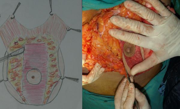
This composite educational resource consists of a medical diagram and an intraoperative clinical photograph illustrating a reduction mammaplasty technique focusing on the resection of medial and lateral tissues while preserving a central pedicle. The anatomical diagram (left) provides a schematic of the breast, highlighting a central rectangular region (pink) containing the nipple-areola complex (NAC) to be preserved as a posterior pedicle, surrounded by peripheral areas designated for resection. The intraoperative photograph (right) displays the surgical application of this technique. It depicts the surgeon's gloved hands using a scalpel to perform a precise resection of the lateral and medial breast parenchyma. The exposed tissue exhibits a characteristic yellow hue indicative of adipose tissue mixed with reddish glandular tissue and superficial vasculature. The central vertical pedicle is clearly visible, appearing more dense and pinkish-red compared to the disrupted peripheral tissues. This visual material is intended for plastic surgery education, demonstrating the creation of a U-shaped central pedicle to maintain neurovascular integrity and lactational function during breast reduction.
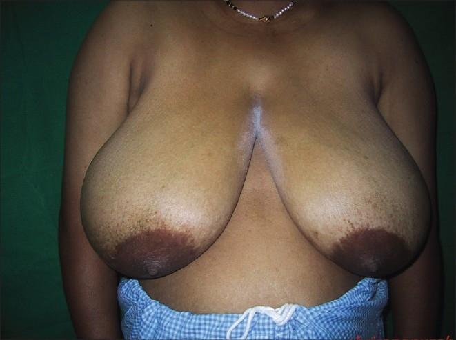
Clinical photograph of a patient with bilateral macromastia (breast hypertrophy), shown in a preoperative state for a reduction mammaplasty using an inverted T resection technique. The breasts are large and pendulous (grade III ptosis), extending significantly below the inframammary crease. The skin shows areas of hyperpigmentation and striae, particularly in the lower poles. The nipple-areola complexes are enlarged and displaced inferiorly due to the weight of the breast tissue. A vertical hypopigmented or perhaps powdered surgical marking area is visible extending from the sternal notch into the intermammary cleft. This image serves as a clinical example of severe breast hypertrophy requiring reconstructive surgery, highlighting the anatomical landmarks such as the breast meridian and nipple projection needed for surgical planning of pedicle-based resections (e.g., medial or inferior pedicle techniques).
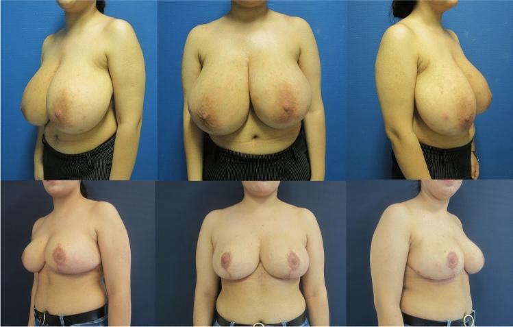
This clinical photograph comparison chart demonstrates the results of a Double-Unit Superomedio-Central (DUS) Pedicled Inverted-T Reduction Mammaplasty in a 20-year-old patient with symptomatic gigantomastia and Regnault Grade II ptosis. The top row shows preoperative views (frontal and bilateral oblique) illustrating significant macromastia, elongated sternal notch-to-nipple-areolar complex (SN-NAC) distances of 34-36 cm, and prominent lower pole fullness. The bottom row presents the status 12 months postoperatively from the same angles. The procedure achieved a substantial reduction in volume (1602g right, 1150g left) and corrected the ptosis by repositioning the NAC to a more anatomical level above the inframammary fold. Key visual changes include improved upper pole projection, a more conical and stable breast shape, and healed surgical scars in the characteristic inverted-T (anchor) pattern. This comparison highlights the efficacy of the DUS pedicle technique in preserving NAC vascularity while managing significant resection weights in young patients.
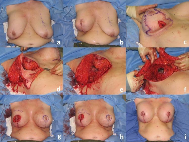
This nine-panel clinical photographic sequence (a-i) documents a reductive augmentation mammaplasty, a plastic surgery procedure combining breast reduction and implant placement. Panels a-c show the initial preoperative state and the 'tailor-tack' mastopexy technique using surgical staples and surgical markings to define the superomedial pedicle and planned incisions. Panels d-f illustrate the intraoperative tissue management: the surgical excision of a significant parenchymal specimen (initial 220g) from the lower pole, followed by thinning and further excision (additional 115g) of the lateral and medial skin flaps to create space for the prosthesis. The exposed breast anatomy demonstrates the relationship between the skin envelope, subcutaneous fat, and glandular tissue. Panels g-h show the re-approximation of tissue with temporary staples to assess symmetry and volume. The final panel (i) displays the postoperative result in a sitting position, demonstrating the final vertical scar closure, nipple-areolar complex repositioning, and improved upper pole fullness achieved via the permanent 470cc silicone implant.
"reduction mammaplasty" AND complications
Reduction Mammaplasty
Definition and Indications
History
Preoperative Work-up
- Mammogram is obtained first in women eligible for screening (per NCI, ACS, ACR annual screening ≥40 years) before any intervention.
- Labs as dictated by age/comorbidities.
- Preoperative standing markings: nipple position is the most critical determinant of aesthetic outcome - it should sit at or just above the inframammary fold (transposed onto the anterior breast surface); overcorrecting too high is the hardest complication to fix. Sternal notch-to-nipple distance is typically 19-22 cm. Breast meridian, inframammary folds, and chest midline are also marked.
Technique
- Skin closure pattern: inverted-T (Wise pattern) vs. vertical.
- Nipple-areolar complex (NAC) pedicle (vascular/nerve supply preserved to the NAC): most common combinations are vertical closure with a superior/superomedial pedicle or inverted-T closure with an inferior pedicle. Other options include central, superomedial, and free-nipple grafting (used in cases of extreme hypertrophy where pedicle length would compromise perfusion, at the cost of sensation and lactation).
- Typical parenchymal resection: 400-2,000 g per breast.
- Mastopexy uses similar pedicle principles but removes minimal parenchyma, instead redraping/lifting ptotic skin to reposition the breast on the chest wall - commonly used for contralateral symmetry procedures.
- Both reduction and mastopexy can be performed without impairing later breast self-exam or mammographic detection of masses.
Outcomes and Satisfaction
Complications
- Overall complication rate 42-50%, with about 15% significant and 5% requiring surgical revision - most revisions are minor and doable under local anesthesia in the office/outpatient setting.
- Standard surgical/anesthetic risks: infection, bleeding, wound issues, anesthesia reactions.
- NAC dysfunction: partial/complete sensory hypesthesia (may be transient or permanent) and inability to lactate. Objective testing often shows persistent loss of pain/pressure/light-touch sensation even when patients subjectively feel normal within weeks. Free nipple grafts have the lowest rate of sensory preservation; preserving the pectoralis fascia improves the odds of retaining innervation.
- Breastfeeding: data are limited, but roughly >50% of mothers can still breastfeed after reduction, often needing supplementation.
- Aesthetic complications (hardest to fully prevent): pre-existing asymmetry commonly persists to some degree regardless of technique/surgeon skill; "bottoming out" (gland descends below nipple over time due to excess skin envelope below the fold); late lateral fullness and upper-pole flatness; nipple malposition relative to the breast mound (too high is difficult to correct; too low is easier). Inframammary scars tend to become more visible over time, while vertical scars often fade, especially in darker skin tones.
Recent Evidence (systematic reviews/meta-analyses, 2023-2025)
- Ran et al., 2025, "Risk factors for complications after reduction mammaplasty: a systematic review and meta-analysis" (PMID: 40452064)
- Liu et al., 2023, "Risk Factors and Complications in Reduction Mammaplasty: A Systematic Review and Meta-analysis" (PMID: 37253843)
- Dušková et al., 2024, "No drains in reduction mammaplasty - a systematic review" (PMID: 38704230)
Lower anterior resection in short which contain every important detail must to know
low anterior resection rectal cancer surgical diagram total mesorectal excision
"low anterior resection" AND anastomotic leak
Lower (Low) Anterior Resection (LAR)
Definition and Indication
Core Oncologic Principle: Total Mesorectal Excision (TME)
- For upper rectal/rectosigmoid tumors, only a tumor-specific (partial) mesorectal excision is needed, with the mesorectum resected 5 cm distal to the tumor - full pelvic floor dissection is not required.
- For mid/low rectal tumors, dissection proceeds to the pelvic floor musculature posterolaterally and to the prostate/rectovaginal septum anteriorly.
- The inferior mesenteric artery is ligated at its base (high ligation), and the inferior mesenteric vein is ligated separately along its own course.
Nerve-Sparing Considerations
- Hypogastric plexus - at risk during high IMA ligation and posterior dissection over the sacral promontory; injury causes retrograde ejaculation.
- Pelvic plexus/nervi erigentes - at risk during lateral stalk division too close to the pelvic sidewall; injury causes erectile dysfunction/impotence and atonic bladder.
- Periprostatic plexus - at risk during anterior dissection; injury causes sexual/bladder dysfunction.
Anastomosis
- Usually an end-to-end colorectal or coloanal anastomosis using a circular stapler (double-staple technique) or hand-sewn for very low/coloanal anastomoses.
- For ultra-low LAR, constructing a colonic J-pouch or a side-to-end anastomosis (colonic reservoir) improves functional outcomes compared with straight end-to-end anastomosis, per multiple RCTs.
- A diverting (protective) loop ileostomy is frequently used for low anastomoses to mitigate consequences of leak.
- Near-infrared perfusion assessment (e.g., ICG/PINPOINT fluorescence, studied in the PILLAR III trial) is increasingly used intraoperatively to check anastomotic perfusion and reduce leak risk.
Minimally Invasive Approaches
- ROLARR (2017): no significant difference between robotic and laparoscopic LAR in conversion, operative time, margins, length of stay, or 30-day morbidity/mortality.
- COLRAR (2023): no significant difference in overall TME quality, but robotic approach had a lower positive circumferential margin rate (0% vs 6.1%); trial underpowered due to early termination.
- A large Chinese multicenter RCT (1,240 patients) found robotic TME had lower positive margin rates, higher complete resection rates, fewer complications, fewer conversions, and shorter length of stay versus laparoscopic TME - the first trial to show a clear advantage, though long-term oncologic outcomes remain pending.
Complications
- Anastomotic leak - the most feared complication; associated with LAR syndrome development but, per some series, not clearly linked to worse oncologic outcome.
- General postoperative risks: bleeding, infection, ileus, stricture.
- Sexual/urinary dysfunction from autonomic nerve injury (see above).
- Low Anterior Resection Syndrome (LARS) - fecal urgency, frequency, incontinence, clustering of bowel movements, and evacuation difficulty from loss of rectal reservoir function and sphincter/nerve disruption; risk correlates with anastomotic height (lower anastomosis = higher LARS risk) and history of anastomotic leak.
Recent Evidence (2023-2024 systematic reviews/meta-analyses)
- Reinforcing sutures at the anastomosis may reduce anastomotic leak rates (Wang et al., 2024, PMID: 38174618).
- Transanal drainage tubes show updated evidence for reducing leak after LAR (Tamura et al., 2024, PMID: 38916755).
- Anastomotic height/level is a significant determinant of LAR syndrome severity (Hashempour et al., 2023, PMID: 37653424).