

photo-1786741733264.jpg
Explain me in detail about all these thyroid cancers
analyse_media
Reading File
Now let me search the medical library for detailed content on all four thyroid cancer types simultaneously.
Finding Sources
Finding Sources
Searching Images
papillary thyroid cancer histology psammoma bodies orphan annie nuclei
Reading File
Finding Sources
Finding Sources
Reading File
Reading File
Finding Sources
Reading File
Reading File
Reading File
Loading Image
Searching Images
thyroid cancer types papillary follicular medullary histology comparison
I now have all the information needed to give a thorough explanation. Here is a complete, detailed breakdown of all four thyroid cancers from your notes:
Thyroid Cancers - Complete Detailed Guide
🔴 1. Papillary Thyroid Cancer (PTC)
Basics
- Most common thyroid cancer (~85% of all thyroid cancers)
- Best prognosis - 5-year survival >98%
- Female predominance (3:1), peak incidence in 3rd-4th decade
Risk Factors & Associations
- Radiation exposure (RT) - most strongly linked to PTC. This includes childhood radiation therapy to the neck.
- Thyroglossal cyst (TGC) - PTC can arise within a thyroglossal duct cyst
- Hashimoto's Thyroiditis (your handwritten note) - chronic lymphocytic inflammation is a known risk factor for both PTC and thyroid lymphoma
Molecular Mutations
- BRAF V600E mutation - the most common driver mutation in PTC (t 10;17 refers to the chromosomal arrangement). This same mutation also occurs in:
- Pilocytic Astrocytoma
- LCH (Langerhans Cell Histiocytosis)
- HCL (Hairy Cell Leukemia)
- Adenocarcinoma of the Colon
- GIST (Gastrointestinal Stromal Tumor)
- Melanoma
Spread
- Lymphatic metastasis is the hallmark - spreads to cervical lymph nodes. Importantly, lymph node involvement does NOT worsen prognosis.
- Distant metastases (lungs most common) in only 1-8% of cases
Histology (what you see under the microscope)
- Orphan Annie nuclei - optically clear, "empty-looking" large nuclei with nuclear grooves
- Psammoma bodies - concentric calcified laminated structures (seen in ~50%)
- Coffee bean nuclei (your handwritten note) - refers to the nuclear groove pattern
- Papillary architecture with fibrovascular cores
- The image in your notes shows classic papillary thyroid cancer histology
Diagnosis
- FNAC (Fine Needle Aspiration Cytology) is the primary diagnostic tool (your note: "dx → FNAC")
- Ultrasound features: microcalcifications, irregular margins, taller-than-wide shape
Treatment
- Surgery: Lobectomy for low-risk tumors <4 cm; total thyroidectomy for larger/high-risk tumors
- RAIU (Radioactive Iodine Uptake scan) - for post-op surveillance
- RAI (I-131 ablation) - radioiodine therapy destroys residual thyroid tissue/mets (only in high-risk; not needed for low-risk)
- TSH suppression with levothyroxine
🟠 2. Follicular Thyroid Cancer (FTC)
Basics
- 2nd most common thyroid cancer (10-15%)
- Female predominance, peak in 5th decade
Risk Factors & Associations
- Iodine-deficient areas (MC in iodine deficiency) - your notes highlight this
- Multinodular Goiter (MNG) - associated background
Molecular Mutations
- RAS gain-of-function (GOF) mutation
- PAX8-PPARγ fusion [t(2;3)] - translocation between chromosomes 2 and 3, creating this fusion oncogene
Key Difference from PTC: Spread
- Hematogenous metastasis (via blood, NOT lymphatics) - goes to bone, lungs, brain
- Lifetime risk of distant metastases: 10-15%
- Lymph node involvement is uncommon (~10%)
Histology
- Follicular architecture (difficult to distinguish from follicular adenoma)
- Diagnosis REQUIRES demonstration of capsular invasion and/or vascular invasion - this CANNOT be done on FNAC alone (a major limitation)
- FNA biopsy reports it as "follicular neoplasm" - cannot say benign vs. malignant
- Frozen section is also unreliable; requires final paraffin sections
Special Variants
- Hurthle Cell Carcinoma: variant of FTC with oncocytic cells (abundant eosinophilic granular cytoplasm); less likely to take up radioiodine (only 10% RAI-avid)
- NIFTP (Noninvasive Follicular Thyroid Neoplasm with Papillary-like nuclear features): reclassified as a non-malignant entity
Treatment
- Total thyroidectomy is preferred (most patients)
- RAI therapy - up to 80% are RAI-sensitive (unlike Hurthle cell)
- Therapeutic lymph node dissection if imaging shows disease
🟣 3. Medullary Thyroid Cancer (MTC)
Basics
- Arises from parafollicular C cells (NOT follicular epithelium) - this is the KEY distinction
- C cells normally produce calcitonin
- ~1-3% of all thyroid cancers
- 25% are familial; 75% are sporadic
Associations
- MEN2 (RET point mutation) - your notes correctly identify this
- MEN2A: MTC + pheochromocytoma + parathyroid hyperplasia
- MEN2B: MTC (very aggressive, presents in infancy) + pheochromocytoma + marfanoid habitus + mucosal neuromas + ganglioneuromatosis
- RET proto-oncogene mutations are the primary driver (germline in familial, somatic in sporadic)
- C-cell hyperplasia precedes MTC in familial cases
Tumor Markers
- Calcitonin (circled in your notes) - the primary tumor marker; elevated preop and used to monitor for recurrence
- CEA (Carcinoembryonic Antigen) - also elevated; used for monitoring
- Dense bodies / amyloid deposits (your note) - histologic hallmark; amyloid staining on FNA biopsy
Clinical Features
- Can secrete ACTH → ectopic Cushing syndrome (your handwritten note "↑ACTH")
- Elevated calcitonin → diarrhea, facial flushing
- Can also secrete PTHrP → TSH-like effects (your note "↑TSH" relates to ectopic hormone secretion)
Spread
- Cervical lymph node metastases are common (~50% at clinical diagnosis)
- Distant metastases: lung (52%), bone (28%), mediastinum (20%)
- "↑ recurrence after removal" - your handwritten note is correct; MTC commonly recurs
Treatment
- Total thyroidectomy with central LN dissection (Level VI - your notes say "central LN dissection")
- No role for RAI or TSH suppression - your handwritten notes are exactly right! (C cells don't respond to iodine or TSH)
- No role for RAIU, RAI ablation, or I-131 (your note: "no role of 1.RAIU, 2.RAI, 3. I-131 ablation")
- RET kinase inhibitors (Vandetanib, Cabozantinib) for metastatic disease
- Genetic counseling + RET germline testing for all first-degree relatives
🔴 4. Anaplastic Thyroid Cancer (ATC)
Basics
- Worst prognosis - your notes correctly state this
- Median survival: 3-6 months after diagnosis; 1-year overall survival only ~20%
- Represents <1% of all thyroid cancers
- Mean age at diagnosis: 65 years, female-to-male ratio 2:1
- All ATCs are classified as Stage IV disease by AJCC (because survival is so poor)
Origin & Pathogenesis
- Undifferentiated tumor of follicular cell origin (same as PTC/FTC)
- Thought to arise from dedifferentiation of PTC - PTC coexists in at least 30% of ATC cases
- Key mutations: TP53, PIK3CA, β-catenin pathway genes
- A history of multinodular goiter or prior thyroidectomy is present in up to 50% of patients
Clinical Presentation
- Rapidly enlarging neck mass (the hallmark)
- Severe local symptoms: neck pain, dyspnea, cough, hemoptysis, dysphagia, hoarseness (RLN invasion)
-
50% have cervical lymphadenopathy at presentation
- 15-50% have distant metastases at presentation (lung, bone, brain, liver, skin)
Histology
The image below from Sabiston's Textbook of Surgery shows the classic H&E appearance:
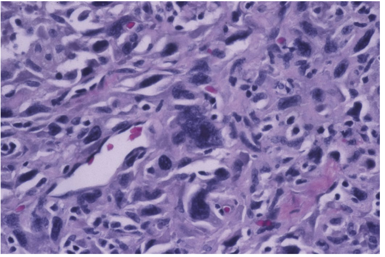
- Mixed patterns of spindle cells, pleomorphic giant cells, and squamoid cells
- Atypical mitoses and extensive necrosis
- Does NOT stain for thyroglobulin (Tg) in most cases
- Cytology: spindled, pleomorphic, and squamoid cells with atypical mitoses
Treatment (per 2021 ATA Guidelines)
- Surgery if resectable (debulking); rarely curative
- External beam radiotherapy + systemic therapy (including targeted therapy)
- Molecular targeted therapies - especially if BRAF V600E positive: BRAF + MEK inhibitor (Dabrafenib + Trametinib) has shown significant benefit
- PET/CT recommended for staging (ATC is intensely PET-avid)
- Brain MRI as part of staging algorithm when clinically indicated
- Multidisciplinary approach is essential
Summary Comparison Table
| Feature | Papillary | Follicular | Medullary | Anaplastic |
|---|---|---|---|---|
| Frequency | ~85% | 10-15% | 1-3% | <1% |
| Cell of origin | Follicular epithelium | Follicular epithelium | Parafollicular C cells | Follicular epithelium |
| Key mutation | BRAF V600E | RAS, PAX8-PPARγ | RET | TP53, PIK3CA |
| Spread | Lymphatic | Hematogenous | Both | Both (early) |
| Tumor marker | Thyroglobulin | Thyroglobulin | Calcitonin + CEA | None reliable |
| RAI therapy | Yes (high-risk) | Yes (80% sensitive) | No | No |
| TSH suppression | Yes | Yes | No | No |
| Prognosis | Excellent | Good | Intermediate | Very poor |
| Histology hallmark | Orphan Annie nuclei, Psammoma bodies | Capsular/vascular invasion | Amyloid deposits, C cells | Pleomorphic giant cells |
Bonus Notes from Your Sheet
Nerve injuries during thyroid surgery:
- RLN (Recurrent Laryngeal Nerve) injury → Vocal cord paralysis → Hoarseness
- ELN (External Laryngeal Nerve), a branch of SLN (Superior Laryngeal Nerve) → Loss of pitch/voice (cannot sing high notes)
GNAS mutation - associated with Toxic Adenoma (not cancer, but important MCQ fact)
Wolf-Chaikoff effect: Excess iodine → blocks organification → Hypothyroidism
Jod-Basedow phenomenon: Iodine supplementation in iodine-deficient patients → Hyperthyroidism
Sources: - Sabiston Textbook of Surgery, 21e; Goldman-Cecil Medicine, 2e; Mulholland & Greenfield's Surgery, 7e
I can see what's written in my book , explain me from the standard textbook abt these cancers
Reading File
Reading File
Reading File
Reading File
Finding Sources
Searching Images
papillary thyroid cancer Orphan Annie nuclei psammoma bodies histology microscopy
Finding Sources
Reading File
Reading File
Reading File
Finding Sources
Finding Sources
Finding Sources
Excellent - Robbins is in the library. Let me pull the full thyroid neoplasm section from Robbins.
Reading File
Reading File
Reading File
Loading Image
Loading Image
Reading File
Reading File
Loading Image
Reading File
Loading Image
Reading File
Loading Image
Here is a full textbook-based explanation drawn directly from Robbins & Cotran Pathologic Basis of Disease (10e), Sabiston Textbook of Surgery (21e), Mulholland & Greenfield's Surgery (7e), and Goldman-Cecil Medicine - with actual histology images from the textbooks.
Thyroid Cancers - From Standard Textbooks
Overview (Robbins & Cotran)
"Most thyroid carcinomas are derived from thyroid follicular epithelium. Most high grade, poorly differentiated, and anaplastic carcinomas arise from well-differentiated PTC or follicular carcinomas through acquisition of additional mutations. These aggressive neoplasms and medullary thyroid carcinomas are the major causes of mortality from thyroid cancer."
- Robbins, p.1005
Incidence breakdown (Mulholland & Greenfield's Surgery, p.4047):
- PTC: 85%
- FTC: 12%
- Poorly differentiated/insular: 3%
- MTC: 1-2%
- ATC: <1%
1. PAPILLARY THYROID CARCINOMA (PTC)
Pathogenesis (Robbins)
Robbins lays out a clear molecular hierarchy for PTC. There are two defining genetic abnormalities:
A. BRAF V600E point mutation (up to 80% of conventional PTCs)
"BRAF encodes a serine/threonine kinase that lies downstream of receptor tyrosine kinases and RAS in growth factor signaling pathways. The presence of BRAF mutation in conventional PTC correlates with reduced expression of thyroid differentiation markers (such as thyroglobulin and thyroid peroxidase) and may be associated with a higher risk of extrathyroidal extension and recurrence. As discussed elsewhere, the BRAFV600E mutation is found in many other cancers including melanoma and hairy cell leukemia."
B. Gene fusions - RET, NTRK, or BRAF fusion genes
"The RET gene is located on chromosome 10q11 and encodes an RTK that is normally not expressed in thyroid follicular cells. PTC rearrangements involving RET create fusion genes that encode constitutively active forms of RET tyrosine kinase. There are more than 20 fusion partners of RET, PTC1 (also known as CCDC6) being the most common."
Environmental Risk Factors (Robbins)
"The major risk factor predisposing to thyroid cancer is exposure to ionizing radiation, particularly during the first 2 decades of life. There was a marked increase in the incidence of conventional PTCs among children exposed to ionizing radiation after the Chernobyl nuclear disaster in 1986."
Morphology - The Histologic Hallmarks (Robbins)
The microscopic hallmarks of papillary carcinoma are:
- Branching papillae with fibrovascular stalks covered by cuboidal epithelial cells
- Orphan Annie eye nuclei - "Nuclei with finely dispersed chromatin and an optically clear or empty appearance, giving rise to the designation ground-glass or Orphan Annie eye nuclei"
- Nuclear grooves (coffee-bean shape) and nuclear pseudoinclusions (cytoplasm invaginates into nucleus)
- Psammoma bodies - "Concentrically calcified structures (psammoma bodies) present in approximately 50% of cases"
Below are the actual Robbins textbook images of PTC - gross specimen (A), branching papillary architecture (B), Orphan Annie nuclei (C), and intranuclear pseudoinclusion/arrow (D):
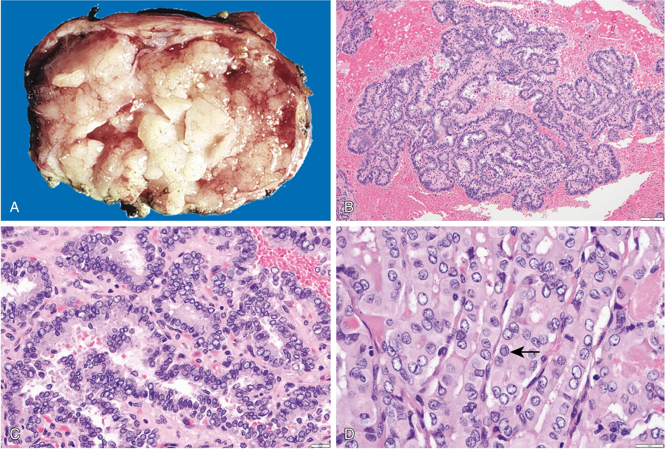
Fig. 24.19 from Robbins: (A) gross papillary structures, (B) well-formed papillae on H&E, (C) high power showing enlarged, overlapping, grooved nuclei with pale/empty chromatin - the "Orphan Annie eye" appearance, (D) intranuclear cytoplasmic pseudoinclusion (arrow)
The Scott-Brown ENT textbook specifically states about psammoma bodies:
"Psammoma bodies (laminated calcospherites) are present in roughly 50% of PTCs histologically, less frequently on FNAC and are virtually pathognomonic. They probably represent mineralized stromal cores."
The image below shows a classic psammoma body - the concentrically laminated, calcified structure:
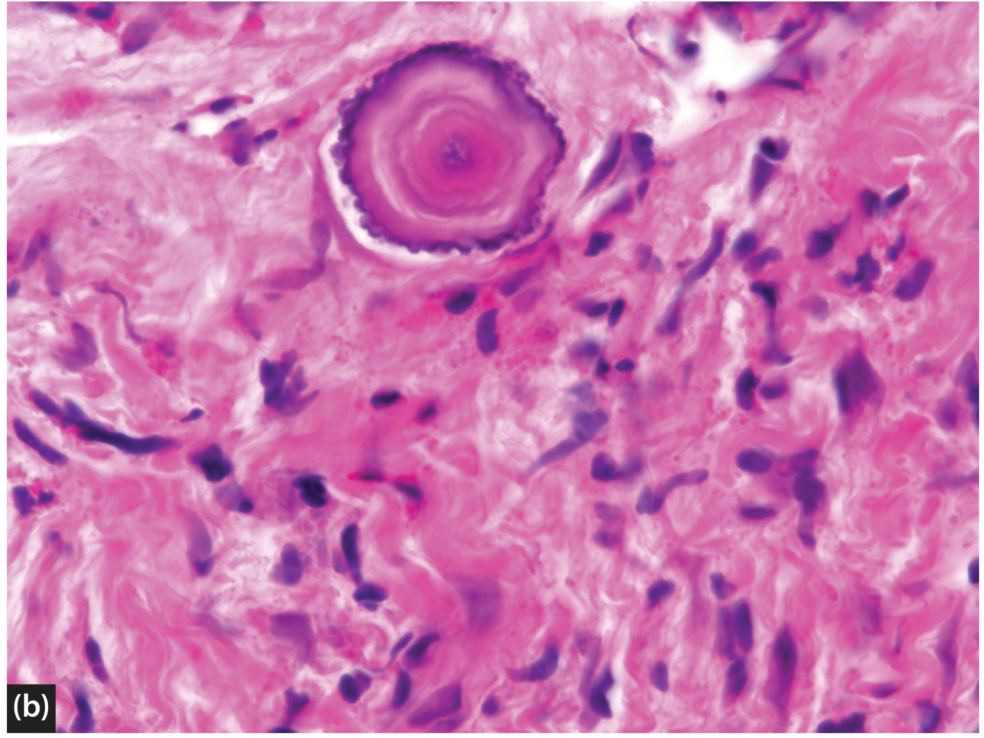
Fig. 58.30 from Scott-Brown's: A psammoma body - the concentric rings of calcification are stromal in location and virtually pathognomonic of PTC
Clinical Features (Robbins)
"Most conventional papillary carcinomas present as asymptomatic thyroid nodules, but the first manifestation may be an enlarged cervical lymph node... Hoarseness, dysphagia, cough, or dyspnea suggests advanced disease."
"Papillary carcinomas are cold masses on scintigraphy, and 'hot' nodules are almost never malignant."
Spread & Prognosis
- Lymphatic spread - cervical lymph node metastases in up to 50% of cases
- Importantly: "Unlike many other cancers, multifocality within the thyroid and/or lymph node involvement does not necessarily convey a poorer prognosis" (Mulholland)
- Hematogenous spread is uncommon
- 10-year survival >95%; prognosis worsens with age >55, extrathyroidal extension, distant metastases
Treatment (Goldman-Cecil)
- Small, low-risk (<1 cm, no high-risk features): active surveillance may be elected
- Lobectomy for 1-4 cm tumors without extrathyroidal extension
- Total thyroidectomy for tumors ≥4 cm, extrathyroidal extension, lymph node or distant metastases
- Post-op RAI (I-131 ablation) for high-risk tumors; no benefit for low-risk
- TSH suppression with levothyroxine
2. FOLLICULAR THYROID CARCINOMA (FTC)
Pathogenesis (Robbins)
"In contrast to papillary carcinomas, follicular neoplasms are often associated with gain-of-function mutations in RAS. A unique (2;3)(q13;p25) translocation has been described in a subset of follicular neoplasms. This translocation creates a fusion gene composed of portions of PAX8, a paired homeobox gene that is important in thyroid development, and the peroxisome proliferator-activated receptor gene (PPARG)."
"Deficiency of dietary iodine (and by extension, goiter) is associated with a higher frequency of follicular neoplasms." (Robbins)
Morphology (Robbins)
FTC is tricky because it looks like normal thyroid tissue. Robbins is explicit:
"Follicular carcinomas are more difficult to diagnose, since the cells closely resemble normal follicular cells and the tumors have a follicular architecture similar to adenomas. Most importantly, the diagnosis of malignancy in follicular lesions rests on the identification of vascular or capsular invasion."
This is why FNA biopsy cannot distinguish follicular adenoma from carcinoma - it can only call it a "follicular neoplasm." Definitive diagnosis requires surgical excision and histologic examination of the capsule.
Epidemiology (Robbins & Mulholland)
- 5-15% of thyroid cancers overall; rises to 25-40% in iodine-deficient regions
- Peak incidence 40-60 years of age
- More common in women
Spread
- Hematogenous spread (the key distinction from PTC) - to bone, lung, brain
- Lifetime risk of distant metastases: 10-15%
- Lymph node metastases are uncommon (~10%)
Treatment (Mulholland)
"Most patients with FTC are treated with total thyroidectomy, with therapeutic lymph node dissection as indicated by preoperative imaging."
- Up to 80% of FTCs are sensitive to RAI (I-131 therapy)
- Hurthle cell variant is RAI-resistant (only 10% avid)
3. MEDULLARY THYROID CARCINOMA (MTC)
What It Is (Robbins)
"Medullary carcinomas of the thyroid are neuroendocrine neoplasms derived from parafollicular cells (C cells). They account for approximately 5% of thyroid neoplasms. Medullary carcinomas, like normal C cells, secrete calcitonin, the measurement of which plays an important role in the diagnosis and postoperative follow-up of patients."
Calcitonin physiology from Robbins:
"Calcitonin is a regulator of calcium metabolism. It is normally produced in response to hypercalcemia, and it reduces serum calcium by inhibiting osteoclast activity and renal tubular reabsorption of calcium."
Genetics (Robbins)
"About 70% of tumors arise sporadically. The remainder occur in the setting of MEN2 syndrome (caused by germline RET mutations). Activating point mutations in the RET proto-oncogene play an important role in the development of both familial and sporadic medullary carcinomas. Syndromic cases tend to occur in younger patients, even during the first decade of life."
MEN2 subtypes (from Mulholland & Greenfield's Surgery, Table 75.6):
| Syndrome | Clinical Features |
|---|---|
| MEN2A | MTC (>90%), Pheochromocytoma (40-60%), Parathyroid hyperplasia (20-30%), cutaneous lichen amyloidosis, Hirschsprung disease |
| MEN2B | MTC (>98%), Pheochromocytoma (40-60%), intestinal and mucosal neuromas (98%), marfanoid habitus, inability to produce tears, prominent corneal nerves |
| Familial MTC | Isolated MTC with typically later age of onset and less aggressive clinical course |
Morphology (Robbins)
"Microscopically, medullary carcinomas are composed of polygonal to spindle-shaped cells, which may form nests, trabeculae, and even follicles. Amyloid deposits derived from calcitonin polypeptides are present in the stroma in many cases. Calcitonin is readily demonstrable within the tumor cells and the stromal amyloid by immunohistochemical methods."
The image below from Robbins (Fig. 24.22B) shows the nested architecture and the characteristic pink amyloid deposits in the stroma:
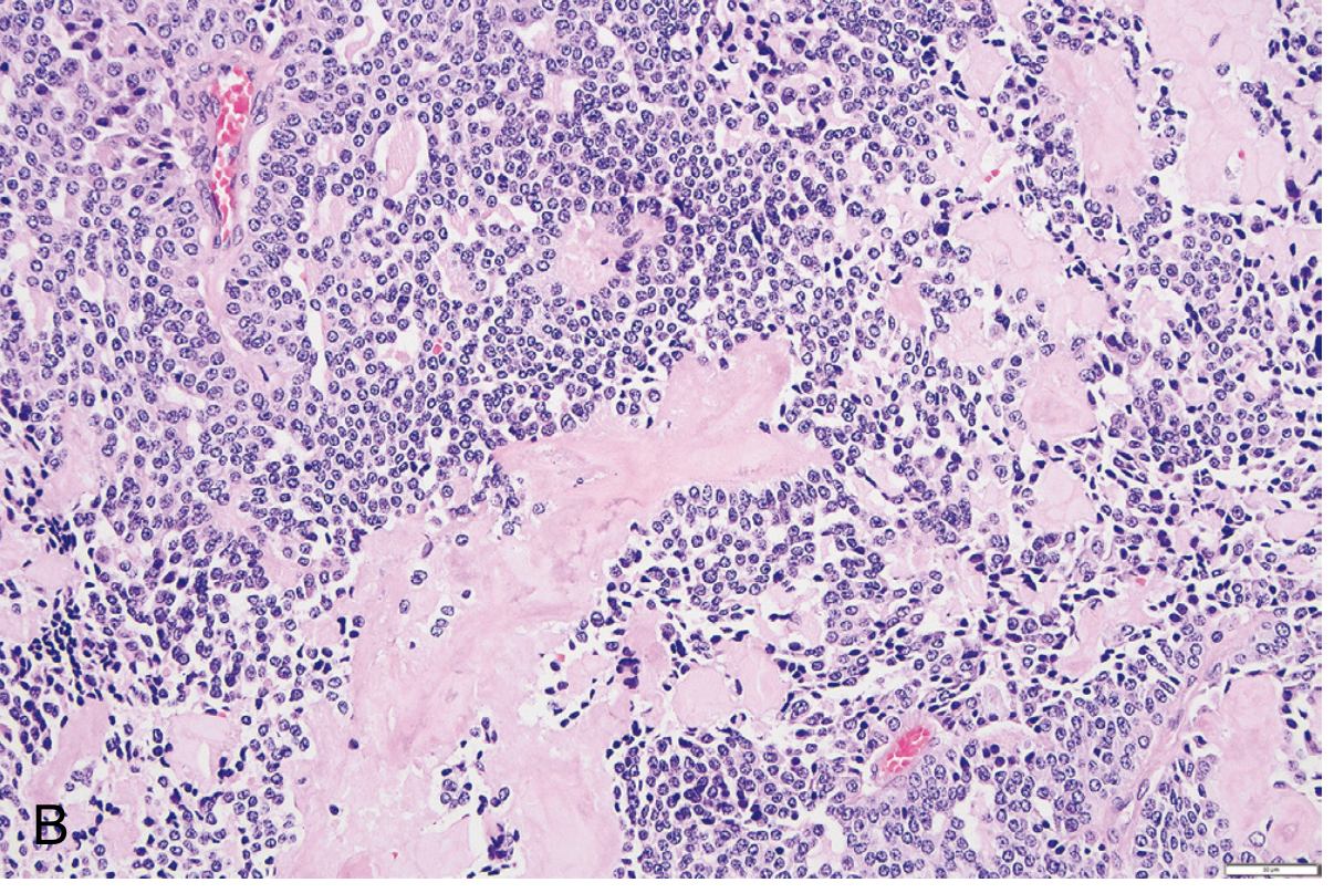
Fig. 24.22B from Robbins: Medullary carcinoma histology - nested polygonal/spindle cells with round nuclei and stippled (salt-and-pepper) chromatin. The pale pink material between nests is amyloid derived from calcitonin polypeptides.
"One of the features of syndromic medullary carcinomas is the presence of C-cell hyperplasia in the surrounding thyroid parenchyma, a feature that is usually absent in sporadic lesions, and that is believed to be a precursor lesion in syndromic cases." (Robbins)
Ectopic Hormone Secretion (Robbins)
"In some instances, the tumor cells elaborate other polypeptide hormones, such as serotonin, corticotropin [ACTH], and vasoactive intestinal peptide (VIP)."
- ACTH secretion → ectopic Cushing syndrome
- VIP secretion → diarrhea
- Calcitonin itself → diarrhea and facial flushing in advanced disease
Spread (Sabiston)
"Nearly half of patients have lymph node metastases if the diagnosis is made clinically rather than based on calcitonin levels. MTC with distant metastases is considered incurable. Nearly 20% of patients with MTC with a palpable neck mass have metastases at diagnosis."
- Most common distant sites: Lung (52%), bone (28%), mediastinum (20%)
- Nodal metastases correlate with tumor size (unlike PTC where size does not correlate)
Calcitonin as Biomarker (Tietz Textbook of Laboratory Medicine)
"Calcitonin is a valuable blood tumor marker for MTC. Basal serum calcitonin and CEA should be measured concurrently. Measurements of calcitonin may also be used to monitor for persistent or recurrent disease after surgery because the concentrations correlate with tumor burden. The MTC growth rate can be determined by measuring serum levels of calcitonin or CEA over multiple time points to determine the rate at which each marker's value doubles."
Treatment (Sabiston & Goldman-Cecil)
- Total thyroidectomy with central neck dissection (Level VI) in all cases
- No role for TSH suppression or RAI - C cells do not respond to TSH or iodine
- Lateral neck dissection guided by preoperative ultrasound and calcitonin levels
- Calcitonin >400 pg/mL warrants imaging for distant metastases
- Targeted therapy for metastatic disease: Vandetanib (RET/EGFR/VEGFR inhibitor) and Cabozantinib (RET/MET/VEGFR2 inhibitor) - both FDA approved
- Newer selective RET inhibitors: Selpercatinib and Pralsetinib showing promising results
Prophylactic Surgery in MEN2 (Mulholland)
"Prophylactic thyroidectomy should be performed within the first year of life for those with RET codon M918T mutations (ATA highest risk) and prior to age 5 for those with RET codon C634 or A883F mutations (ATA high risk)."
4. ANAPLASTIC THYROID CARCINOMA (ATC)
What It Is (Robbins & Sabiston)
"ATC is an extremely aggressive undifferentiated tumor of follicular cell origin. ATC is uncommon, comprising approximately 1% of all thyroid cancers. The mean age at diagnosis is 65 years, with a 2:1 female-to-male incidence ratio." (Sabiston)
"High grade, poorly differentiated, and anaplastic (undifferentiated) carcinomas can arise de novo, or, much more commonly, by transformation of a papillary or follicular thyroid carcinoma. In addition to having driver mutations seen in well-differentiated thyroid cancers, other common genetic 'hits' - point mutations of TP53 and the promoter region of TERT - are essentially restricted to aggressive carcinomas and likely have central roles in their genesis and behavior." (Robbins)
Evidence of Dedifferentiation from PTC (Sabiston)
"ATC is thought to arise from DTC of follicular cell origin (particularly PTC), based on the coexistence of PTC in at least 30% of cases as well as longitudinal case studies demonstrating dedifferentiation and transformation from DTC to ATC over time."
Clinical Presentation (Goldman-Cecil & Sabiston)
"Most patients present with a rapidly enlarging anterior neck mass, which may be painful and usually causes compressive symptoms. The trachea may be partially obstructed. Systemic symptoms may include weight loss, fatigue, and fever of unknown origin. Examination shows a large, firm, fixed, nodular goiter." (Goldman-Cecil)
Local symptoms are severe: dyspnea, dysphagia, hoarseness (RLN invasion), cough, hemoptysis
- Over 50% have cervical lymphadenopathy at presentation
- 15-50% have distant metastases at presentation (lung, bone, brain, skin, liver, kidneys, pancreas, heart, adrenal glands)
Histology (Sabiston)
"Cytologic features of ATC include mixed patterns of spindled, pleomorphic giant, and squamoid cells with mitotic figures, atypical mitoses, and extensive necrosis. ATCs typically do not secrete or stain for Tg."
The H&E image below from Sabiston (Fig. 73.27) shows the classic appearance:
Fig. 73.27 from Sabiston: Anaplastic thyroid cancer. H&E staining showing marked nuclear pleomorphism, oval to spindle-shaped cells, and multinucleated tumor cells - contrasting sharply with the well-organized architecture of PTC or FTC.
Staging (Sabiston)
"According to the AJCC TNM system, all ATCs are considered Stage IV disease because it is the most lethal type of thyroid cancer, with 1-year overall survival of 20%."
Unlike other thyroid cancers, ATC requires PET/CT (not CT alone) for staging because of its intense FDG avidity.
Prognosis (Goldman-Cecil)
"Metastatic anaplastic cancer is invariably fatal. The median survival in patients with anaplastic cancer is 5 to 6 months, and the 1-year overall survival is only 20%."
Treatment - Modern Era (Mulholland & Sabiston)
The discovery of targetable mutations has changed management:
"The discovery of the targetable BRAF V600E mutation in 25% to 45% of ATCs has provided at least some hope for life-prolonging therapy. In 2018, the dual-agent BRAF/MEK inhibitors dabrafenib and trametinib became the first FDA-approved targeted agents for ATC, based on the results of a phase II open-label trial that showed an overall response rate of 69% (1-year estimate of OS 80%)." (Mulholland)
- BRAF V600E positive: Dabrafenib + Trametinib (BRAF/MEK inhibitor combination) - FDA approved 2018
- Other mutations (ALK, NTRK, RET fusions): targeted therapies available
- Surgical resection if tumor is downstaged sufficiently
- Checkpoint inhibitors (e.g., pembrolizumab) at progression
- Palliative: chemotherapy (taxane or platinum-based) + external beam radiotherapy
Key Concepts Summary (from Robbins Key Concepts box)
- "Conventional papillary carcinomas harbor either BRAF V600E point mutations or fusion genes (RET, NTRK, BRAF)."
- "Follicular neoplasms of all subtypes are characterized by oncogenic RAS mutations and PAX8-PPARG fusions."
- "Psammoma bodies are a characteristic feature of papillary carcinomas; although these neoplasms often metastasize by way of lymphatics, the prognosis is excellent."
- "Medullary carcinomas are derived from parafollicular C cells and secrete calcitonin; amyloid deposits in the stroma are a characteristic feature."
Sources: Robbins & Cotran Pathologic Basis of Disease, 10e | Sabiston Textbook of Surgery, 21e | Mulholland & Greenfield's Surgery, 7e | Goldman-Cecil Medicine, International Ed. | Scott-Brown's Otorhinolaryngology, Vol. 1 | Tietz Textbook of Laboratory Medicine, 7e
This is a shared conversation. Sign in to Orris to start your own chat.