
What are vaginal fornix
vaginal fornix anatomy diagram cervix
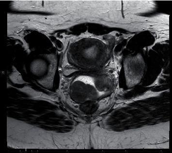
This diagnostic image is an axial T2-weighted MRI of the female pelvis, demonstrating a prominent mass in the superior vaginal fornix. The image shows a clear anatomical distinction between the cervix and the pathological lesion. On the patient's left side, the cervix is visible as a rounded structure with heterogeneous signal intensity. On the patient's right side, there is a large, somewhat circumscribed vaginal mass exhibiting complex internal characteristics. The mass displays central high signal intensity, suggestive of necrosis or fluid content, surrounded by a lower-signal-intensity rim. While the borders are relatively well-defined, there is evidence of displacement of adjacent pelvic structures and blurring of the anterior margin, raising the possibility of focal extension. The surrounding pelvic anatomy, including the musculature and hip joints, is visible as landmarks. This image is clinically significant for the assessment of gynecological smooth muscle tumors, such as leiomyosarcoma or cellular leiomyoma, and aids in surgical planning by mapping the mass relative to the cervix and vaginal walls.
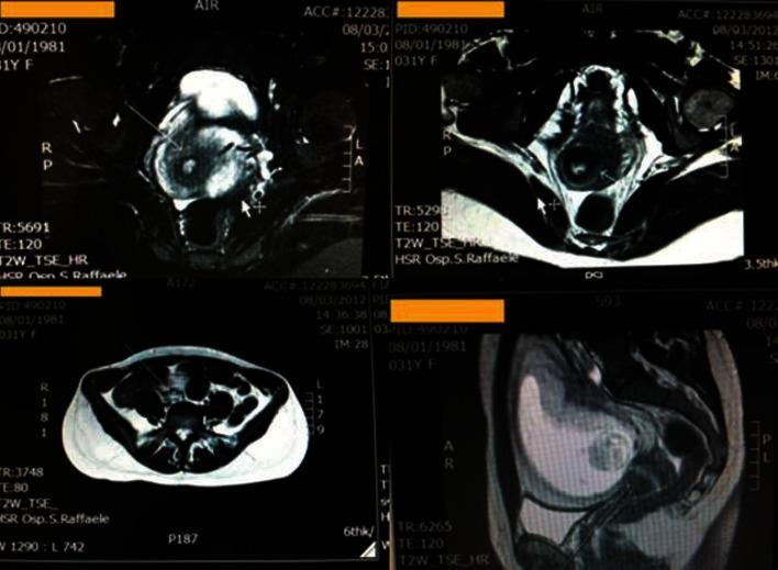
This diagnostic image set consists of pelvic Magnetic Resonance Imaging (MRI) scans in axial and sagittal planes, likely T2-weighted sequences (T2W TSE HR). The images focus on the female pelvic anatomy, specifically the uterus and cervix in a pregnant patient. A mild hyperintense signal is visible in the posterior-left and anterior aspects of the cervix, indicated by arrows. This signal abnormality extends toward the outer cervical border. The surrounding parametrium shows no signs of infiltration, maintaining clear fat planes. The left vaginal fornix appears minimally thickened without abnormal signal intensity. Other pelvic structures, including the ovaries and the gravid uterus, show normal morphology for gestational age without evidence of lymphadenopathy in the pelvic or lumbar-aortic regions. These findings were documented in a clinical case of primary cervical schwannoma, highlighting the importance of MRI in evaluating localized cervical lesions and their relationship to adjacent structures during pregnancy.
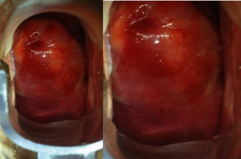
This clinical photograph displays a colposcopic view of the cervix. The cervix appears significantly enlarged and occupies the majority of the vaginal vault, consistent with a bulky or 'stony' clinical presentation. The surface of the cervix and the adjacent posterior vaginal fornix exhibit a distinct 'discolored area' characterized by a dark red, erythematous appearance. Numerous fine, dark red punctations are visible across the surface, representing abnormal vascular patterns. While the surface texture remains relatively smooth without overt epithelial laceration or ulceration, the atypical vascularity and discoloration are prominent. The squamocolumnar junction and cervical os are not clearly demarcated due to the presence of the lesion. This image is used to demonstrate clinical findings in the gynecologic evaluation of abnormal cervical masses, where differential diagnoses include cervical fibroids or rarer entities such as extramedullary leukemic infiltration of the female genital tract.
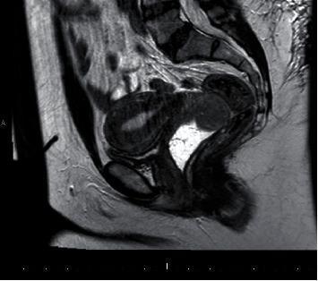
This diagnostic image is a T2-weighted sagittal MRI of the female pelvis, emphasizing the gynecological tract. The image utilizes gel distension of the vagina to enhance contrast and delineate vaginal anatomy. A well-defined mass of intermediate signal intensity is visible in the apical vaginal region, specifically at the superior vaginal fornix. The mass is positioned adjacent to the cervix and the inferior portion of the uterus. The uterus itself displays findings consistent with adenomyosis, characterized by a thickened junctional zone and heterogeneous myometrial signal. The hyperintense (bright) signal of the vaginal gel provides a clear interface against the intermediate-signal mass, assisting in the assessment of the lesion's morphology and its relationship to the vaginal walls. This imaging study is used in the clinical evaluation of pelvic masses, specifically for characterizing vaginal smooth muscle tumors and determining their extent relative to the bladder and rectum in a sagittal plane.
Vaginal Fornix
Definition
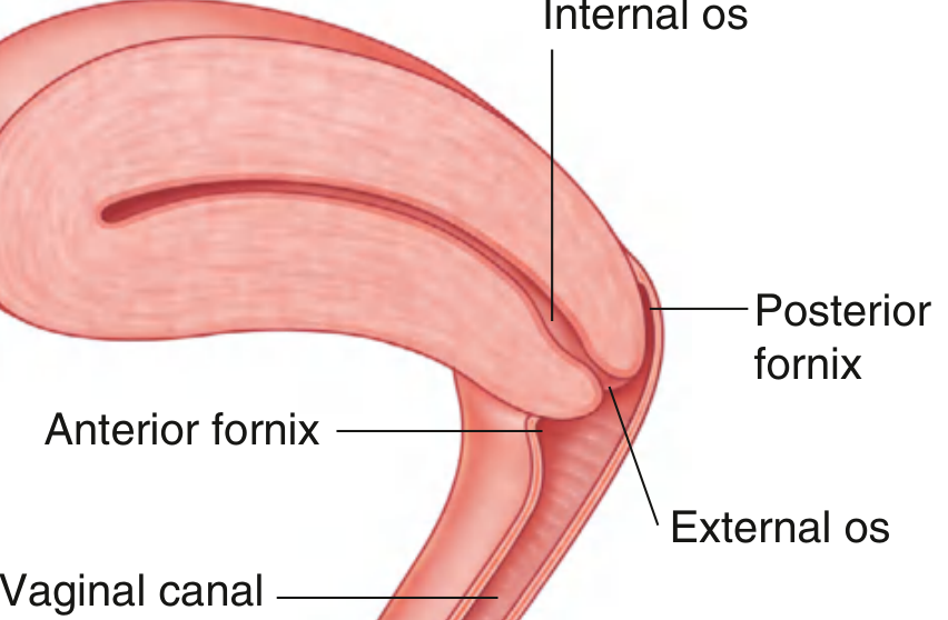
Subdivisions
| Part | Position | Key Relation |
|---|---|---|
| Posterior fornix | Behind the cervix | Deepest; lies directly against the rectouterine pouch (pouch of Douglas) |
| Anterior fornix | In front of the cervix | Related to the base of the bladder |
| Lateral fornices (×2) | On either side | Related to the parametrium; ureters pass nearby |
Key Relations
- Posterior fornix - separated from the peritoneal cavity only by the thin vaginal wall and the peritoneum of the rectouterine pouch (pouch of Douglas). This is why fluid collections (blood, pus, ascites) in the peritoneal cavity can be drained via the posterior fornix (culdocentesis).
- Anterior fornix - related to the base (fundus) of the urinary bladder and, inferiorly, to the urethra embedded in the anterior vaginal wall.
- Lateral fornices - the ureters pass approximately 2 cm lateral to the cervix before entering the bladder, making them vulnerable to injury in surgery near this area.
Clinical Importance
-
Culdocentesis - a needle is inserted through the posterior fornix into the rectouterine pouch to aspirate blood or pus (e.g., in suspected ectopic pregnancy rupture or pelvic abscess).
-
Pap smear / speculum examination - when a speculum is inserted, the physician can directly visualize the cervix, the fornices, and the external os. Specimens are taken from around the cervical os and fornices.
-
Vaginal injuries - penetrating injuries through the posterior fornix or lateral walls can enter the abdominal cavity directly, as the peritoneum is close by. - Essentials of Forensic Medicine & Toxicology, 36th ed.
-
Pessary fitting - the length of the vagina is estimated by measuring from the posterior fornix to the introitus, as this guides the size of pessary to use.
-
Vaginal cancer staging - small stage I cancers located at the posterior vaginal fornix may be treated with upper vaginectomy.
-
Semen pooling - during intercourse, semen is deposited in the vaginal vault/fornix, from where spermatozoa enter the external cervical os.