
First of all, write about the nerve system of heart. Uh, and then it must be... Describe it as... It must, uh, include all the things in simplifying, uh, terms and also all the main parts must be included in... So that any student can understand. Eat properly. And can answer any question related to this.
cardiac nerve supply autonomic innervation heart diagram
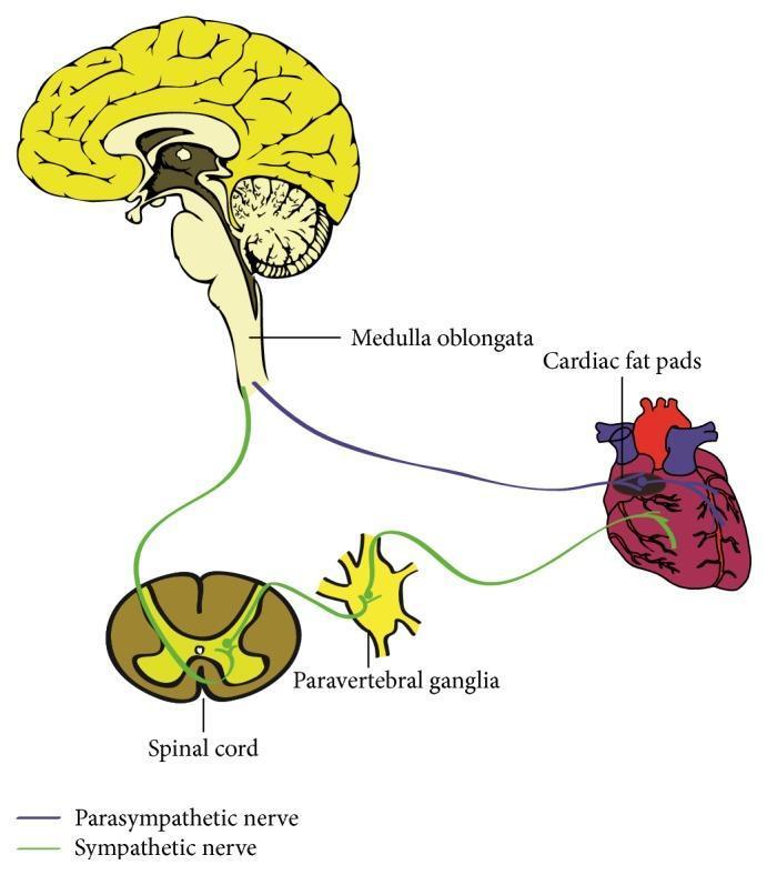
This medical anatomical diagram illustrates the autonomic innervation of the human heart, detailing both the parasympathetic and sympathetic pathways. The parasympathetic pathway is represented by a purple line originating from the medulla oblongata within the brainstem; these preganglionic fibers travel via the vagus nerve to synapse at the cardiac fat pads (ganglionated plexi) located on the epicardial surface. The sympathetic pathway is depicted in green, showing the circuit from the spinal cord to the paravertebral ganglia. From these ganglia, postganglionic sympathetic fibers project to the base of the heart and distribute into the myocardium. Key anatomical structures labeled include the medulla oblongata, cardiac fat pads, spinal cord, and paravertebral ganglia. The diagram serves as an educational tool for understanding the neural regulation of cardiac performance, specifically the origin and trajectory of autonomic fibers that modulate heart rate and contractility.
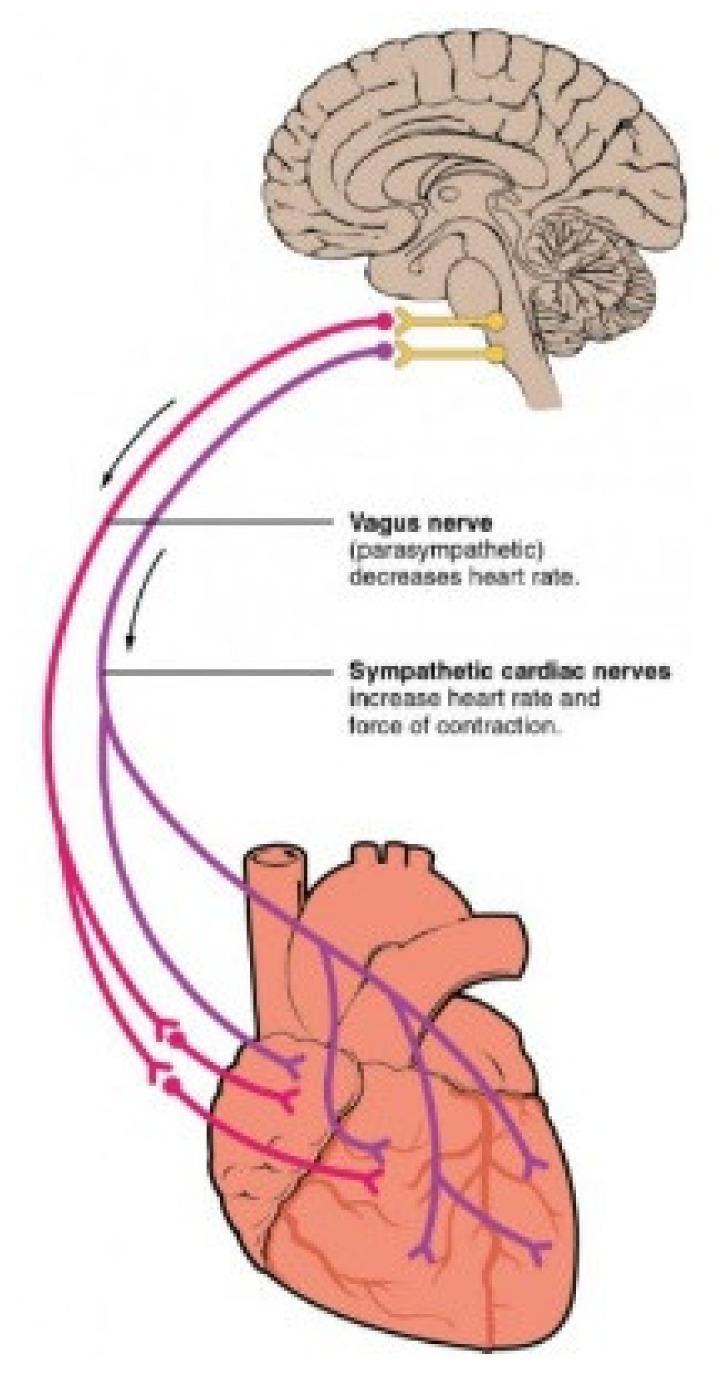
This medical illustration depicts the Autonomic Nervous System (ANS) pathways connecting the brainstem to the heart. A sagittal section of the human brain shows the medulla oblongata as the origin for both parasympathetic and sympathetic innervation. The Vagus nerve (parasympathetic), color-coded in purple, is shown descending from the brainstem to the heart, where it branches to innervate the atria and ventricles; text annotations indicate its role in decreasing heart rate. The sympathetic cardiac nerves, color-coded in red, originate from the same region and follow a parallel descending path, branching across the cardiac tissue; text labels specify their role in increasing both heart rate and the force of contraction. The diagram illustrates the functional antagonism between the two branches of the ANS in cardiac regulation, highlighting the primary innervation sites at the sinoatrial node, atrioventricular node, and the myocardium.
This medical anatomical diagram illustrates the autonomic nerve supply to the human pelvic viscera, specifically demonstrating the 'concentrated type' of innervation pattern. The illustration depicts a lateral view of the pelvic cavity with the rectum (1) and urinary bladder (2) as primary targets of innervation. Key structures include the sacral sympathetic trunk (3), shown as prominent vertical dark cords, and the nervi erigentes (4) or pelvic splanchnic nerves arising from the sacral plexus. The inferior hypogastric plexus (5) is shown as a network of nerves and ganglia (red highlights) distributing to the bladder and rectum. This concentrated variation is characterized by a simplified neural architecture: fewer, larger ganglia and less frequent, weaker connections between the sympathetic trunk and the visceral plexuses compared to the segmental type. The diagram is intended for neuroanatomical education, highlighting variations in pelvic autonomic pathways relevant to surgical considerations in proctology and urology.
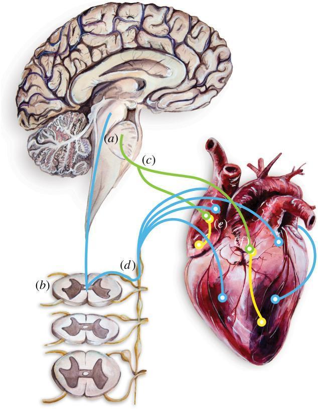
This medical illustration depicts the centers of cardiac neurotransmission, highlighting the bidirectional communication between the central nervous system and the heart. The diagram includes a sagittal section of the human brain focusing on the brainstem (a), which contains autonomic centers. Below, cross-sections of the spinal cord (b) are shown. The heart is illustrated with its major vessels, where intrinsic ganglionated plexi (e) are located within basal fat deposits (f). Nerve pathways are represented by colored lines: green lines (c) signify the vagus nerves connecting the brainstem directly to the heart, while blue lines (d) show pathways projecting from the brainstem through the spinal cord to the sympathetic chain ganglia and finally to the cardiac tissue. These pathways facilitate both efferent (motor) and afferent (sensory) signals, regulating cardiac function, vasculature, and the specialized conduction system. This illustration is an educational tool for understanding neuro-cardiac physiology and the pathophysiology of conditions such as Catecholaminergic Polymorphic Ventricular Tachycardia (CPVT).
nerve supply of heart cardiac innervation autonomic sympathetic parasympathetic explained
https://www.kenhub.com/en/library/anatomy/innervation-of-the…
The Nerve Supply of the Heart - A Complete Student Guide
Sources: Color Atlas of Human Anatomy Vol. 2 (Internal Organs); Braunwald's Heart Disease - A Textbook of Cardiovascular Medicine; Kenhub; CV Physiology
1. Overview - The Big Picture
| Layer | What it is | What happens if removed |
|---|---|---|
| Extrinsic | Nerves coming from the brain/spinal cord to the heart | Heart rate changes are impaired (e.g., after heart transplant) |
| Intrinsic | Nerves/conduction tissue built inside the heart itself | Heart still beats on its own! |
Key point for exams: A transplanted heart still beats because the intrinsic system (SA node, AV node, conduction tissue) keeps working even without external nerve connections.
2. The Two Divisions - Sympathetic vs. Parasympathetic
| Feature | Sympathetic ("Fight or Flight") | Parasympathetic ("Rest and Digest") |
|---|---|---|
| Main nerve/origin | Spinal cord (T1-T5) via sympathetic chain | Vagus nerve (CN X) from brainstem |
| Neurotransmitter | Norepinephrine (adrenaline) | Acetylcholine |
| Receptor on heart | Beta-1 adrenoceptors | Muscarinic receptors (M2) |
| Heart rate | ↑ Increases (tachycardia) | ↓ Decreases (bradycardia) |
| Force of contraction | ↑ Increases (positive inotropy) | ↓ Decreases |
| Conduction speed | ↑ Faster through AV node | ↓ Slower through AV node |
| Coronary arteries | Dilation | Constriction |
| When active | Exercise, stress, fear | Rest, sleep, eating |
3. The Sympathetic Pathway - Step by Step
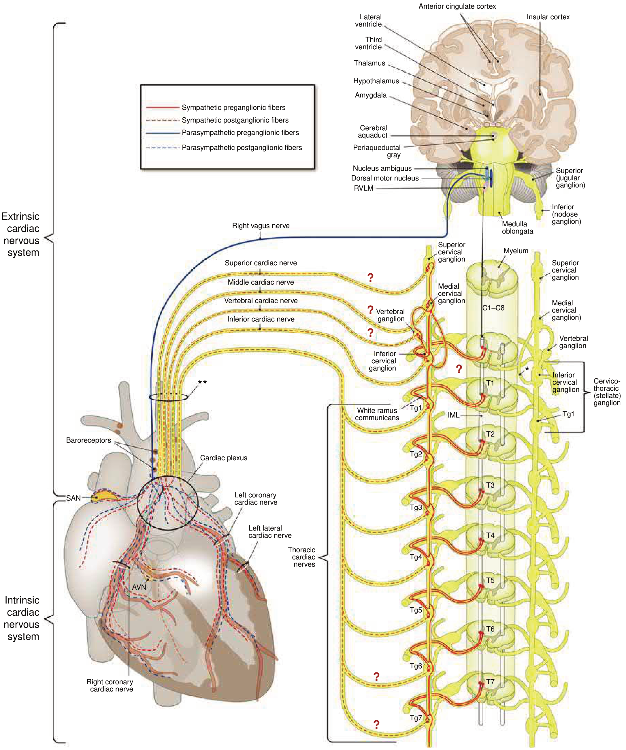
- Superior cervical cardiac nerve (from superior cervical ganglion)
- Middle cervical cardiac nerve (from middle cervical ganglion)
- Inferior cervical cardiac nerve (from cervicothoracic/stellate ganglion)
Memory tip: Sympathetic = Thorax → T1-T5. Think "T for Turbo" - it turbocharges the heart.
4. The Parasympathetic Pathway - Step by Step
- Dorsal motor nucleus of the vagus
- Nucleus ambiguus
- Superior and inferior cervical cardiac branches of the vagus
- Thoracic cardiac branches of the vagus
Important distinction for exams:
- Sympathetic = long preganglionic → synapse in chain ganglion → short postganglionic to heart
- Parasympathetic = long preganglionic → synapse ON/NEAR heart → short postganglionic to target
5. Which Nerve Goes Where? (Regional Distribution)
| Structure | Sympathetic | Parasympathetic (Vagus) |
|---|---|---|
| SA Node (pacemaker) | Right > Left | Right vagus (dominant) |
| AV Node | Both sides | Left vagus (dominant) |
| Atrial muscle | Yes | Yes (well innervated) |
| Ventricular muscle | Yes (throughout) | Sparse (barely innervated) |
| Coronary arteries | Yes (mainly dilation) | Yes (constriction) |
| Conduction system | Yes | Yes (mainly SA + AV nodes) |
Exam tip: Right vagus = SA node. Left vagus = AV node. Easy to remember: Right starts, Left conducts.
6. The Cardiac Plexus - The Junction Box
| Part | Location | Fibers |
|---|---|---|
| Superficial (anterior) part | Below the aortic arch, in front of the right pulmonary artery | Mainly from the left cardiac nerves |
| Deep (posterior) part | Behind the aortic arch, in front of the tracheal bifurcation | From both sides |
- The conduction system (SA node, AV node, bundle of His)
- The coronary vasculature
- The myocardium of the atria and ventricles
7. Sensory (Afferent) Fibers - How the Heart Reports Back
A. Sympathetic Afferents (Pain fibers)
- Travel back through the sympathetic cardiac nerves
- Their cell bodies (perikarya) lie in the cervical and thoracic spinal ganglia (C3-C4, T1-T7)
- These carry pain signals from the heart
B. Parasympathetic Afferents (Reflex fibers)
- Travel back through the vagus nerve cardiac branches
- Carry signals from baroreceptors (sense blood pressure) and stretch receptors in the heart walls
- Important for heart rate reflexes (e.g., carotid sinus reflex)
8. Referred Pain - Why Heart Attack Hurts in Your Left Arm
- Left shoulder and neck region (C3-C4 dermatomes)
- Left arm, specifically the ulnar (inner) side (T1 dermatome)
- Sometimes the jaw, upper back, or epigastrium
Memory tip: When the heart is in distress, the pain travels the same nerve highways (T1-T5) and confuses the brain into thinking the arm/shoulder/neck hurts.
9. Intrinsic Nerve Supply - The Heart's Own Internal Wiring
| Structure | Role |
|---|---|
| SA Node (Sinoatrial node) | Natural pacemaker - generates the heartbeat at ~60-100 bpm |
| AV Node (Atrioventricular node) | Receives signal from SA node, delays it slightly, then passes to ventricles |
| Bundle of His | Carries signal from AV node down the septum |
| Left and Right Bundle Branches | Split to supply each ventricle |
| Purkinje Fibers | Spread the signal through ventricular muscle for synchronized contraction |
| Intrinsic cardiac ganglia | Small nerve cell clusters embedded in the heart wall (mostly in fat pads on atria) |
10. Higher Brain Control
- Cardioacceleratory center - activates sympathetic outflow
- Cardioinhibitory center - activates parasympathetic (vagal) outflow
- Baroreceptors (aortic arch, carotid sinus) - blood pressure feedback
- Higher brain centers (hypothalamus, amygdala, insular cortex, anterior cingulate cortex) - emotional/stress responses
This is why emotions like fear, excitement, or anxiety cause your heart to race - the amygdala and hypothalamus activate the sympathetic system via the medulla.
11. Quick Summary Table - Everything at a Glance
| Feature | Sympathetic | Parasympathetic |
|---|---|---|
| Origin | T1-T5 spinal cord (IML) | Medulla oblongata (dorsal motor nucleus + nucleus ambiguus) |
| Pre-ganglionic length | Short | Long |
| Where synapse occurs | Cervical + thoracic chain ganglia | Cardiac ganglia on/near heart (epicardial fat pads) |
| Post-ganglionic length | Long | Short |
| Neurotransmitter | Norepinephrine | Acetylcholine |
| Receptor | Beta-1 | M2 (muscarinic) |
| Heart rate | Increases | Decreases |
| Contractility | Increases | Decreases |
| AV conduction | Faster | Slower |
| Dominant over | SA node (both, R > L) | SA node (right vagus); AV node (left vagus) |
| Afferent function | Pain from heart (referred pain) | Baroreceptor/stretch receptor signals |
12. Clinical Correlations (Commonly Tested)
| Condition | Mechanism | Result |
|---|---|---|
| Vagus nerve damage | Loss of parasympathetic braking | Resting tachycardia |
| Sympathetic nerve damage | Loss of accelerator | Inability to increase HR with exercise (bradycardia) |
| Heart transplant | Both extrinsic nerves cut | Heart beats via intrinsic SA node only; no response to atropine (blocks vagus) |
| MI (heart attack) pain | Sympathetic afferents → T1-T5 | Referred pain to left arm/jaw/shoulder |
| Carotid sinus massage | Activates vagal afferents | Slows HR - used to treat SVT |
| Beta-blockers | Block beta-1 receptors | Reduce sympathetic effect → ↓ HR, ↓ force |
| Atropine | Blocks M2 muscarinic receptors | Blocks parasympathetic → ↑ HR (used in bradycardia) |