

1000425871.jpg
Diagnosis of this
The diagnosis here is Waldenström's Macroglobulinemia (WM), and the answer to the question is A) Cytoplasmic invaginations.
Searching sources...
Reading File
Finding Sources
Finding Sources
Reading File
Reading File
Reading File
Loading Image
Loading Image
Diagnosis: Waldenström's Macroglobulinemia (Lymphoplasmacytic Lymphoma)
Answer: A) Cytoplasmic invaginations
The "intranuclear" eosinophilic inclusions described in this case are Dutcher bodies - and the answer to the trick question is that they are not truly intranuclear. They are cytoplasmic invaginations into the nucleus, creating the optical illusion of a nuclear inclusion on light microscopy.
What are Dutcher Bodies?
Dutcher bodies are PAS-positive, eosinophilic pseudoinclusions that appear to sit inside the nucleus of plasma cells or plasmacytoid lymphocytes. However, ultrastructurally (by electron microscopy), they are actually cytoplasmic invaginations into the nuclear membrane - pockets of immunoglobulin-laden cytoplasm that herniate into the nuclear compartment. They are NOT truly intranuclear structures, and they are NOT nucleolar hypertrophy, viral aggregates, or ribosomal precipitates.
Compare them to Russell bodies, which are the same material (accumulated IgM) but located in the cytoplasm proper.
Histological correlate from Robbins & Kumar
Here is the textbook image of lymphoplasmacytic lymphoma with Dutcher bodies (arrows = pink pseudonuclear inclusions):
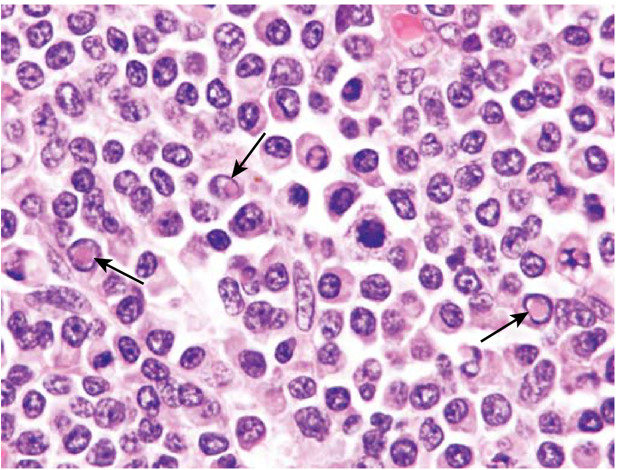
Lymphoplasmacytic lymphoma. Tumor cells show varying degrees of plasmacytic differentiation. Pink pseudonuclear inclusions (Dutcher bodies, arrows) are caused by retention of immunoglobulin. - Robbins & Kumar Basic Pathology
Clinical Summary: Waldenström's Macroglobulinemia
| Feature | Detail |
|---|---|
| Underlying neoplasm | Lymphoplasmacytic lymphoma (LPL) |
| Monoclonal protein | IgM (pentameric - large, stays intravascular) |
| Driver mutation | MYD88 L265P (~95% of cases) |
| Key symptom | Hyperviscosity syndrome (blurred vision, nosebleeds, CNS symptoms) |
| Bone marrow biopsy | Mixture of lymphocytes, plasmacytoid lymphocytes, and plasma cells with mast cell hyperplasia |
| Hallmark inclusions | Dutcher bodies (pseudointranuclear) and Russell bodies (cytoplasmic) |
| Unlike myeloma | No lytic bone lesions, no renal failure from light chains (rare) |
Why the other options are wrong
- B) Nucleolar hypertrophy - Enlarged nucleoli are seen in other aggressive tumors (e.g., Burkitt lymphoma, DLBCL), not in LPL.
- C) Viral capsid aggregates - These would imply a viral infection (e.g., CMV, Herpesvirus); there is no viral etiology here.
- D) Ribosomal precipitates - Not a recognized pathological entity in this context.
The answer is firmly A - cytoplasmic invaginations, because Dutcher bodies, despite appearing intranuclear on routine H&E staining, are cytoplasmic immunoglobulin deposits that have herniated into (but not truly entered) the nucleus.
Source: Robbins, Cotran & Kumar Pathologic Basis of Disease - Lymphoplasmacytic Lymphoma; Robbins & Kumar Basic Pathology, p. 572
This is a shared conversation. Sign in to Orris to start your own chat.