
Clinical anatomy for axillary artery
Reading File
This is a visual/anatomy topic. Let me run the three-layer search in parallel.
Finding Sources
Finding Sources
Excellent results. Now let me read the most comprehensive sources in parallel - Gray's Anatomy for Students, the Imaging Atlas, and the THIEME Atlas.
Reading File
Reading File
Reading File
Good data. Now let me read more from Gray's (branches detail) and the Imaging Atlas (clinical relations and anastomoses), plus fetch the diagrams.
Reading File
Reading File
Loading Image
Loading Image
I now have excellent content from Gray's Anatomy for Students and the Imaging Atlas, along with two high-quality anatomical diagrams. Let me also fetch the angiogram image and one more diagram from the Imaging Atlas for extra visual support, and simultaneously check for clinical/surgical relevance from Fischer's.
Loading Image
Loading Image
Reading File
I now have rich content from multiple authoritative sources. Let me compile a comprehensive answer.
Axillary Artery - Clinical Anatomy
Overview
The axillary artery is the main arterial trunk supplying the upper limb and axillary walls. It is the direct continuation of the subclavian artery, beginning at the lateral margin of the first rib and ending at the lower border of teres major, where it becomes the brachial artery.
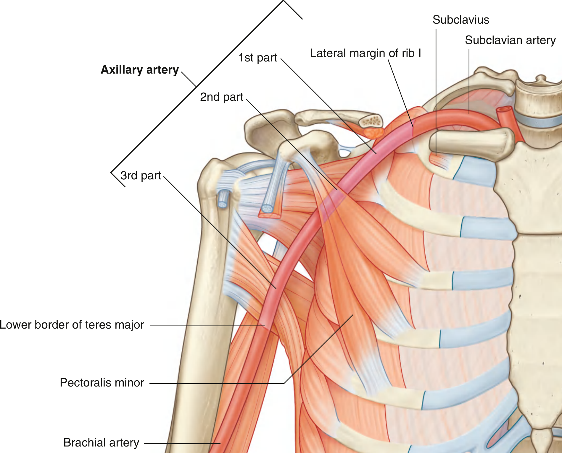
Fig. 7.49 - Three-part division of the axillary artery - Gray's Anatomy for Students
Three Parts (Divided by Pectoralis Minor)
The pectoralis minor crosses anteriorly over the vessel and divides it into three parts. A useful mnemonic: 1 branch, 2 branches, 3 branches (the number of branches matches the part number).
| Part | Position relative to pectoralis minor | Number of branches |
|---|---|---|
| 1st part | Proximal to (medial to) pectoralis minor | 1 branch |
| 2nd part | Posterior to (deep to) pectoralis minor | 2 branches |
| 3rd part | Distal to (lateral to) pectoralis minor | 3 branches |
The artery lies within the axillary sheath proximally, in close relation to the cords and branches of the brachial plexus throughout its course.
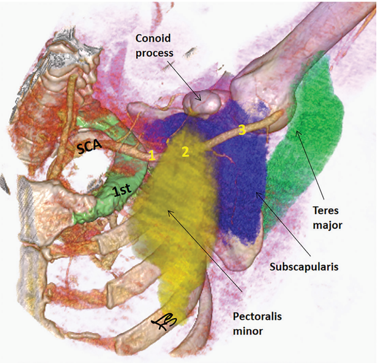
Fig. 4.16 - Axillary artery segments on CTA - Imaging Anatomy Atlas Vol. 3
Six Branches
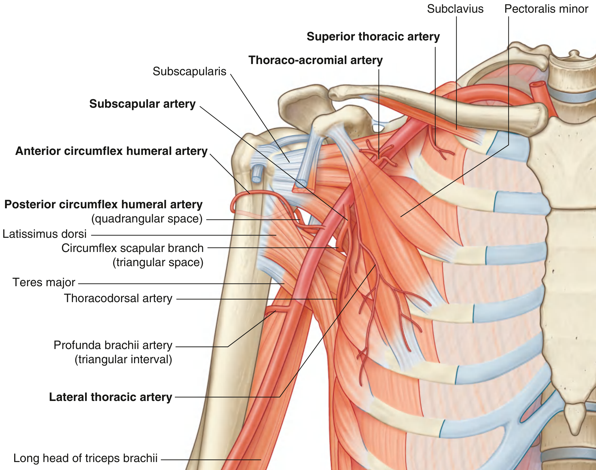
Fig. 7.50 - Branches of the Axillary Artery - Gray's Anatomy for Students
1st Part (1 branch)
Superior thoracic artery
- Small vessel from the anterior surface of the 1st part
- Supplies the upper medial and anterior axillary walls
2nd Part (2 branches)
Thoraco-acromial artery
- Short trunk from the anterior surface of the 2nd part, just proximal to the medial margin of pectoralis minor
- Curves around the superior border of the muscle, pierces the clavipectoral fascia
- Divides into four branches: pectoral, deltoid, clavicular, and acromial
- Pectoral branch contributes to breast supply; deltoid branch accompanies the cephalic vein in the deltopectoral triangle
Lateral thoracic artery
- Arises posterior to the lateral (inferior) margin of pectoralis minor
- Follows the muscle margin to the thoracic wall
- Supplies medial and anterior axillary walls; in women, branches emerge around the inferior border of pectoralis major to supply the breast
3rd Part (3 branches)
Subscapular artery (largest branch)
- Originates from the posterior surface of the 3rd part
- Follows the inferior margin of subscapularis briefly, then divides into two terminal branches:
- Circumflex scapular artery - passes through the triangular space (bounded by subscapularis above, teres major below, long head of triceps medially); enters the infraspinous fossa and anastomoses with suprascapular and dorsal scapular arteries
- Thoracodorsal artery - follows the lateral border of the scapula toward the inferior angle; supplies posterior and medial axillary walls
Anterior circumflex humeral artery (ACHA)
- Small; arises from the lateral side of the 3rd part
- Passes anterior to the surgical neck of the humerus
- Supplies the glenohumeral joint and head of humerus
- Anastomoses with posterior circumflex humeral artery
Posterior circumflex humeral artery (PCHA)
- Originates immediately posterior to the ACHA origin
- Exits the axilla with the axillary nerve through the quadrangular space (bounded by teres minor above, teres major below, long head of triceps medially, surgical neck of humerus laterally)
- Winds around the surgical neck of humerus
- Supplies surrounding muscles, glenohumeral joint
- Anastomoses with ACHA, profunda brachii, suprascapular, and thoraco-acromial arteries
Angiographic Appearance
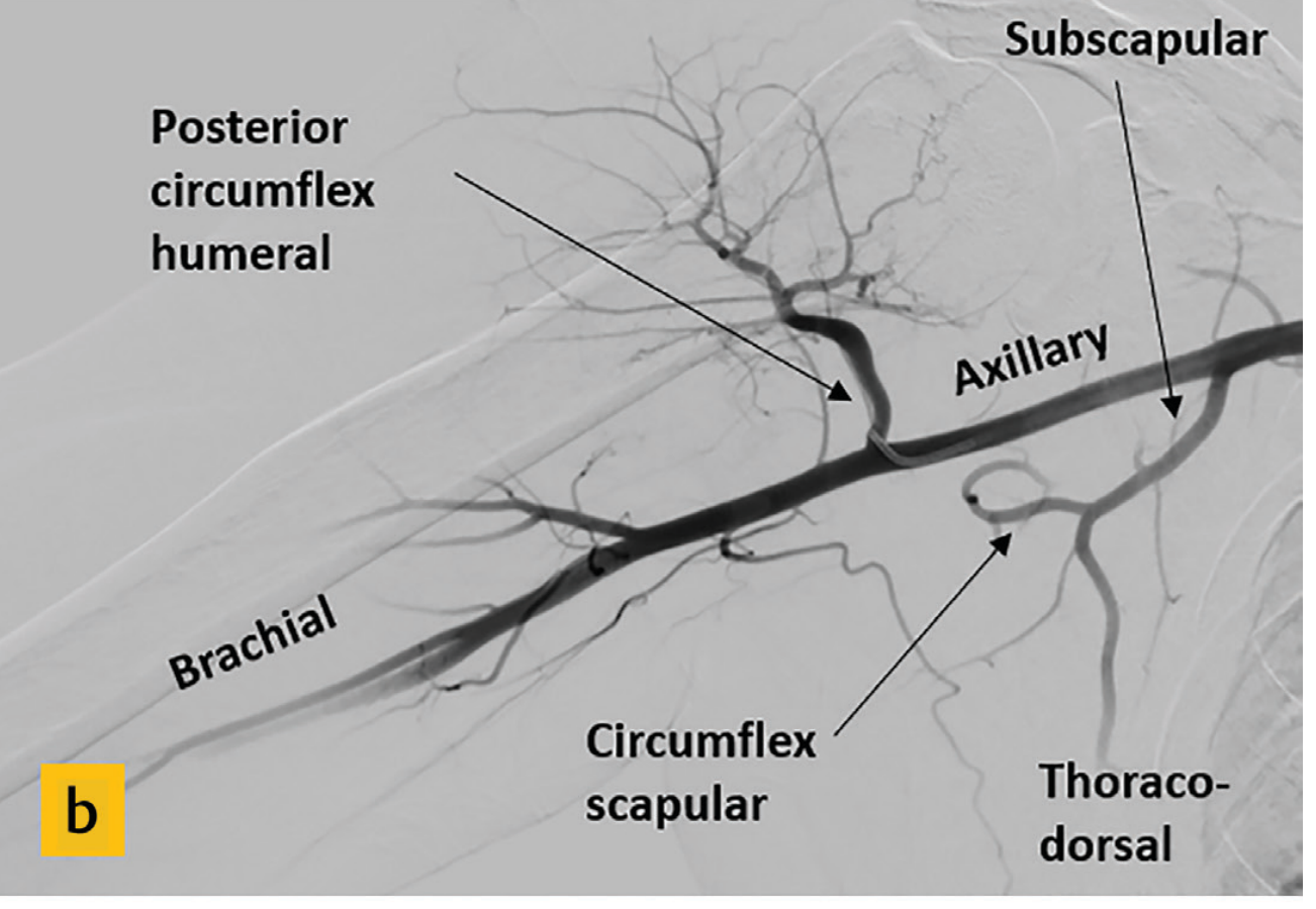
Fig. 4.17b - DSA angiogram showing axillary artery branches - Imaging Anatomy Atlas Vol. 3
Relations in the Axilla
The axillary artery is surrounded by the contents of the axilla:
- The axillary vein lies medial and anterior to the artery throughout
- The brachial plexus cords wrap around the 2nd part: lateral cord lies lateral, medial cord lies medial, posterior cord lies posteriorly
- The axillary lymph nodes and fat surround the neurovascular bundle
- Anterior wall: pectoralis major + pectoral fascia (superficial layer); pectoralis minor + clavipectoral fascia (deep layer)
- Posterior wall: subscapularis (anteriorly), teres major + latissimus dorsi (inferiorly)
- Medial wall: thoracic wall with serratus anterior
- Lateral wall: intertubercular sulcus of the humerus
Peri-scapular Anastomotic Network (Clinical Importance)
A rich anastomotic network around the scapula provides collateral circulation when the axillary artery is occluded. The key vessels involved include:
- Transverse cervical artery (dorsal scapular branch) -- 1st part of subclavian
- Suprascapular artery -- thyrocervical trunk
- Subscapular artery (circumflex scapular branch) -- 3rd part of axillary
This network connects the 1st part of the subclavian to the 3rd part of the axillary artery, so occlusion proximal to the subscapular origin can still be compensated.
Branching Variations (Surgically Important)
Branching patterns are highly variable, particularly involving the subscapular and posterior circumflex humeral arteries (~66% of people show a variant):
- High origin of subscapular artery from the 2nd part: 29-36%
- Common trunk of subscapular + PCHA (from 2nd or 3rd part): 12-42%
- Axillary origin of the thoracodorsal artery (without arising from subscapular): 3-10%
- Superficial brachial artery: early bifurcation of the axillary into a superficial and deep brachial artery
These variations are important in axillary dissection (breast surgery), free flap planning (latissimus dorsi, serratus anterior), and interpreting vascular imaging.
Clinical Correlations
| Situation | Relevance |
|---|---|
| Anterior shoulder dislocation | 3rd part of axillary artery is vulnerable (least protected, no overlying muscle); can be torn especially in elderly patients |
| Axillary node dissection (breast cancer) | Surgical borders: axillary vein superiorly, latissimus dorsi laterally, chest wall medially - artery lies just deep to vein |
| Quadrangular space syndrome | PCHA + axillary nerve compression - causes shoulder pain and deltoid wasting |
| PCHA embolization | Avascular necrosis of the humeral head (PCHA supplies a significant portion of humeral head via anterolateral ascending branch) |
| Axillary artery aneurysm | Can cause thromboembolism to distal upper limb or compress brachial plexus |
| Chronic lung disease (TB, bronchiectasis) | Hypertrophy of thoracoacromial, lateral thoracic, subscapular, and thoracodorsal arteries due to transpleural collateralization |
Sources: Gray's Anatomy for Students, p. 853-855 | Imaging Anatomy Text and Atlas Vol. 3 (Bones, Joints, Vessels and Nerves), p. 154-156 | Fischer's Mastery of Surgery 8th ed., p. 1552-1553 | THIEME Atlas of General Anatomy and Musculoskeletal System
This is a shared conversation. Sign in to Orris to start your own chat.