
Thyroid gland, shortly histology and detail physiology with relevant flow chart and diagram, and clinical physiology
thyroid gland histology follicle colloid
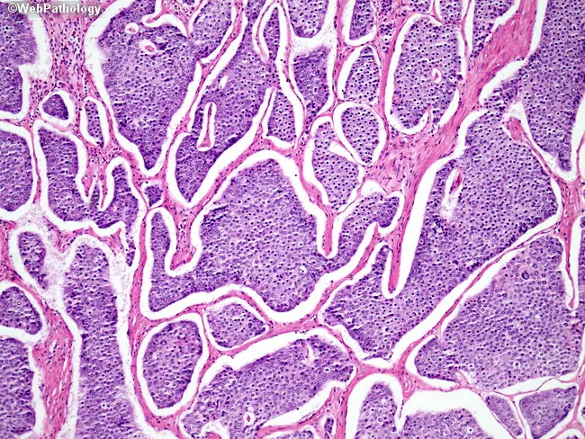
This is a light micrograph of thyroid gland tissue prepared for histopathology and stained with Hematoxylin and Eosin (H&E). The sample reveals multiple spherical to irregular follicles of varying size separated by wispy fibrous septa. The colloid-filled lumens are pink to pale pink, while the surrounding follicular epithelium is a uniform single layer of cuboidal to low-columnar cells with round to oval nuclei and inconspicuous nucleoli. No nuclear grooves, inclusions, or pseudoinclusions characteristic of papillary carcinoma are evident. The stromal background is relatively scant with no dense lymphocytic infiltrate or Hurthle cell metaplasia apparent. The architecture is reminiscent of normal thyroid parenchyma or benign nodular colloid goiter pattern, where follicle size varies and colloid predominates. There are no mitotic figures or cytologic atypia observed. This image demonstrates classic histology of endocrine gland tissue with preserved colloid and regular follicle lining, suitable as reference for normal thyroid architecture in educational, diagnostic, and research contexts. Clinically, such histology supports benign thyroid pathology when correlated with imaging and labs. In malignant differentials, absence of papillary nuclear features or tall columnar cells argues against papillary thyroid carcinoma; however, definitive diagnosis should consider capsule invasion and margin status in surgical specimens.
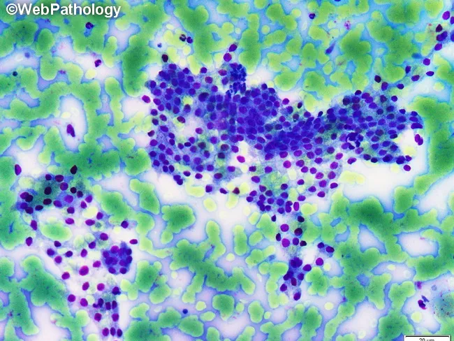
This is a light microscopy histology image of thyroid gland tissue prepared with a Romanowsky-type stain (e.g., Diff-Quik/Wright-Giemsa). The specimen shows normal-appearing follicular epithelium arranged in intact macrofollicles with abundant colloid occupying the luminal spaces. Follicular cells are evenly spaced with uniform, round to oval nuclei and scant to moderate cytoplasm, consistent with well-differentiated thyroid epithelium. The colloid background is pale blue and prominent, reflecting thyroglobulin-containing material released from follicles. The architectural pattern emphasizes large follicle units with a single layer of cuboidal to low-columnar epithelium surrounding expansive colloid pools. No prominent nuclear features of papillary carcinoma are evident, and there is no marked cellular atypia or mitotic activity in the visible fields. This image illustrates classic histology of benign thyroid tissue or colloid-rich goiter, useful for teaching follicular morphologies, thyroid folliculogenesis, and colloid dynamics. Clinically, such samples are evaluated in the context of thyroid nodules and goiter workups, with correlation to serum thyroid function tests and, when applicable, ultrasound or FNA findings. Potential educational uses include cytology-histology correlation, differential diagnosis of thyroid disease, and artifact recognition in Romanowsky-stained preparations. Images are suitable for medical education, pathology review, and database indexing. Correlative data improve student comprehension and diagnostic recall greatly.
hypothalamic pituitary thyroid axis diagram TRH TSH
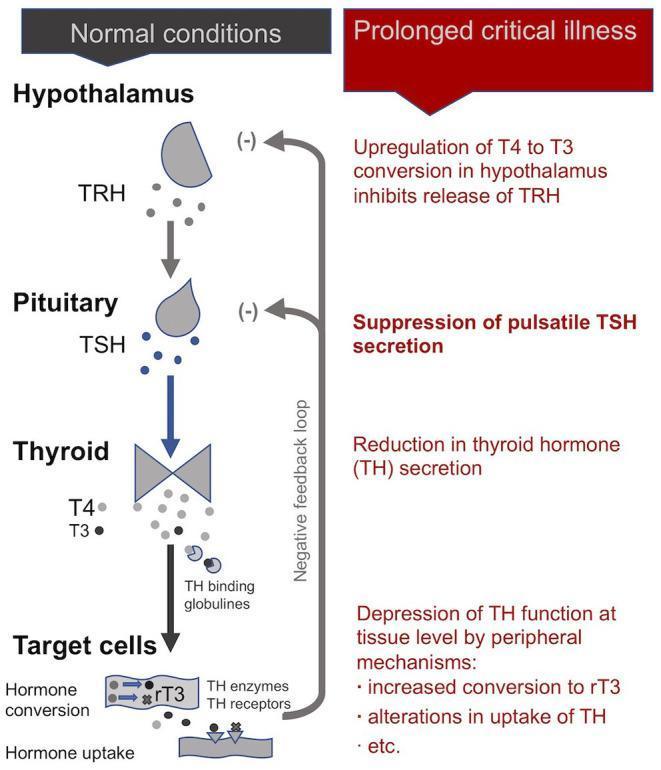
This pathophysiology diagram illustrates the Hypothalamic-Pituitary-Thyroid (HPT) axis, comparing normal physiological conditions with the alterations observed during prolonged critical illness. The 'Normal conditions' section shows the standard cascade: the Hypothalamus secretes TRH, which stimulates the Pituitary to release TSH, leading the Thyroid gland to produce T4 and T3. These hormones reach target cells via TH binding globulins for hormone conversion and uptake, with a negative feedback loop inhibiting TRH and TSH secretion. In contrast, the 'Prolonged critical illness' section details the central and peripheral suppression of the axis. Key pathological features include: upregulation of T4 to T3 conversion in the hypothalamus (inhibiting TRH release), suppression of pulsatile TSH secretion by the pituitary, reduced thyroid hormone secretion, and depression of thyroid function at the tissue level. Peripheral mechanisms shown include increased conversion to inactive rT3 and altered hormone uptake. This comparison illustrates the endocrine maladaptation typical of Non-Thyroidal Illness Syndrome (NTIS) in intensive care settings.
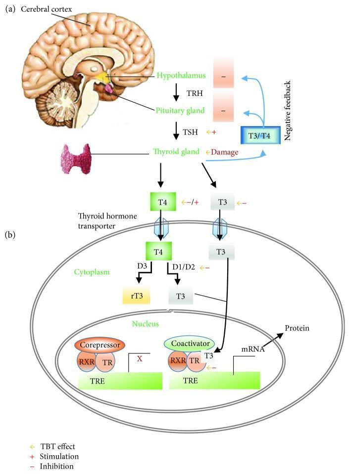
A two-part medical illustration depicting the pathophysiology of the Hypothalamic-Pituitary-Thyroid (HPT) axis and cellular thyroid hormone action. Section (a) shows the systemic HPT axis, beginning with the cerebral cortex and hypothalamus. The hypothalamus releases thyrotropin-releasing hormone (TRH), which stimulates the pituitary gland to secrete thyroid-stimulating hormone (TSH). TSH subsequently acts on the thyroid gland to stimulate the production of T4 and T3 hormones, which exert negative feedback on the hypothalamus and pituitary. Section (b) details the intracellular mechanism of action within a target cell. T4 and T3 enter the cell via thyroid hormone transporters. In the cytoplasm, deiodinases (D1/D2) convert T4 to T3, while D3 converts T4 to inactive reverse T3 (rT3). T3 enters the nucleus, binding to the thyroid hormone receptor (TR) which heterodimerizes with the retinoid X receptor (RXR). This complex binds to the thyroid hormone response element (TRE) on DNA, displacing corepressors and recruiting coactivators to initiate gene transcription (mRNA) and subsequent protein synthesis. The diagram also highlights potential sites of endocrine disruption (e.g., by Tributyltin/TBT), including receptor inhibition and deiodinase interference.
Graves disease exophthalmos goiter clinical photo
"thyroid function" AND physiology
Thyroid Gland: Histology, Physiology, and Clinical Correlations
1. Histology (brief)
- Follicular cells (thyrocytes): A single layer of cuboidal-to-low-columnar epithelial cells surrounding a central lumen. They become taller/columnar when hyperactive (TSH-stimulated) and flatten when inactive.
- Colloid: The pink, proteinaceous material filling the follicular lumen, composed mainly of thyroglobulin - the storage form of thyroid hormone.
- Parafollicular (C) cells: Neural-crest-derived cells scattered singly or in clusters between follicles (concentrated at the junction of the upper 1/3 and lower 2/3 of each lobe). They secrete calcitonin, unrelated to the iodine-handling machinery of follicular cells, and are the cell of origin of medullary thyroid carcinoma.
- Follicle size varies (small, active follicles vs. large, colloid-distended quiescent ones), and this variation is normal.
2. Detailed Physiology
A. Hormone synthesis and secretion (step-by-step)
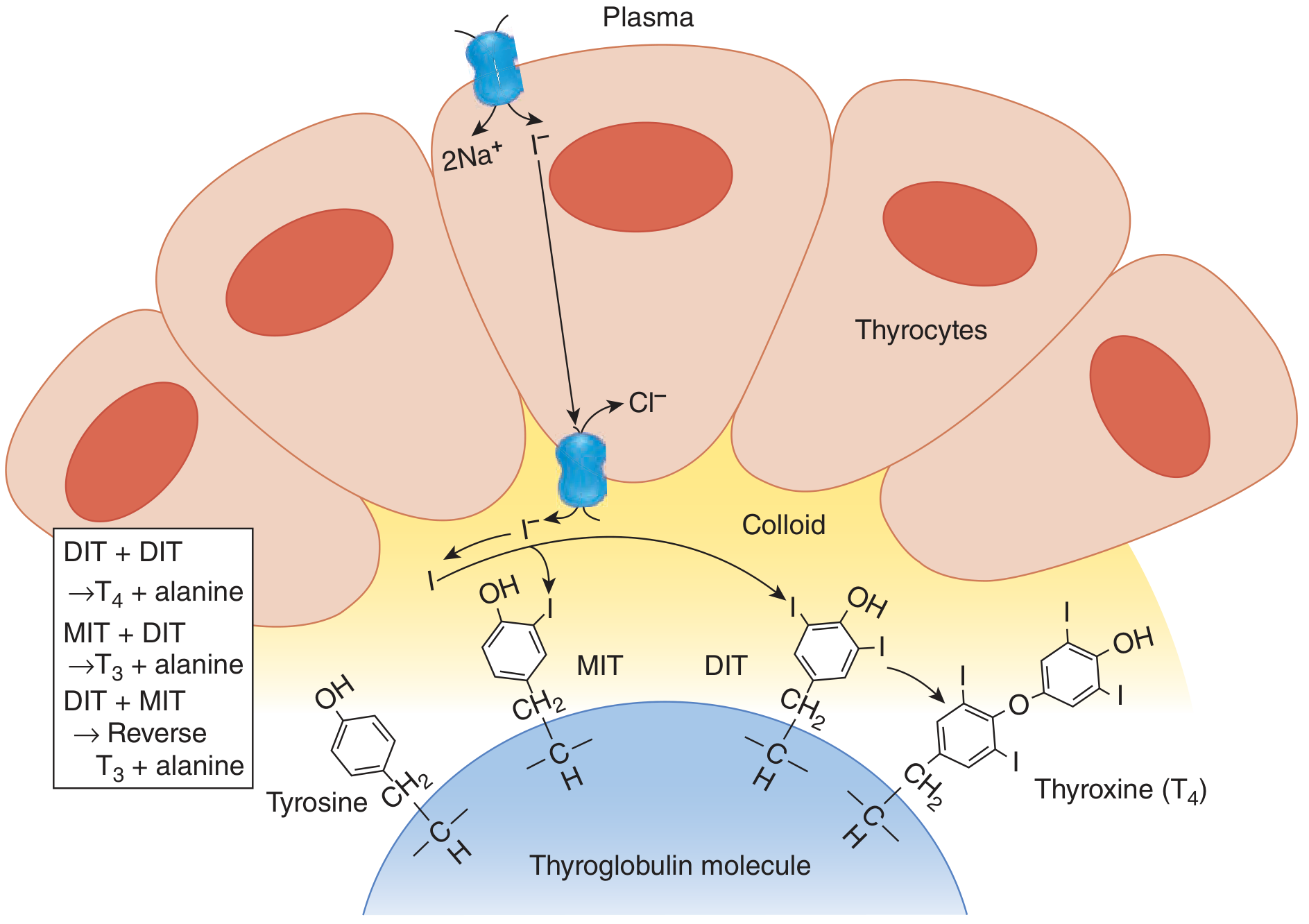
- Iodide trapping: Dietary iodide (need ≥100 μg/day) is actively transported into the thyrocyte across the basal membrane by the sodium-iodide symporter (NIS), which co-transports Na⁺ (down its gradient, maintained by Na⁺/K⁺-ATPase) with I⁻ (against its gradient). The gland concentrates iodide 20-40x plasma levels. NIS is stimulated by TSH.
- Iodide efflux into colloid: Iodide moves to the apical membrane and exits into the follicular lumen, aided by the transporter pendrin (mutated in Pendred syndrome: goiter + sensorineural deafness).
- Organification: At the apical border, thyroid peroxidase (TPO) oxidizes iodide to iodine and attaches it to tyrosine residues on thyroglobulin (a large glycoprotein with ~123 tyrosines, only 4-8 of which get iodinated), forming monoiodotyrosine (MIT) and diiodotyrosine (DIT).
- Coupling: TPO couples two iodotyrosines within the thyroglobulin backbone: MIT + DIT → T3; DIT + DIT → T4. Hormone remains bound within thyroglobulin, stored in colloid - a reservoir large enough to supply the body for ~2 months even without dietary iodide.
- Release: On TSH stimulation, thyrocytes endocytose colloid, fuse it with lysosomes, and proteolytically cleave thyroglobulin, releasing free T4 (~90% of secreted hormone) and T3 (~10%) into the circulation. Leftover MIT/DIT are deiodinated and the iodine recycled.
B. Flow chart - Hypothalamic-Pituitary-Thyroid (HPT) axis
Cerebral cortex / stress, cold, other stimuli
            │
            ▼
      HYPOTHALAMUS  →  releases TRH (thyrotropin-releasing hormone)
            │
            ▼  (portal vessels)
  ANTERIOR PITUITARY (thyrotrophs)  →  releases TSH (thyroid-stimulating hormone)
            │
            ▼  (systemic circulation)
      THYROID GLAND (follicular cells)
            │  TSH binds TSH-receptor → ↑NIS, ↑TPO, ↑colloid endocytosis
            ▼
     Secretes T4 (~90%) + T3 (~10%) into blood
            │
            ▼
  Peripheral tissues (liver, kidney, muscle, brain):
     T4 --5'-deiodinase (D1/D2)--> T3 (active form)
     T4 --5-deiodinase (D3)-----> reverse T3 (inactive)
            │
            ▼
  T3 binds nuclear Thyroid Hormone Receptor (TR-α/β) → heterodimerizes with RXR
  → binds Thyroid Response Elements (TRE) on DNA → alters gene transcription
            │
            ▼
   Metabolic / developmental effects on virtually every tissue
            │
            └──────── NEGATIVE FEEDBACK ────────┐
                 (T4/T3 suppress TRH & TSH release)
C. Transport and peripheral metabolism
- Circulating T4/T3 are >99% bound to thyroxine-binding globulin (TBG), transthyretin, and albumin; only the free fraction is biologically active.
- T4 is largely a prohormone; peripheral 5'-monodeiodination (D1 in liver/kidney, D2 in pituitary/brain/brown fat) converts it to the far more potent T3. Alternative deiodination (D3) produces biologically inactive reverse T3 (rT3) - upregulated in illness/starvation ("non-thyroidal illness/euthyroid sick syndrome"), a mechanism for tissue energy conservation.
D. Mechanism and systemic effects of thyroid hormone
- Basal metabolic rate: ↑O2 consumption, heat production, mitochondrial biogenesis (calorigenic effect).
- Cardiovascular system: ↑heart rate and contractility (↑α-myosin heavy chain, SERCA, β-adrenergic receptors), ↓systemic vascular resistance from cutaneous vasodilation, ↑cardiac output.
- Nervous system: essential for CNS maturation (cerebral cortex, basal ganglia, cochlea) in fetal/neonatal life; in adults, modulates mentation and catecholamine sensitivity.
- Growth and skeletal maturation: synergizes with growth hormone; essential for normal bone growth and epiphyseal maturation.
- Metabolism: ↑carbohydrate absorption and turnover, ↑lipolysis/cholesterol clearance, ↑protein synthesis and degradation (net catabolic in excess).
- Skin/connective tissue: in hypothyroidism, glycosaminoglycan accumulation causes myxedema (puffy, non-pitting skin).
- Reproduction: required for normal menstrual cyclicity and fertility, though not directly metabolically active on the uterus.
3. Clinical Physiology
| Disruption | Mechanism | Clinical picture |
|---|---|---|
| Iodine deficiency | Insufficient substrate for organification → ↓T3/T4 → loss of feedback → ↑TSH → gland hyperplasia | Endemic goiter, hypothyroidism |
| Autoimmune thyroiditis (Hashimoto) | Anti-TPO/anti-thyroglobulin antibodies destroy follicular cells | Primary hypothyroidism: fatigue, cold intolerance, weight gain, bradycardia, myxedema, ↑TSH/↓fT4 |
| Graves disease | TSH-receptor stimulating antibodies (TSI) mimic TSH, driving continuous NIS/TPO activity independent of feedback | Thyrotoxicosis: tachycardia, weight loss, heat intolerance, tremor, goiter, ophthalmopathy; ↓TSH/↑fT4-fT3 |
| Toxic nodular goiter / adenoma | Autonomous, TSH-independent hormone secretion from a nodule | Hyperthyroidism with suppressed TSH, focal uptake on scintigraphy |
| Pendred syndrome | Pendrin defect impairs iodide efflux to colloid | Goiter + sensorineural hearing loss, mild hypothyroidism |
| Congenital NIS defect | Failure of iodide trapping | Congenital hypothyroidism (screened at birth to prevent cretinism/intellectual disability) |
| Non-thyroidal illness (sick euthyroid) syndrome | Critical illness ↑D3 activity, converting T4 preferentially to inactive rT3; central TRH/TSH suppression | Low T3, variably low T4, inappropriately normal/low TSH - a physiologic adaptation, not true hypothyroidism |
| Amiodarone-induced thyroid dysfunction | High iodine load from the drug can precipitate either Jod-Basedow hyperthyroidism (autonomous tissue) or Wolff-Chaikoff hypothyroidism (excess iodide transiently blocks organification) | Either thyrotoxicosis or hypothyroidism depending on gland reserve |