
How to remember blood supply to middle ear
Reading File
The middle ear receives blood from multiple branches of the external carotid artery (mainly), with a small contribution from the internal carotid artery. A popular mnemonic is "AAASPIT" (or similar). Let me search the textbooks for the exact list.
Searching sources...
Finding Sources
Finding Sources
Excellent - there's a dedicated table in Scott-Brown's and a diagram in Cummings. Let me read all of these in parallel.
Reading File
Reading File
Reading File
I have the full table from Scott-Brown's. Now let me fetch the Cummings anatomical diagram image and search for any mnemonics.
Loading Image
Reading File
Searching the Web
mnemonic blood supply middle ear tympanic cavity arteries remember
I now have everything I need to give a thorough, well-organized answer with a memory aid. Here it is:
Blood Supply to the Middle Ear - How to Remember It
The Mnemonic: "AIMS + SP"
The 6 named tympanic arteries all end in the word "tympanic" or supply the tympanic region. Group them by their parent vessel:
Master Table (from Scott-Brown's Otolaryngology, Table 46.2)
| Branch | Parent Artery | What It Supplies |
|---|---|---|
| Anterior tympanic | Maxillary a. | TM, malleus & incus, anterior tympanic cavity |
| Superior tympanic | Middle meningeal a. | Malleus & incus, tensor tympani |
| Petrosal branch | Middle meningeal a. | Roof of mastoid, roof of epitympanum |
| Inferior tympanic | Ascending pharyngeal a. | Mesotympanum |
| Stylomastoid a. | Posterior auricular a. | Posterior tympanic cavity, stapedius muscle |
| Mastoid a. | Stylomastoid a. | Mastoid air cells |
| Canal of pterygoid (Vidian a.) | Artery of pterygoid canal | Meso- and hypotympanum |
| Tympanic arches | Internal carotid a. | Meso- and hypotympanum |
Memory Strategy: "ASPI + SM + CT"
Think of it region by region:
1. External Carotid Artery branches (the main source)
Use the phrase: "A Surgeon's Persistent Instrument Stops Massive Complications"
| First letter | Artery |
|---|---|
| A | Anterior tympanic (from maxillary a.) |
| S | Superior tympanic (from middle meningeal a.) |
| P | Petrosal branch (from middle meningeal a.) |
| I | Inferior tympanic (from ascending pharyngeal a.) |
| S | Stylomastoid a. (from posterior auricular a.) |
| M | Mastoid branch (from stylomastoid a.) |
2. Internal Carotid Artery (1 contribution)
- Tympanic arches - the only ICA contribution; they supply the meso- and hypotympanum.
3. Artery of pterygoid canal (Vidian)
- Supplies meso- and hypotympanum alongside ICA tympanic arches.
Visual Aid (Cummings Otolaryngology, Fig. 126.11)
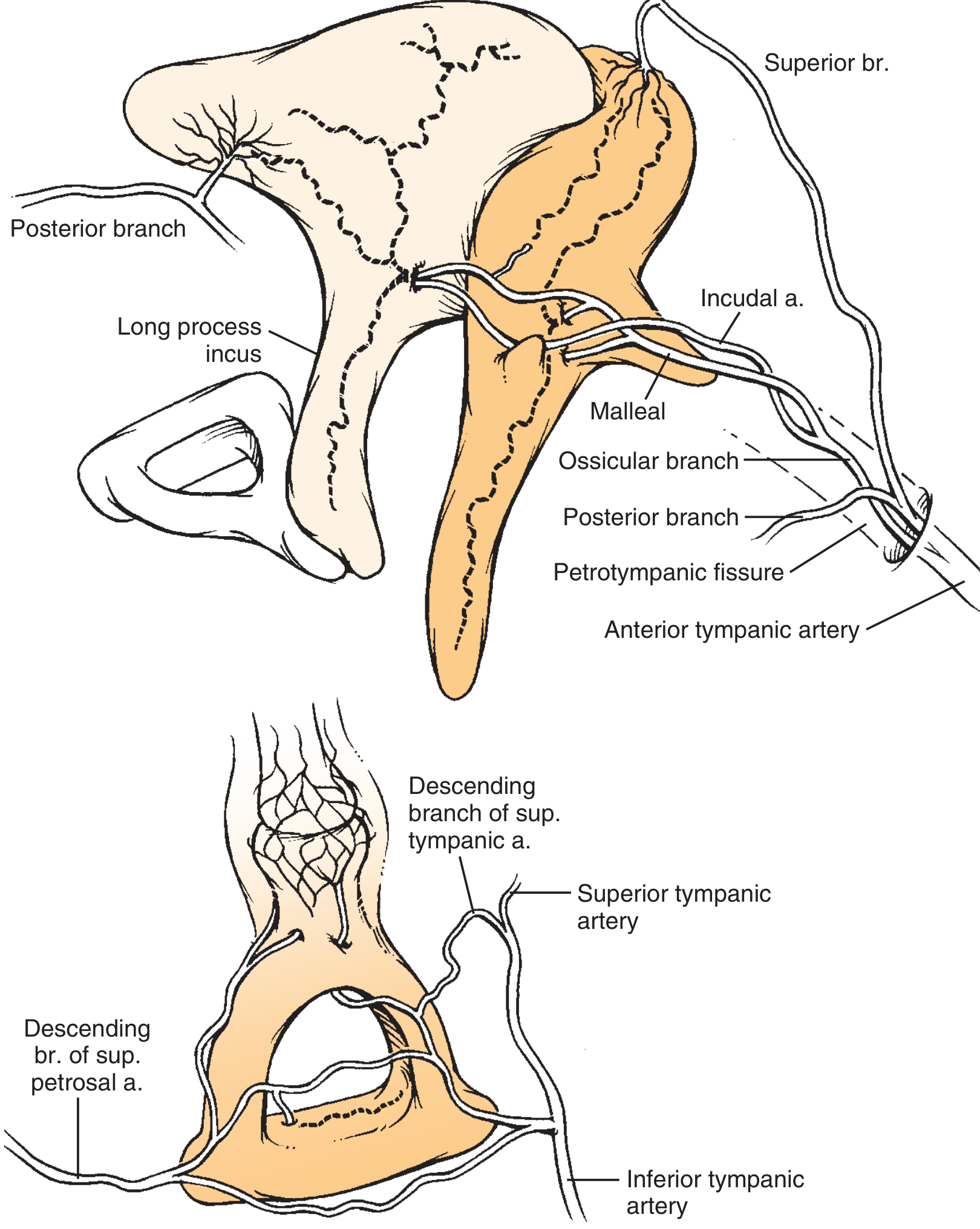
Blood supply of the middle ear ossicles. Note the anterior tympanic artery entering through the petrotympanic fissure, superior tympanic artery, and inferior tympanic artery. The long process of the incus has only a single nutrient vessel - this is clinically important as it is prone to avascular necrosis in chronic otitis media.
Region-Based Memory Hook (for exams)
| Region | Artery to Remember |
|---|---|
| Tympanic membrane (external) | Deep auricular a. (from maxillary) |
| Tympanic membrane (internal) | Anterior tympanic a. (from maxillary) |
| Epitympanum roof | Superior tympanic + Petrosal (both from middle meningeal) |
| Mesotympanum | Inferior tympanic (ascending pharyngeal) + Vidian + ICA tympanic arches |
| Posterior cavity + stapedius | Stylomastoid a. |
| Mastoid air cells | Mastoid br. (from stylomastoid) |
High-Yield Clinical Points
-
Middle meningeal artery supplies the roof of the tympanum via 2 branches (superior tympanic + petrosal). Epidural haematomas from middle meningeal injury can therefore cause blood in the middle ear (haemotympanum).
-
Long process of the incus has only a single nutrient vessel (the incudal artery, from the anterior tympanic). This makes it the ossicle most vulnerable to avascular necrosis in chronic ear disease - Cummings, Fig. 126.11.
-
ICA gives tympanic arches - the only contribution of the internal carotid artery to the middle ear. This is a high-yield one-liner for MCQs.
-
Ascending pharyngeal → inferior tympanic = the pathway by which nasopharyngeal infections can affect middle ear vascularity.
Simplest Exam Mnemonic
If you need just one sentence: "Max Gave Inferior Patients Stylish Mastoid Crowns Today"
| Word | Artery | Parent |
|---|---|---|
| Max | Anterior tympanic | Maxillary |
| Gave | Superior tympanic | Middle meningeal |
| Inferior | Inferior tympanic | Ascending pharyngeal |
| Patients | Petrosal | Middle meningeal |
| Stylish | Stylomastoid | Posterior auricular |
| Mastoid | Mastoid branch | Stylomastoid |
| Crowns | Canal of pterygoid | Vidian a. |
| Today | Tympanic arches | Internal carotid |
- Scott-Brown's Otorhinolaryngology Head & Neck Surgery, Vol. 2, Table 46.2
- Cummings Otolaryngology, Fig. 126.11
This is a shared conversation. Sign in to Orris to start your own chat.