Head and Neck
Reading File
Asking for Preferences
Finding Sources
Searching Images
head and neck anatomy overview
Reading File
Loading Image
Loading Image
Finding Sources
Finding Sources
Reading File
Loading Image
Loading Image
Here is a quick overview of Head & Neck Anatomy, sourced from Gray's Anatomy for Students and Bailey & Love's Short Practice of Surgery.
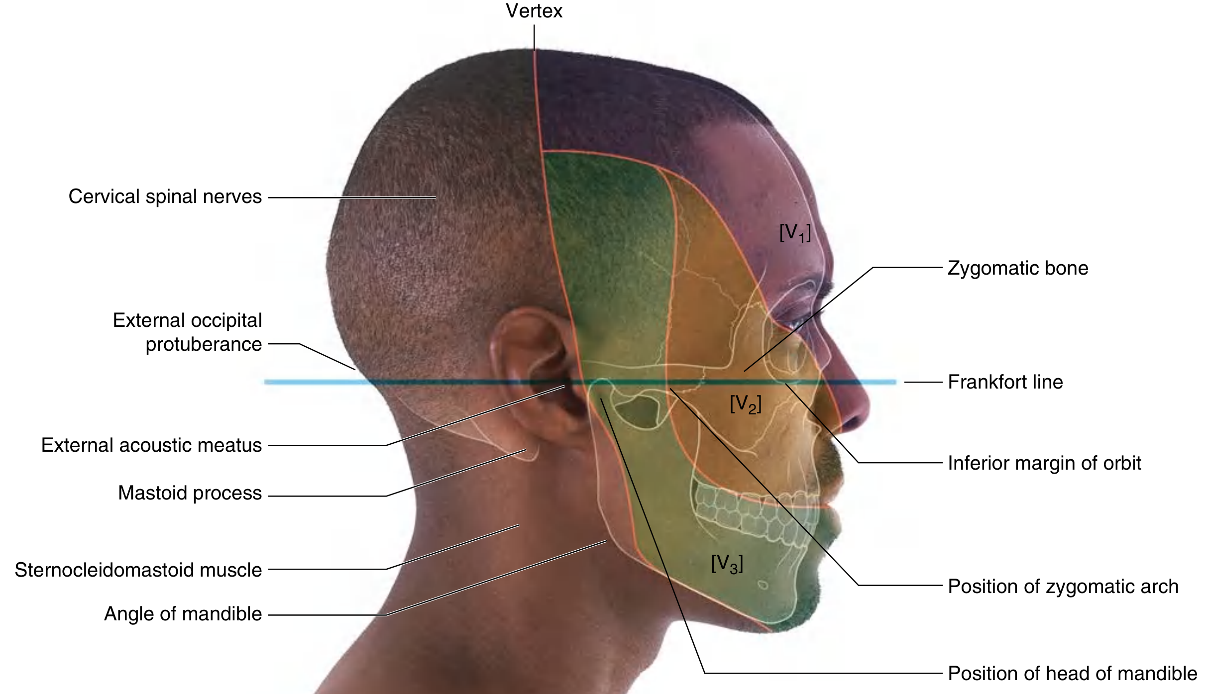
🗺 Surface Anatomy & Landmarks
The head is in the anatomical position (Frankfort plane) when the inferior margins of the bony orbits and the superior margins of the external acoustic meatuses are in the same horizontal plane.
Key palpable landmarks:
| Landmark | Location / Note |
|---|---|
| Head of mandible | Anterior to external ear; moves on jaw opening |
| Zygomatic arch | Extends from TMJ to zygomatic bone |
| Mastoid process | Posterior to external acoustic meatus; SCM attachment |
| External occipital protuberance | Midline posteriorly; marks scalp innervation transition |
| Vertex | Highest point of skull; transition from CN V (anterior) to cervical spinal nerve (posterior) innervation |
📐 Key Vertebral Levels
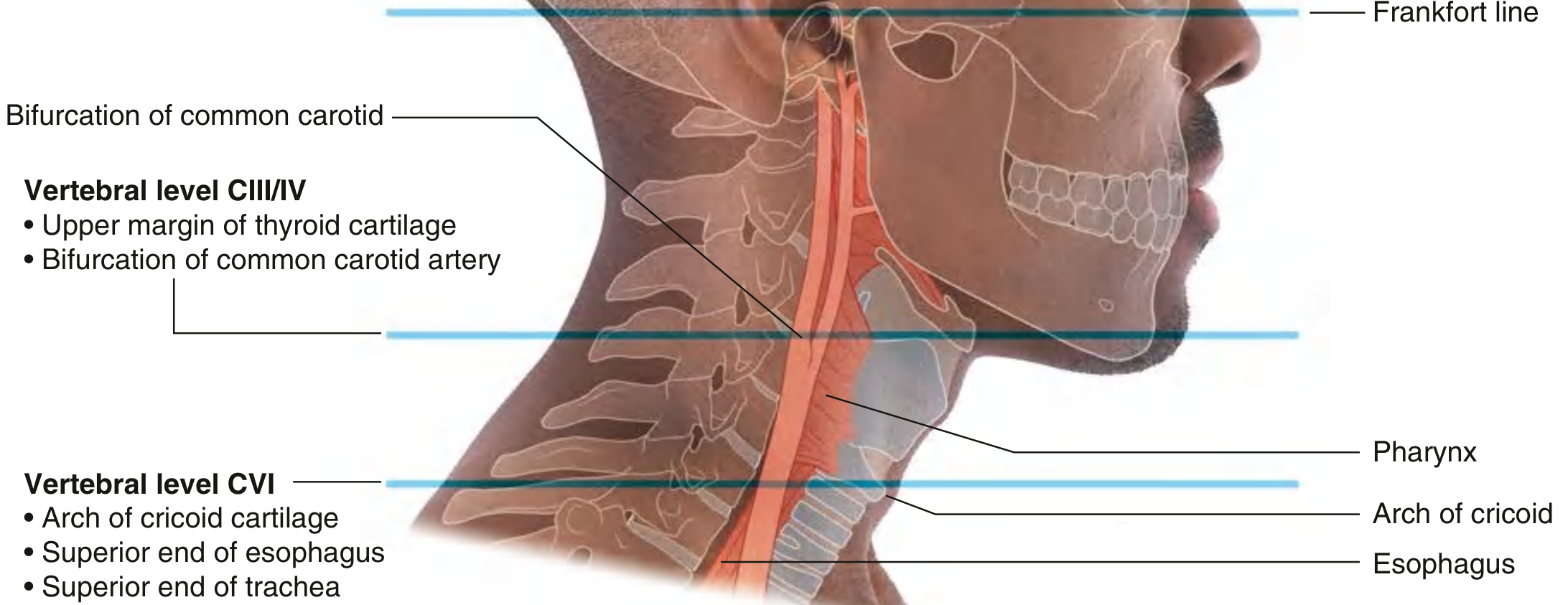
| Level | Landmark |
|---|---|
| CIII/CIV | Bifurcation of common carotid artery; upper margin of thyroid cartilage |
| CVI | Pharynx → Esophagus; Larynx → Trachea; inferior margin of cricoid cartilage |
🔺 Triangles of the Neck
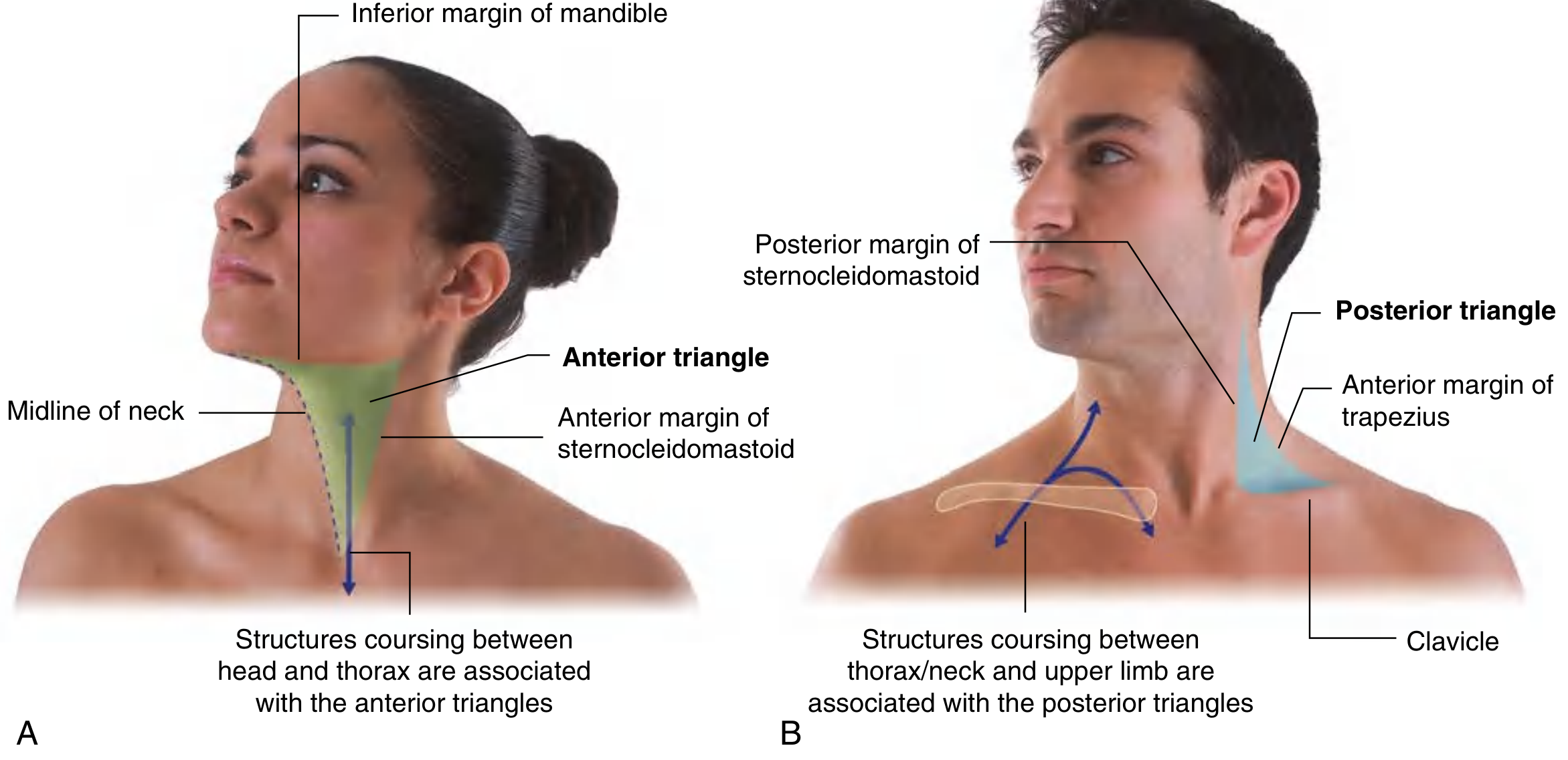
Anterior Triangle
- Base: inferior margin of mandible
- Anterior border: midline of neck
- Posterior border: anterior border of SCM
- Apex: suprasternal notch
- Contents: airway, digestive tract, thyroid/parathyroid glands, carotid vessels, cranial nerves
Posterior Triangle
- Base: middle third of clavicle
- Medial border: posterior border of SCM
- Lateral border: anterior border of trapezius
- Apex: posteroinferior to mastoid process
- Contents: nerves and vessels supplying the upper limb (brachial plexus, subclavian vessels, accessory nerve CN XI)
🗣 Pharynx Regions (Sagittal View)
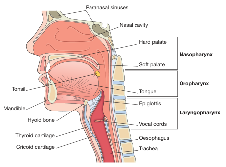
| Region | Boundaries |
|---|---|
| Nasopharynx | Posterior to nasal cavity; above soft palate |
| Oropharynx | Posterior to oral cavity; soft palate to epiglottis |
| Laryngopharynx (Hypopharynx) | Epiglottis to C6; leads to esophagus posteriorly and larynx anteriorly |
⚡ Cricothyroid Ligament (Emergency Access Point)
The median cricothyroid ligament lies between the thyroid and cricoid cartilages in the midline. It is the site of emergency cricothyrotomy when the upper airway (above the vocal folds) is obstructed. It is located by palpating the thyroid notch → laryngeal prominence → inferior surface of thyroid cartilage → soft dip = cricothyroid membrane.
Sources: Gray's Anatomy for Students (9780323934237), Bailey & Love's Short Practice of Surgery 28th ed.
This is a shared conversation. Sign in to Orris to start your own chat.