
DHEAS ?
Reading File
DHEAS (Dehydroepiandrosterone sulfate) is the sulfated form of DHEA, the most abundant circulating androgen precursor in the body. It is produced almost exclusively by the adrenal cortex (zona reticularis), making it a specific marker of adrenal androgen output. It has a long half-life (~10 hours) and stable serum levels, making it more useful clinically than DHEA itself.
Searching sources...
Finding Sources
Searching PubMed
DHEAS dehydroepiandrosterone sulfate clinical significance
Good results. Let me pull in the key content from several of these sources.
Reading File
Reading File
Reading File
Now let me pull the Tietz lab medicine and Goodman & Gilman content for reference ranges and pharmacology:
Reading File
Reading File
I now have comprehensive content from multiple authoritative sources. Here is the full answer:
DHEAS (Dehydroepiandrosterone Sulfate)
What It Is
DHEAS is the sulfated ester of DHEA (dehydroepiandrosterone), a C-19 steroid. It is produced almost exclusively in the zona reticularis of the adrenal cortex via the enzyme SULT2A1 (dehydroepiandrosterone sulfotransferase, gene on chromosome 19q13.33), which sulfates DHEA to form DHEAS.
- DHEAS circulates at levels 100-fold higher than free DHEA because tight albumin binding greatly extends its half-life (hours vs. ~30 minutes for free DHEA)
- It is the most abundant circulating steroid in the body
- Relative androgenicity: DHT = 300, Testosterone = 100, Androstenedione = 10, DHEAS = 5 (weak androgen)
Biosynthesis and Regulation
| Step | Detail |
|---|---|
| Substrate | Cholesterol → pregnenolone → DHEA (via CYP17A1) |
| Sulfation | DHEA + sulfate → DHEAS (via SULT2A1) |
| Regulator | ACTH (via MC2R) - primary regulator, but adrenal androgens do not mirror cortisol exactly; additional modulators exist |
| Peripheral conversion | Steroid sulfatases in gonads/peripheral tissues convert DHEAS back to DHEA → testosterone or estradiol |
The zona reticularis is the last adrenocortical zone to develop, emerging at adrenarche. DHEA/DHEAS production peaks during fetal development, wanes ~1 year after birth, then reappears around age 8-10 years as adrenarche approaches. Levels peak in the third decade of life, then progressively decline with aging.
- Tietz Textbook of Laboratory Medicine, 7th Ed.
- Goodman & Gilman's Pharmacological Basis of Therapeutics
Life-Course Pattern of DHEAS Levels
| Life Phase | DHEAS Level |
|---|---|
| Newborn | High (fetal zone activity) |
| Ages 2-4 years | Very low |
| Adrenarche (~age 8-10) | Begins to rise |
| Third decade | Peak |
| Adult life | Steady decline |
| Post-menopause | Continues to decline (testosterone levels maintained) |
Clinical Significance
1. Adrenarche Marker
DHEAS rise at adrenarche heralds the first appearance of pubic hair (pubarche) and is the major androgen source prior to gonadarche.
2. Hyperandrogenism Evaluation
DHEAS is the most useful single marker to distinguish adrenal vs. ovarian sources of androgen excess:
- Normal/mildly elevated DHEAS + elevated testosterone → likely ovarian source (e.g., PCOS)
- Markedly elevated DHEAS → adrenal source (e.g., CAH, adrenal tumor)
- DHEAS >18.5 μmol/L (>7000 μg/L) → strongly suggests an adrenal androgen-secreting tumor (per Harrison's 22E)
- Modest DHEAS elevations can occur in PCOS as well (not exclusively an adrenal marker)
- Elevated serum DHEAS or 17-hydroxyprogesterone identifies an adrenal source of excess androgen in acne workup (per Fitzpatrick's/Dermatology 5e)
3. Dexamethasone Suppression Test
Since adrenal androgens are readily suppressed by low-dose glucocorticoids:
- 0.5 mg dexamethasone q6h × 4 days (or overnight 1 mg dex test)
- Suppression of DHEAS/testosterone into normal range → adrenal source
- Incomplete suppression → ovarian source
4. Adrenal Insufficiency
Patients with adrenal insufficiency have markedly low DHEAS. However, adrenal androgen replacement is not required for survival - though DHEA supplementation has been studied with inconclusive results regarding libido, cognition, and well-being.
5. Precursor for Sex Steroids
DHEAS provides the precursor for >50% of circulating testosterone in women through peripheral conversion. In pregnancy, fetal DHEAS is the substrate for placental estrogen synthesis (DHEAS → estradiol; 16-OH-DHEAS → estriol).
Key Clinical Conditions
| Condition | DHEAS Level | Notes |
|---|---|---|
| PCOS | Mildly elevated or normal | Ovarian source predominates |
| Nonclassic CAH (21-hydroxylase deficiency) | Elevated | Also check 17-OHP |
| Adrenal androgen-secreting tumor | Markedly elevated (>7000 μg/L) | CT/MRI to localize |
| Adrenal insufficiency | Very low | No replacement needed for survival |
| Normal aging / menopause | Progressively declining | Testosterone maintained longer |
| ACTH-dependent Cushing's | May be elevated | DHEAS follows ACTH drive |
| Exogenous glucocorticoids | Suppressed | Suppresses ACTH → zona reticularis |
Reference (Interpretation)
- Preferred test: Serum DHEAS (stable, no diurnal variation, no need for 24h urine)
- Levels are age- and sex-dependent (peak ~300-500 μg/dL in young adults, declining with age)
- Because of its adrenal specificity, elevated DHEAS is more diagnostically useful than free DHEA for localizing excess androgen production
Sources: Harrison's Principles of Internal Medicine 22E | Berek & Novak's Gynecology | Tietz Textbook of Laboratory Medicine 7th Ed. | Goodman & Gilman's Pharmacological Basis of Therapeutics | Fitzpatrick's Dermatology Vol. 1-2
Medial canali form dystrophy of nails ?
Median canaliform dystrophy (also called median canal dystrophy or solenonychia) is a nail disorder characterized by a midline longitudinal split or canal in the nail plate, most commonly affecting the thumbs. It results from a defect in the nail matrix.
Searching sources...
Reading File
Finding Sources
Finding Sources
Excellent hits. Let me read the full sections from Andrews' and Dermatology 5e, and fetch the nail images.
Reading File
Reading File
Reading File
Let me now get the rest of the Andrews' section and fetch the nail images.
Reading File
Loading Image
Loading Image
I now have comprehensive content and the clinical images. Here is the full answer:
Median Canaliform Dystrophy of Nails
(Dystrophia Unguis Mediana Canaliformis / Heller's Median Canaliform Dystrophy / Solenonychia)
Definition
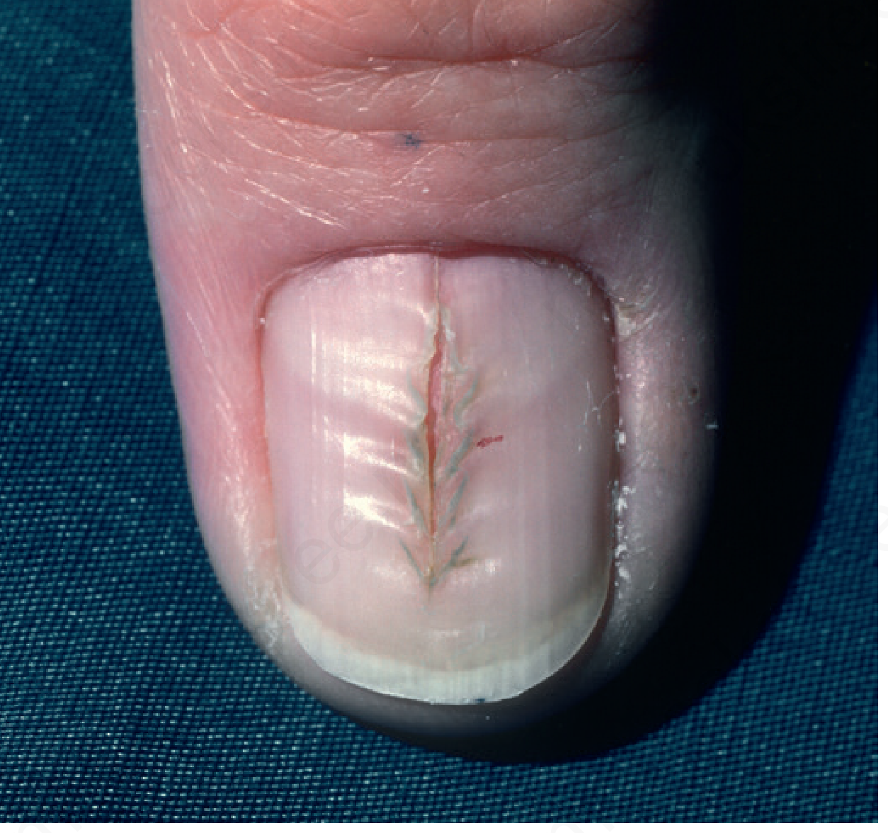
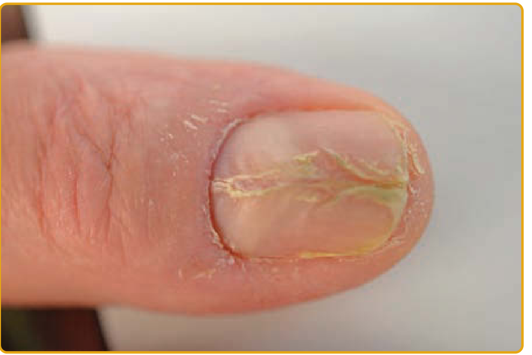
A nail disorder characterized by a central (midline) longitudinal splitting or canal formation in the nail plate. The split typically starts at the proximal nail plate (cuticle region) and extends distally as the nail grows, producing an appearance classically described as a "fir tree" or "Christmas tree" or "inverted fir tree" pattern - with oblique furrows running proximally on both sides of a central split.
Clinical Image
Classic appearance - midline vertical canal with fir-tree branching pattern (Andrews' Diseases of the Skin):
Heller's median canaliform dystrophy of the thumbnail (Fitzpatrick's Dermatology):
Key Features
| Feature | Detail |
|---|---|
| Nail affected | Thumbs (most common), occasionally other fingers |
| Pattern | Midline longitudinal split/canal, inverted fir tree pattern |
| Direction | Starts proximally at cuticle, extends distally with nail growth |
| Oblique furrows | Run proximally on both sides of the central split |
| Synonyms | Solenonychia, Heller's dystrophy, Dystrophia unguis mediana canaliformis |
Etiology and Associations
Primary cause: Trauma to the nail matrix (zona proximalis)
Specific causes and associations include:
- Repetitive mechanical trauma - e.g., repeated pressing/rubbing of the cuticle area; repeated typing with nail tip on a PDA
- Habit-tic behavior - important to distinguish from habit-tic deformity (see below)
- Nail matrix papilloma or glomus tumor - can produce a tube-like (solenos) channel distal to it
- Drug-induced - isotretinoin, ritonavir (antiretroviral)
- Familial cases - rare
- Idiopathic - many cases have no identifiable cause
Distinction from Habit-Tic Deformity
This is a classic exam distinction:
| Feature | Habit-Tic Deformity | Median Canaliform Dystrophy (Heller) |
|---|---|---|
| Mechanism | Nervous rubbing/pushing back of the cuticle midportion with the index fingernail | Trauma (often idiopathic or PDA use, drug) |
| Nail surface | Multiple midline transverse ridges (washboard/Beau's lines) | Central longitudinal split |
| Pattern | Washboard (multiple transverse grooves) | Inverted fir tree (longitudinal canal + oblique branches) |
| Lunula | Unusually long; cuticle absent | Usually normal |
| Classification | Autoaggressive/psychological | Separate entity (though also likely traumatic) |
Both are considered separate entities, though both are probably caused by trauma to the nail matrix. - Dermatology 2-Volume Set 5e
Diagnosis
- Clinical - the characteristic fir-tree pattern on the midline of the nail plate is diagnostic
- Dermoscopy/onychoscopy can help confirm
- If a subungual tumor (papilloma, glomus tumor) is suspected as the cause, MRI or surgical exploration may be warranted
- Exclude drug history (isotretinoin, ritonavir)
Treatment
Treatment is often unsatisfactory; many cases persist for years:
- Avoidance of trauma - first-line; identify and eliminate triggering habit or activity
- Occlusive therapy - occlusion of the nail with bandages
- Topical tacrolimus 0.1% ointment - reported success (Kim BY et al, J Dermatol 2010; 37:573)
- Behavioral interventions - if habit-related
- Serotonin reuptake inhibitors (fluoxetine, paroxetine, sertraline) and N-acetylcysteine - for cases with obsessive-compulsive component
- Surgical - if an underlying matrix tumor (papilloma, glomus tumor) is found - Andrews' Diseases of the Skin
Sources: Andrews' Diseases of the Skin - Clinical Dermatology | Fitzpatrick's Dermatology Vol. 1-2 | Dermatology 2-Volume Set 5e (Bolognia)
This is a shared conversation. Sign in to Orris to start your own chat.