
gbs grading and prognosis criteria
Reading File
GBS (Guillain-Barre Syndrome) grading uses the Hughes Disability Scale, and prognosis relies on clinical scoring systems like EGOS and mEGOS. Let me pull the detailed information from the medical library and recent literature simultaneously.
Searching sources...
Finding Sources
Searching PubMed
"Guillain-Barre syndrome" AND prognosis AND scoring
Reading File
Finding Sources
Finding Sources
I found the EGOS reference in Rosen's Emergency Medicine. Let me now read the detailed GBS grading/prognosis content from both Bradley & Daroff's Neurology and Rosen's.
Reading File
Reading File
Reading File
Finding Sources
Let me read the detailed prognosis and treatment sections from Bradley & Daroff's Neurology, which contains the most comprehensive GBS coverage:
Reading File
Excellent - I now have comprehensive GBS grading and prognosis data. Let me also search for the IGOS (International GBS Outcome Study) scoring details and the exact Hughes Disability Scale:
Finding Sources
Searching the Web
Hughes disability scale GBS grading EGOS mEGOS ISNCSCI scoring criteria 2024
The PMC article has the updated 2025 GBS Disability Scale. Let me extract it alongside the management flowchart image:
Reading Web Page
https://pmc.ncbi.nlm.nih.gov/articles/PMC12573220
Loading Image
I now have all the key information needed for a comprehensive answer. Here is the complete GBS grading and prognosis summary:
Guillain-Barre Syndrome (GBS): Grading & Prognosis
1. Hughes GBS Disability Scale (Functional Grading)
The standard clinical grading tool, used as a primary outcome measure since 1978 and recently updated (Hughes et al., 2025, J Peripher Nerv Syst):
| Grade | Description |
|---|---|
| 0 | Healthy, no symptoms or signs |
| 1 | Minor symptoms or signs, able to run 10 m |
| 2 | Able to walk 5 m unaided (cannot run 10 m), with or without walking aid |
| 3 | Able to walk 5 m only with 1-2 sticks/canes or help of 1 person |
| 4 | Bedbound or chairbound - unable to walk 5 m even with aid |
| 5 | Requires invasive (mechanical) ventilation for any part of the day or night |
| 6 | Death |
A score of >=3 is generally considered "unable to walk unaided" - the key threshold used in treatment trials. Grades 0-2 = ambulatory; Grades 3-6 = non-ambulatory.
2. GBS Subtypes (Affects Prognosis)
| Subtype | Key Features | Prognosis |
|---|---|---|
| AIDP (most common in West) | Demyelinating; classic ascending weakness | Good; full recovery in majority |
| AMAN | Pure motor axonal; common in China | Variable; can be good if conduction failure (reversible) rather than axonal degeneration |
| AMSAN | Motor + sensory axonal | Severe; poor recovery |
| Miller-Fisher | Ophthalmoplegia, ataxia, areflexia | Generally good prognosis |
- Bradley & Daroff's Neurology in Clinical Practice, p. 2663
3. Prognostic Scoring Systems
A. Erasmus GBS Outcome Score (EGOS) - used at Day 14
Predicts inability to walk independently at 6 months. Combines three factors:
| Variable | Score |
|---|---|
| Age at onset: <40 yrs | 0 |
| Age 40-60 yrs | 0.5 |
| Age >60 yrs | 1 |
| Preceding diarrhea: Yes | 1 |
| Preceding diarrhea: No | 0 |
| GBS Disability Score at Day 14: Grade 1 | 1 |
| Grade 2 | 2 |
| Grade 3 | 3 |
| Grade 4 | 4 |
| Grade 5 | 5 |
Total EGOS range: 1-7
- EGOS 1-2: ~<10% unable to walk at 6 months
- EGOS 5-7: ~60-80% unable to walk at 6 months
B. Modified EGOS (mEGOS) - used at Day 7 (earlier)
Uses MRC sum score instead of GBS Disability Score (more objective, can be used 1 week earlier in admission):
| Variable | Score |
|---|---|
| Age <40 | 0 |
| Age 40-60 | 0.5 |
| Age >60 | 1 |
| Preceding diarrhea: Yes | 1 |
| Preceding diarrhea: No | 0 |
| MRC sum score 0-20 | 6 |
| MRC sum score 21-40 | 4 |
| MRC sum score 41-50 | 2 |
| MRC sum score 51-60 | 0 |
Total mEGOS range: 0-9
-
Higher score = worse prognosis
-
Rosen's Emergency Medicine, p. 1519
4. Clinical Predictors of Poor Prognosis
The following predict <20% probability of walking independently at 6 months (Bradley & Daroff's, p. 2669):
- Age >60 years
- Preceding diarrheal illness (especially Campylobacter jejuni - 25-50% of cases)
- Recent CMV infection
- Rapid progression - reaching maximum deficit in <7 days
- Requirement for ventilatory support
- Low distal CMAP amplitudes (≤20% of lower limit of normal) or inexcitable nerves on NCS
- Hyponatremia
- The AMAN/AMSAN subtype with true axonal degeneration (vs. reversible conduction failure)
- Higher CSF neurofilament and tau protein levels
5. Overall Outcome Data
| Timepoint | Data |
|---|---|
| Mortality | ~1-5% (modern era); was 33% before positive-pressure ventilation |
| Ventilator requirement | 9-30% of hospitalized patients |
| Full recovery by 12 months | ~70% |
| Full recovery by 24 months | ~82% |
| Residual motor weakness at 1 year | ~20% |
| Recurrence (re-GBS) | ~5% |
6. Management Decision Flowchart
The following diagram from Bradley & Daroff's summarizes GBS management based on clinical severity:
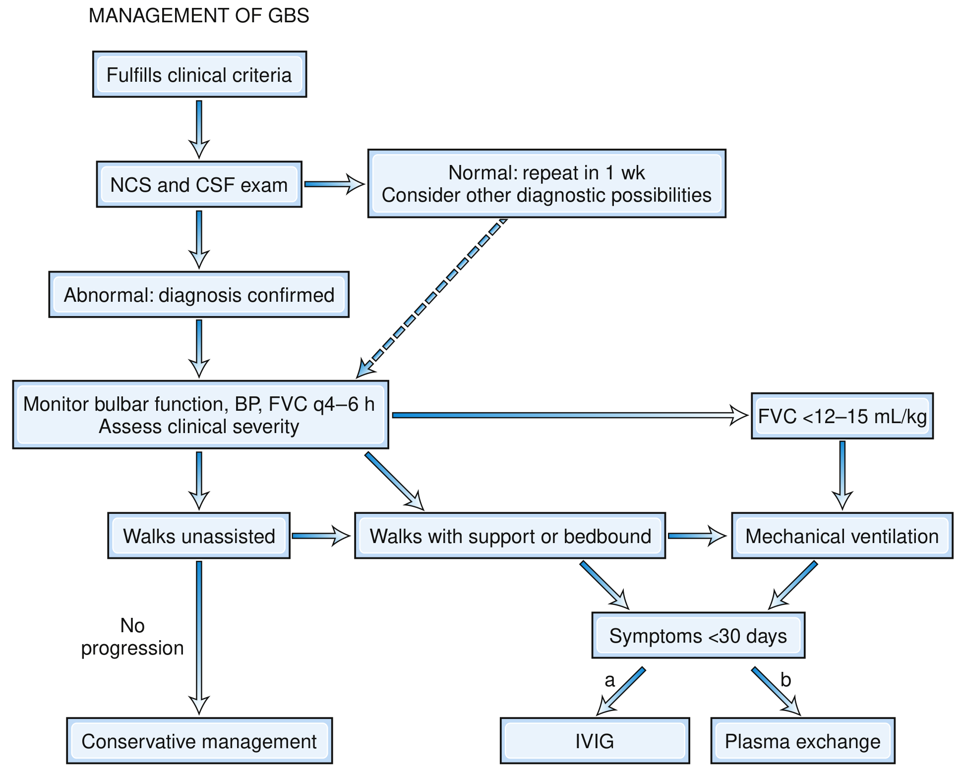
Key points: Patients who walk unaided with no progression - conservative management. Patients who walk with support or are bedbound AND symptoms <30 days - IVIG or plasma exchange (equally efficacious; IVIG preferred for ease of administration). FVC <12-15 mL/kg - mechanical ventilation.
7. CIDP vs. GBS: Progression Rule
If a patient initially diagnosed with GBS:
-
Deteriorates after 8 weeks, OR
-
Has >2 treatment-related fluctuations (especially beyond 1 month)
-
Has prominent sensory signs, nerve enlargement on ultrasound, or new demyelinating features on EDX months later
-
Consider acute-onset CIDP rather than GBS.
Sources:
- Bradley and Daroff's Neurology in Clinical Practice, pp. 2663-2669
- Rosen's Emergency Medicine: Concepts and Clinical Practice, p. 1519
- Hughes et al., 2025 - GBS Disability Scale update, J Peripher Nerv Syst 30(4):e70064
Groove pancreatitis
Reading File
Groove pancreatitis is a rare form of segmental chronic pancreatitis affecting the groove between the pancreatic head, duodenum, and common bile duct. It is strongly associated with alcohol and is often mistaken for pancreatic head carcinoma. Let me pull detailed textbook content and recent literature simultaneously.
Searching sources...
Finding Sources
Finding Sources
Searching PubMed
"groove pancreatitis" AND diagnosis
Excellent - I found dedicated sections in Current Surgical Therapy, Grainger & Allison's Radiology, and Clinical GI Endoscopy. Let me read all of them in parallel.
Reading File
Reading File
Reading File
Reading File
Reading File
Now let me fetch the CT images and also pull any radiology-specific details about imaging subtypes:
Loading Image
Finding Sources
Let me fetch the key paraduodenal pancreatitis CT/MRI images from Grainger & Allison:
Loading Image
Reading File
I now have comprehensive content from multiple authoritative textbooks. Here is the complete review:
Groove (Paraduodenal) Pancreatitis
Definition & Anatomy
Groove pancreatitis (now preferred term: paraduodenal pancreatitis) is a distinct form of chronic pancreatitis affecting the "groove" - the space between:
- The pancreatic head
- The descending (2nd) part of the duodenum
- The common bile duct
It specifically involves the region between the major and minor pancreatic papillae, and typically spares the main pancreatic parenchyma (in its groove-predominant form).
- Current Surgical Therapy 14e, p. 669
- Grainger & Allison's Diagnostic Radiology, p. 689
Epidemiology & Risk Factors
- Predominantly affects middle-aged males (M >> F)
- Strongly associated with chronic alcohol abuse and tobacco use
- Also associated with heterotopic pancreatic tissue in the duodenal wall and peptic ulcer disease (duodenal scarring causing minor papilla obstruction)
Pathogenesis
The proposed mechanism involves obstruction of the accessory pancreatic duct (duct of Santorini) and minor papilla, leading to:
- Ductal hypertension in the groove area
- Recurrent inflammation and fibrosis
- Cystic degeneration in the duodenal wall and groove
- Progressive scarring that can cause biliary and duodenal obstruction
Classification (Subtypes)
Four subtypes are now described based on imaging morphology (Grainger & Allison, p. 689-690):
| Type | Description |
|---|---|
| Type 1A (Solid, groove-predominant) | Mass in the groove between duodenum and pancreatic head; pancreas largely unaffected |
| Type 1B (Solid, pancreas-involving) | Cancer-mimicking mass extending into pancreatic head |
| Type 2A (Cystic, groove-predominant) | Classic form with cysts in the groove/duodenal wall |
| Type 2B (Cystic, segmental/pancreas-involving) | Cysts in groove + pancreatic head involvement |
On imaging, two broad patterns are recognized:
- Groove-predominant form - plate-like hypoattenuating lesion between pancreatic head and duodenum; pancreas relatively preserved
- Segmental (pancreas-involving) form - extends into the pancreatic head; most difficult to differentiate from cancer
Clinical Features
| Feature | Detail |
|---|---|
| Pain | Chronic epigastric pain, often postprandial |
| Weight loss | Common, due to food avoidance |
| Nausea/vomiting | From gastric outlet obstruction |
| Jaundice | Obstructive - from CBD compression |
| Gastric outlet obstruction | Duodenal wall fibrosis/cysts causing stenosis |
| Tumor markers | CA 19-9 and CEA typically negative (key differentiator from cancer) |
- Rosen's Emergency Medicine, p. 1519; Current Surgical Therapy 14e, p. 669
Imaging
CT Findings
- Plate-like or sheet-like hypodense/poorly enhancing lesion in the groove between pancreatic head and duodenum
- Cystic changes in the duodenal wall (pathognomonic when present)
- Thickening of the 2nd part of the duodenum with luminal narrowing
- Duodenal wall cysts
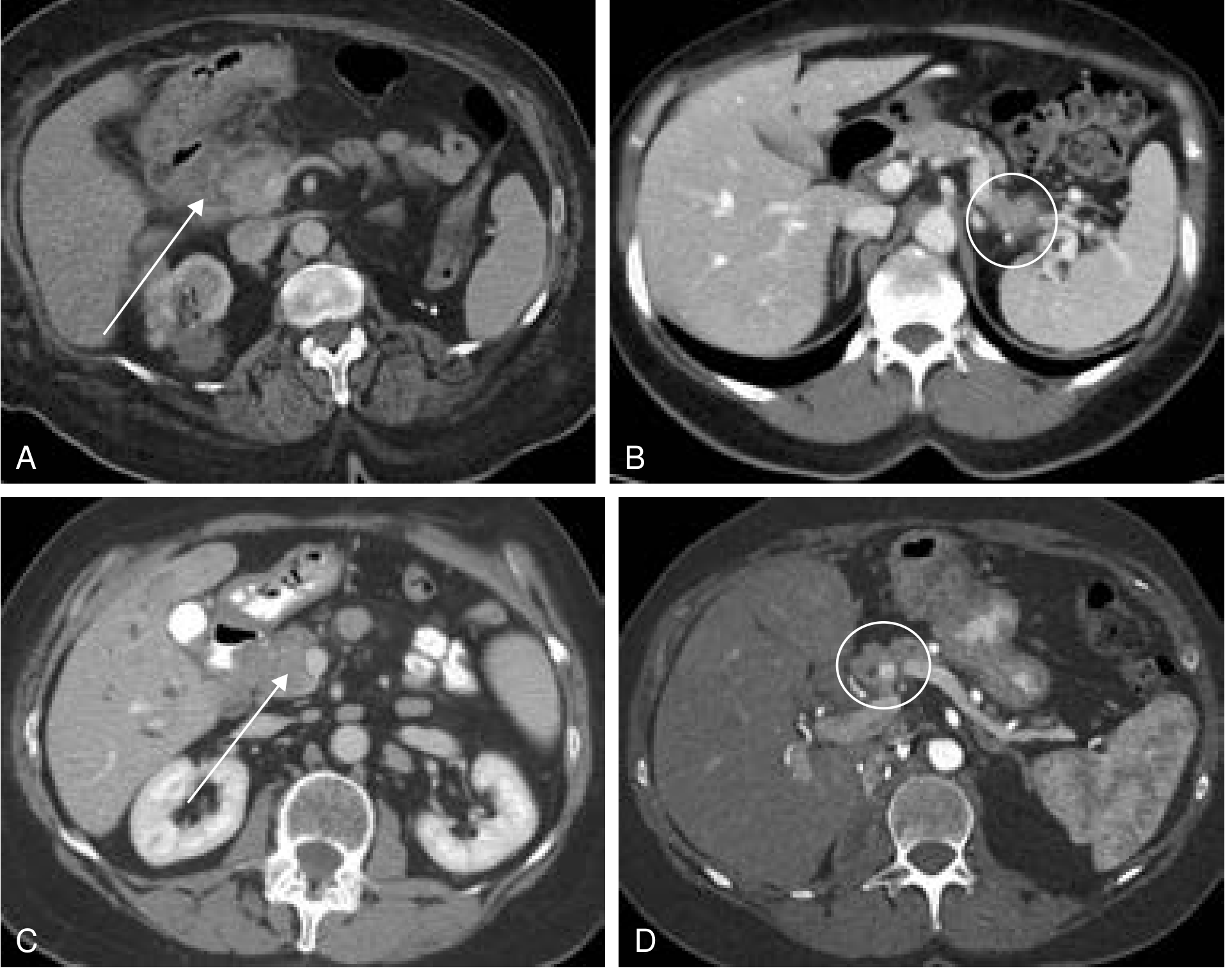
- May show a 2.9-cm hypodense fluid collection (arrow) with inflammatory stranding of the periduodenal fat
CT image (A - groove pancreatitis; B - AIP type I; C - pancreatic lymphoma; D - renal metastasis):
MRI / MRCP (Modality of Choice)
- T2W: High-signal cysts in the groove and duodenal wall; multiple small cysts are pathognomonic
- T1W Gd-enhanced: Hypovascular mass in groove (no enhancement = fibrosis)
- MRCP: Shows cystic lesions in the groove clearly, pancreatic duct often normal or minimally dilated (critical distinguishing point from PDAC)
- Duct-penetrating sign: Main pancreatic duct traverses (not obstructed by) the mass - favors inflammatory over malignant
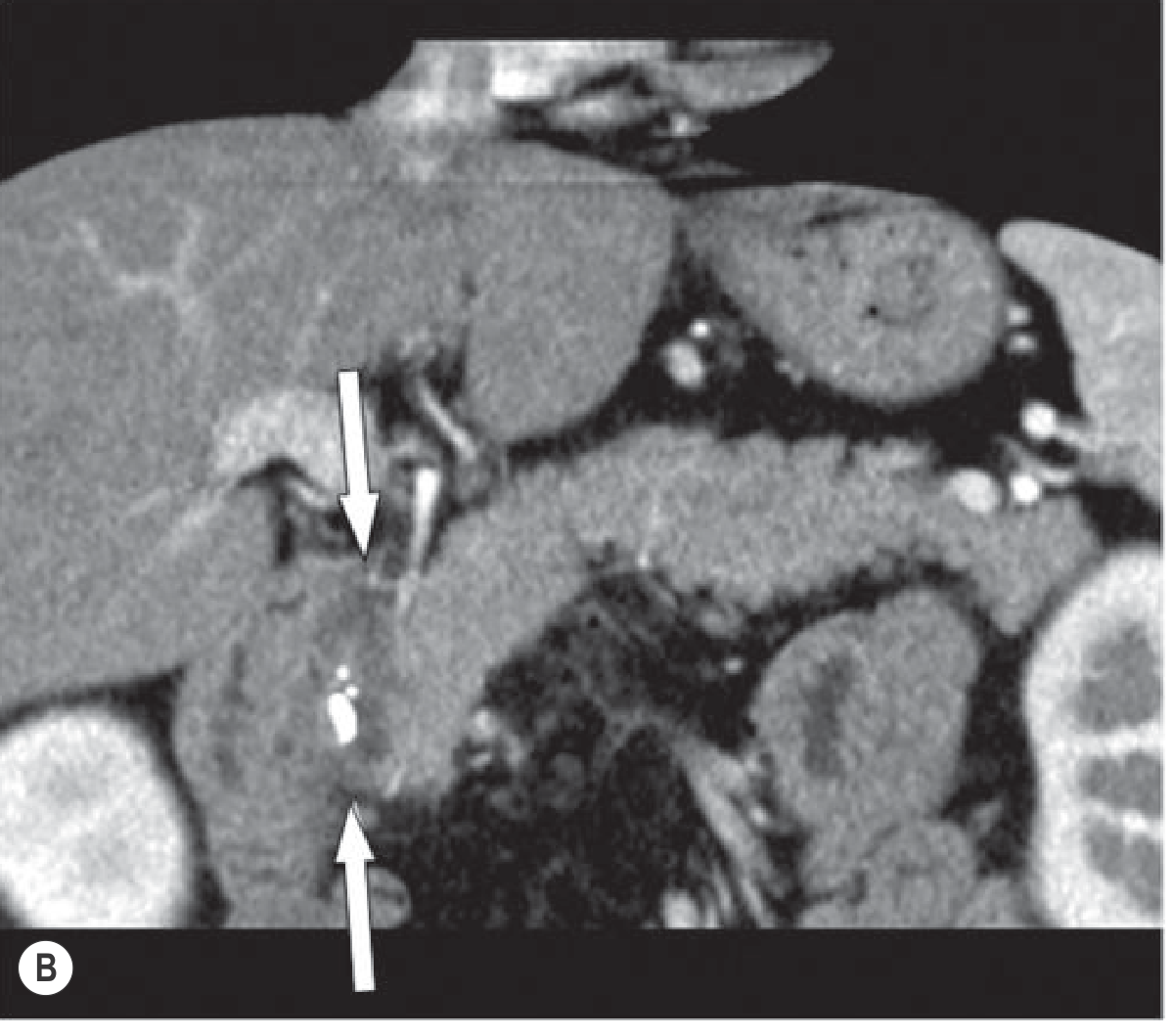
CT: Paraduodenal pancreatitis (groove-predominant form), showing the low-density mass between pancreatic head and duodenum:
- Grainger & Allison's Diagnostic Radiology, pp. 689-694
EUS
- EUS-guided FNA/biopsy is often required to confirm diagnosis and exclude malignancy
- Useful when imaging features overlap with pancreatic head adenocarcinoma
- Main pancreatic duct and CBD may show strictures on ERCP (72.5% and 56.9% of cases, respectively)
Histopathology
- Fibrosis with myofibroblast proliferation in the groove
- Cystic spaces lined by Brunner gland-like epithelium or ductal epithelium
- Heterotopic pancreatic tissue in the duodenal wall
- Inflammatory infiltrate (lymphocytes, plasma cells)
- No dysplasia (important - distinguishes from malignancy, though cytology can be misleading)
Differential Diagnosis
The most important differential is pancreatic ductal adenocarcinoma (PDAC):
| Feature | Groove Pancreatitis | PDAC |
|---|---|---|
| CA 19-9 | Normal/mildly elevated | Often markedly elevated |
| Duodenal wall cysts | Present (pathognomonic) | Absent |
| Duct-penetrating sign | Present | Absent (duct abruptly cut off) |
| Enhancement | Poor (fibrosis) | Hypoenhancing but different pattern |
| Main PD | Normal or mildly dilated | Obstructed upstream |
| EUS-FNA | No malignant cells | Malignant cells |
| Response to conservative Rx | Improves | Progresses |
Other differentials: autoimmune pancreatitis (IgG4), duodenal GIST, periampullary adenoma, duodenal carcinoma
Treatment
Conservative (First-line)
- Alcohol and tobacco cessation (essential)
- Analgesics for pain control
- Nutritional support (often significant weight loss)
- Subcutaneous octreotide (reduces pancreatic secretion; used in some protocols)
- >70% clinical success with conservative + endoscopic therapy in dedicated series
Endoscopic Therapy
Used when there are strictures or fluid collections:
- Pancreatic duct stenting (for PD stricture)
- CBD stenting (for biliary obstruction)
- EUS-guided transduodenal drainage of periduodenal fluid collections
- Stricture of PD present in ~72.5%, CBD in ~56.9% of cases
- Clinical GI Endoscopy, p. 1519
Surgical (Whipple / Pancreatoduodenectomy)
Indications:
- Failure of conservative management
- Gastric outlet obstruction not responding to endoscopy
- Biliary obstruction refractory to stenting
- Cannot exclude malignancy despite biopsy (cytology can be misleading)
- Duodenum-preserving pancreatic head resection (DPPHR) is an alternative with less morbidity when feasible
Resection is "not uncommon" even in biopsy-proven groove pancreatitis because cytology may be misleading and the concern for cancer drives surgical decision-making. - Current Surgical Therapy 14e, p. 669
Key Teaching Points
- Classic patient: Middle-aged alcoholic male with chronic epigastric pain, weight loss, and gastric outlet obstruction
- Pathognomonic finding: Cystic changes in the duodenal wall on T2W MRI
- Most dangerous pitfall: Misdiagnosis as pancreatic head carcinoma - leads to unnecessary Whipple
- MRCP > CT for characterizing the cysts and the duct-penetrating sign
- Negative tumor markers + cysts in the groove/duodenal wall = strongly favor groove pancreatitis
- Surgery may still be needed even with proven benign diagnosis when obstruction is refractory
Sources:
- Current Surgical Therapy 14e (Cameron), p. 669
- Grainger & Allison's Diagnostic Radiology, pp. 689-694
- Clinical Gastrointestinal Endoscopy (Expert Consult), 3e, p. 1519
- Fischer's Mastery of Surgery, 8e
Could u retry
Could you clarify what you'd like me to retry? For example:
- Re-explain groove pancreatitis in a different format (e.g., shorter summary, table-only, exam-style)?
- Search for more/different content (e.g., more imaging details, pathology focus, surgical management)?
- Something specific that didn't display correctly?
This is a shared conversation. Sign in to Orris to start your own chat.