
Can you help me make notes on arteries of upper limb and reference can be from sapiens textbook.
Reading File
Now let me search the library for the Sapiens textbook and arteries of the upper limb content.
Finding Sources
Finding Sources
I don't see a "Sapiens" textbook directly. Let me search more specifically for it and also look for anatomy books that may cover upper limb arteries.
Finding Sources
The library doesn't have a "Sapiens" anatomy textbook specifically. The closest authoritative anatomy references available are Gray's Anatomy for Students and the THIEME Atlas (General Anatomy and Musculoskeletal System). Let me check all anatomy books for upper limb artery content and pull detailed notes from the best sources - I'll use Gray's Anatomy for Students as the primary reference (which is widely used like Sapiens) and supplement with THIEME Atlas.
Finding Sources
Finding Sources
Gray's Anatomy for Students has the best dedicated sections. Let me now read all the relevant sections simultaneously - axillary artery, brachial artery, and the upper limb arteries overview from Gray's, plus the THIEME Atlas.
Reading File
Reading File
Reading File
Good content coming through. Now let me read the forearm arteries (radial and ulnar) and the hand arteries from Gray's Anatomy for Students.
Finding Sources
Reading File
Finding Sources
Finding Sources
Reading File
Reading File
I now have comprehensive content from Gray's Anatomy for Students. Let me also fetch the key anatomical diagrams to include in the notes.
Loading Image
Loading Image
Loading Image
I now have comprehensive content and all key diagrams. Let me also fetch the branches of axillary artery diagram and check the ulnar artery branches in the forearm.
Loading Image
Reading File
I now have everything I need to compile thorough, well-structured notes. Here are your comprehensive notes on the Arteries of the Upper Limb, referenced from Gray's Anatomy for Students:
Arteries of the Upper Limb
Reference: Gray's Anatomy for Students
Overview - Sequential Flow
The arterial supply to the upper limb runs as a single main channel that changes name at key anatomical landmarks:
Subclavian artery → Axillary artery → Brachial artery → Radial artery + Ulnar artery → Palmar arches → Digital arteries
1. Axillary Artery
Definition: The subclavian artery becomes the axillary artery at the lateral margin of rib I. It becomes the brachial artery at the inferior margin of the teres major muscle.
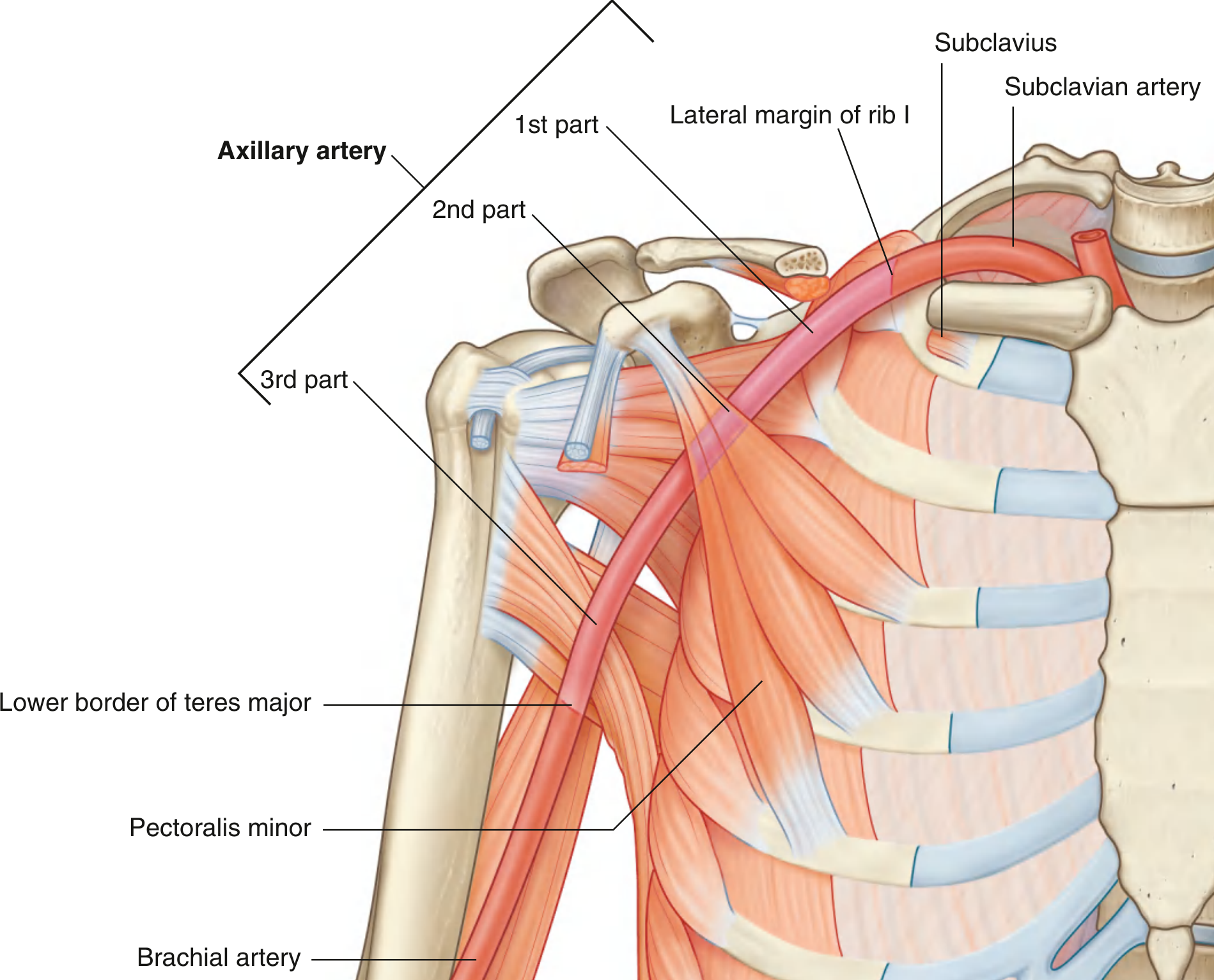
Parts of the Axillary Artery
The pectoralis minor divides it into 3 parts:
| Part | Relation to Pectoralis Minor | Branches |
|---|---|---|
| 1st | Proximal (medial) to it | 1 - Superior thoracic artery |
| 2nd | Posterior to it | 2 - Thoraco-acromial + Lateral thoracic arteries |
| 3rd | Distal (lateral) to it | 3 - Subscapular + Anterior circumflex humeral + Posterior circumflex humeral arteries |
Mnemonic: "1st part = 1 branch, 2nd part = 2 branches, 3rd part = 3 branches"
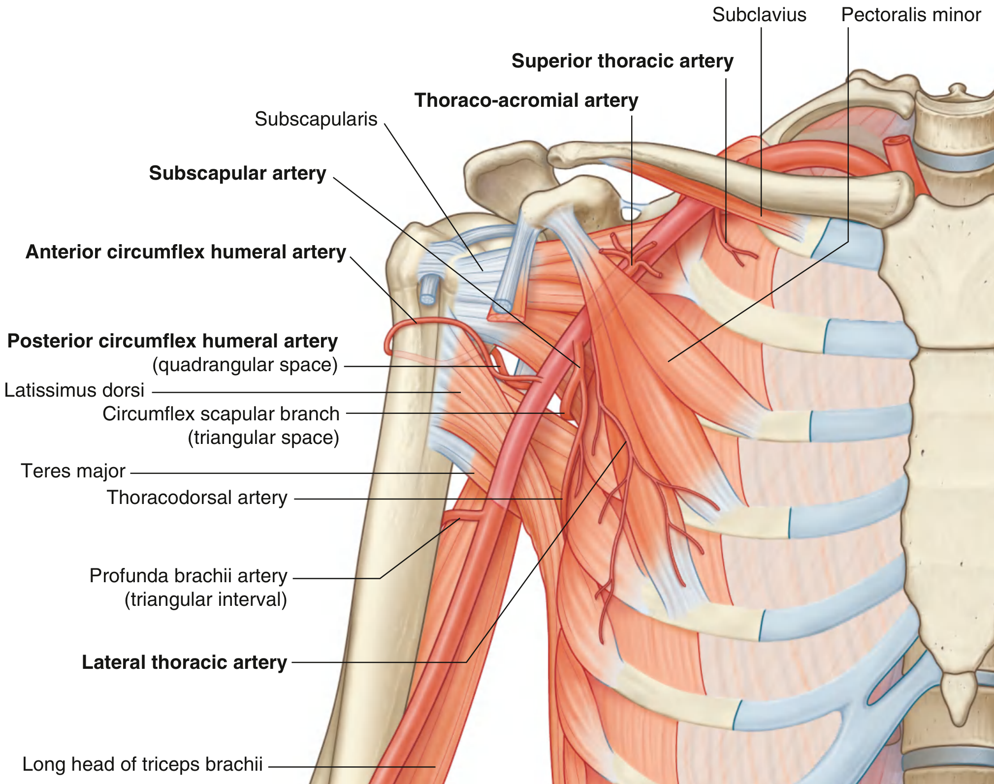
Branches in Detail
1st Part Branch:
- Superior thoracic artery - small; from anterior surface of 1st part; supplies upper regions of medial and anterior axillary walls
2nd Part Branches:
- Thoraco-acromial artery - short; from anterior surface of 2nd part just posterior to the superior margin of pectoralis minor; curves around the muscle, penetrates clavipectoral fascia, divides into four branches:
- Pectoral branch (also supplies breast)
- Deltoid branch (passes with cephalic vein in clavipectoral triangle)
- Clavicular branch
- Acromial branch
- Lateral thoracic artery - arises from anterior surface of 2nd part, follows the margin of pectoralis minor to the thoracic wall; supplies medial and anterior walls of axilla; contributes to breast supply in women
3rd Part Branches:
- Subscapular artery - largest branch of the axillary artery; major blood supply to posterior wall of axilla; divides into:
- Circumflex scapular artery - passes through the triangular space; anastomoses with suprascapular artery and dorsal scapular artery (scapular anastomosis network)
- Thoracodorsal artery - follows lateral border of scapula; supplies posterior and medial walls of axilla
- Anterior circumflex humeral artery - small; passes anterior to surgical neck of humerus; anastomoses with posterior circumflex humeral artery; supplies glenohumeral joint and head of humerus
- Posterior circumflex humeral artery - larger; passes through the quadrangular space with the axillary nerve; curves around surgical neck of humerus; supplies surrounding muscles and glenohumeral joint; anastomoses with anterior circumflex humeral, profunda brachii, suprascapular, and thoraco-acromial arteries
2. Brachial Artery
Definition: The major artery of the arm. Begins as continuation of the axillary artery at the lower border of teres major; terminates just distal to the elbow joint by dividing into radial and ulnar arteries.
Position in the arm:
- In proximal arm - lies on the medial side
- In distal arm - moves laterally to lie midway between the medial and lateral epicondyles of the humerus
- Crosses anteriorly to the elbow joint; lies immediately medial to the tendon of biceps brachii
- Palpable along its entire length; in proximal regions can be compressed against the medial side of the humerus
Clinical note: The brachial artery is the site for blood pressure measurement - the sphygmomanometer cuff compresses it against the humerus; Korotkoff sounds are auscultated over the brachial artery in the cubital fossa.
Branches of the Brachial Artery
- Profunda brachii artery - the largest branch; passes into the posterior compartment through the triangular interval (bounded by shaft of humerus, inferior margin of teres major, lateral margin of long head of triceps); travels with the radial nerve in the radial groove; supplies posterior compartment muscles; terminates as two collateral vessels contributing to the anastomotic network around the elbow
- Superior ulnar collateral artery - contributes to elbow anastomosis
- Inferior ulnar collateral artery - contributes to elbow anastomosis
- Nutrient arteries to the humerus - through a foramen in the anteromedial surface of the humeral shaft
- Muscular branches to adjacent muscles
3. Radial Artery (Lateral Division)
Origin: From brachial artery at approximately the neck of the radius.
Course in the forearm:
- Passes along the lateral aspect of the forearm
- Deep to brachioradialis in the proximal half
- Related to the superficial branch of the radial nerve on its lateral side in the middle third
- In the distal forearm - medial to the brachioradialis tendon, covered only by deep fascia, superficial fascia, and skin
- Lies immediately lateral to the flexor carpi radialis tendon in the distal forearm → this is where the radial pulse is felt
Distal course: Leaves the forearm, passes around the lateral side of the wrist, over the floor of the anatomical snuffbox, and penetrates the posterolateral hand between the bases of metacarpals I and II (passing between the two heads of the first dorsal interosseus and then the adductor pollicis) to form the deep palmar arch.
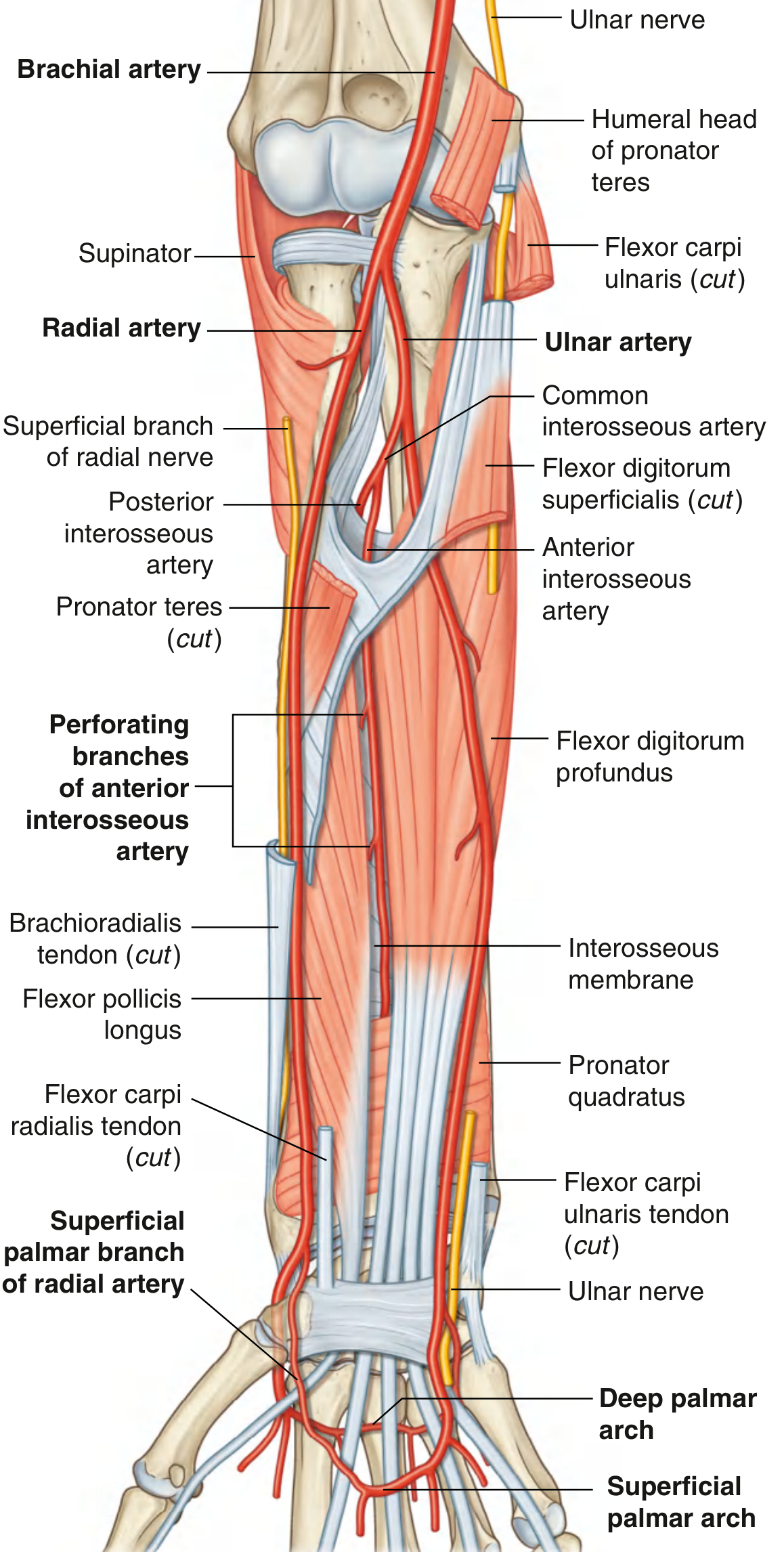
Branches of the Radial Artery
| Location | Branch | Supply/Notes |
|---|---|---|
| Forearm | Radial recurrent artery | Anastomotic network around elbow joint |
| Forearm | Muscular branches | Muscles on lateral side of forearm |
| Distal forearm | Palmar carpal branch | Anastomotic network for carpal bones/joints |
| Distal forearm | Superficial palmar branch | Passes through/superficial to thenar muscles; anastomoses with superficial palmar arch of ulnar artery |
| Wrist (posterolateral) | Dorsal carpal branch | Forms dorsal carpal arch → dorsal metacarpal arteries → dorsal digital arteries |
| Wrist (posterolateral) | First dorsal metacarpal artery | Supplies thumb and lateral index finger (major supply) |
| Palm (deep) | Deep palmar arch | Main terminal branch; passes medially between metacarpals and long flexor tendons |
4. Ulnar Artery (Medial Division)
Origin: From brachial artery at the neck of the radius (same point as radial artery). The ulnar artery is larger than the radial artery.
Course in the forearm:
- Leaves cubital fossa by passing deep to pronator teres
- Passes in the fascial plane between flexor carpi ulnaris and flexor digitorum profundus
- In the distal forearm, often tucked under the anterolateral lip of the flexor carpi ulnaris tendon - not easily palpable
- The ulnar nerve is immediately medial to the ulnar artery in the distal forearm
Entry to hand: Passes lateral to the pisiform bone, superficial to the flexor retinaculum, and arches across the palm as the superficial palmar arch (often the major supply to medial 3½ digits).
Branches of the Ulnar Artery
| Location | Branch | Supply/Notes |
|---|---|---|
| Forearm | Ulnar recurrent artery (anterior + posterior) | Anastomotic network around elbow joint |
| Forearm | Muscular branches | Surrounding muscles |
| Forearm | Common interosseous artery | Divides into anterior and posterior interosseous arteries |
| Forearm | Dorsal carpal branch | Carpal joints and bones |
| Forearm | Palmar carpal branch | Carpal joints and bones |
| Hand | Deep palmar branch | Arises medial to ulnar artery at pisiform; curves around hook of hamate to join deep palmar arch |
Common Interosseous Artery - Important Branch
- Anterior interosseous artery - travels along anterior aspect of interosseous membrane; supplies deep muscles of the anterior compartment, radius and ulna; perforating branches supply deep posterior compartment muscles; terminates by joining posterior interosseous artery in distal forearm
- Posterior interosseous artery - passes dorsally over proximal margin of interosseous membrane into posterior compartment; supplies posterior forearm muscles
5. Palmar Arches
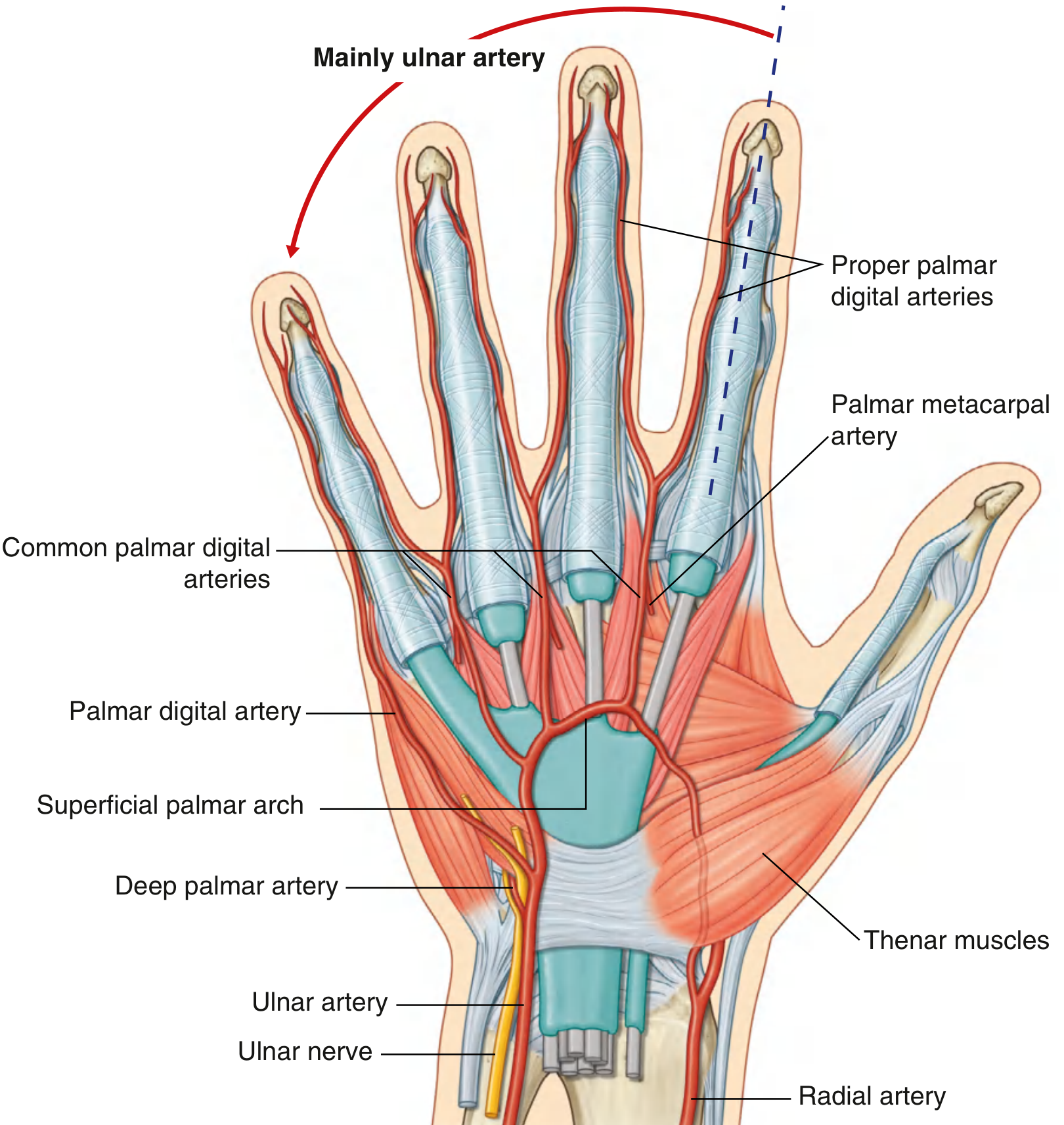
Superficial Palmar Arch
- Formed mainly by: Ulnar artery
- Completed by: Superficial palmar branch of the radial artery (lateral side)
- Position: Superficial to long flexor tendons, just deep to palmar aponeurosis
- Branches:
- Palmar digital artery to the medial side of the little finger
- Three common palmar digital arteries - supply lateral side of little finger, both sides of ring and middle fingers, medial side of index finger; they are joined by palmar metacarpal arteries from the deep palmar arch before bifurcating into the proper palmar digital arteries
Deep Palmar Arch
- Formed mainly by: Radial artery (terminal branch)
- Completed by: Deep palmar branch of the ulnar artery (medial side)
- Position: Deep to long flexor tendons, between metacarpal bones
- Branches:
- Palmar metacarpal arteries - join the common palmar digital arteries of the superficial arch
6. Summary Table - Arterial Tree of the Upper Limb
Subclavian artery
└── Axillary artery (lateral margin of rib I)
├── 1st part: Superior thoracic artery
├── 2nd part: Thoraco-acromial artery
│ Lateral thoracic artery
└── 3rd part: Subscapular artery
├── Circumflex scapular artery
└── Thoracodorsal artery
Anterior circumflex humeral artery
Posterior circumflex humeral artery
└── Brachial artery (lower border of teres major)
├── Profunda brachii artery
│ └── (radial groove with radial nerve)
├── Superior ulnar collateral artery
├── Inferior ulnar collateral artery
└── Nutrient arteries to humerus
┌──────────────────────────┐
▼ ▼
Radial artery Ulnar artery
(lateral forearm) (medial forearm)
│ │
│ Common interosseous
│ ├── Anterior interosseous
│ └── Posterior interosseous
│ │
▼ ▼
Deep palmar arch ←──── ──→ Superficial palmar arch
│ │
Palmar metacarpal arteries Common palmar digital arteries
└──────────────────┘
Proper palmar digital arteries
(to each finger)
7. Clinical Correlations
Trauma to arteries of the upper limb (Gray's Anatomy for Students):
-
Fracture of rib I: The subclavian artery is fixed to the superior surface of rib I. Rapid deceleration injury can fracture rib I and compromise the distal subclavian or first part of the axillary artery. However, the extensive anastomotic network around the scapula (formed by branches of subclavian and axillary arteries) means the arm is rarely rendered completely ischemic.
-
Anterior dislocation of humeral head: May compress the axillary artery, causing vessel occlusion. Surgical reconstruction may be required. The axillary artery is intimately related to the brachial plexus, which may also be damaged.
-
Transection of radial or ulnar artery: Both are relatively subcutaneous (common injury - hand forced through plate glass). Due to the dual blood supply to the hand via the palmar arches, either artery can usually be ligated without significant ischemic consequence.
-
Radial pulse: Felt by palpating the radial artery against underlying bone immediately lateral to the flexor carpi radialis tendon in the distal forearm.
-
Profunda brachii and radial nerve: Travel together in the radial groove. A fracture of the mid-shaft of the humerus risks injury to both.
Note on "Sapiens" textbook: The library does not contain a textbook specifically titled "Sapiens." These notes are compiled from Gray's Anatomy for Students (ISBN: 9780323934237), which is a standard and highly authoritative anatomy reference covering the same content as most recommended anatomy texts. If you have a specific Sapiens (BD Chaurasia's Human Anatomy or similar) edition, please let me know and I can tailor the notes to match that text's organization.
Arteries of upper limbs
The notes I just compiled above are a full, detailed reference on the Arteries of the Upper Limb from Gray's Anatomy for Students. Here's a quick recap of everything covered:
Arteries of the Upper Limb - Quick Reference
Main Arterial Chain
Subclavian → Axillary → Brachial → Radial + Ulnar → Palmar Arches → Digital Arteries
1. Axillary Artery
- Begins at lateral margin of rib I, ends at lower border of teres major
- Divided into 3 parts by pectoralis minor:
| Part | Branches (1-2-3 rule) |
|---|---|
| 1st | Superior thoracic artery |
| 2nd | Thoraco-acromial artery, Lateral thoracic artery |
| 3rd | Subscapular artery (→ circumflex scapular + thoracodorsal), Anterior circumflex humeral, Posterior circumflex humeral |
2. Brachial Artery
- Begins at lower border of teres major, ends just distal to elbow (divides into radial + ulnar)
- Key branches:
- Profunda brachii - largest branch; travels with radial nerve in radial groove (triangular interval)
- Superior and inferior ulnar collateral arteries
- Nutrient arteries to humerus
3. Radial Artery
- Lateral forearm → lateral wrist → anatomical snuffbox → enters palm between metacarpals I and II
- Forms deep palmar arch (mainly radial)
- Key branches: radial recurrent, palmar carpal, superficial palmar branch, dorsal carpal arch
- Radial pulse palpated lateral to flexor carpi radialis tendon
4. Ulnar Artery
- Medial forearm → passes lateral to pisiform → superficial to flexor retinaculum
- Forms superficial palmar arch (mainly ulnar)
- Key branches: ulnar recurrent, common interosseous (→ anterior + posterior interosseous), carpal branches, deep palmar branch
5. Palmar Arches
| Arch | Formed By | Completed By | Branches |
|---|---|---|---|
| Superficial | Ulnar artery (mainly) | Superficial palmar branch of radial | Common palmar digital arteries → proper palmar digital arteries |
| Deep | Radial artery (mainly) | Deep palmar branch of ulnar | Palmar metacarpal arteries |
Clinical Points
- Rib I fracture → subclavian/axillary artery injury (scapular anastomosis protects the limb)
- Anterior shoulder dislocation → axillary artery compression + brachial plexus injury
- Mid-shaft humerus fracture → profunda brachii + radial nerve at risk
- Radial/ulnar artery transection → dual supply via palmar arches usually prevents major ischemia
- Blood pressure → measured by compressing brachial artery against humerus
The detailed version with diagrams and full branch descriptions is in my previous response above. Let me know if you'd like notes on any specific artery in more depth, or if you want a downloadable version!
This is a shared conversation. Sign in to Orris to start your own chat.