

IMG_20260714_071310.jpg
Please interpret the results
This is an abdominal ultrasound report. Let me read the skill file and interpret the findings for you.
Reading File
The report is clearly visible from the image. Here is a thorough interpretation:
Abdominal Ultrasound Report - Interpretation
What the Report Contains
The scan covers the kidneys (partially visible at top), urinary bladder, and prostate.
Findings Broken Down
Kidneys (partially visible - top of report)
- Both kidneys appear normal in size
- One kidney: 9.9 x 4.0 cm
- Other kidney: 9.6 x 3.7 cm
- (Normal kidney size is roughly 9-12 cm in length)
- Normal echotexture - the tissue looks healthy on ultrasound
- Corticomedullary junction is clear - the inner and outer portions of the kidney are well-differentiated (a sign of healthy kidney tissue)
- No calculi (kidney stones) detected
- No hydronephrosis - the kidney collecting system is not swollen or blocked
Urinary Bladder
- Well distended - adequately filled, allowing proper evaluation
- Normal echotexture
- No calculi (no bladder stones)
- No wall thickening - bladder wall is of normal thickness
- No internal echoes - no blood clots, debris, or tumors inside
Prostate
- Normal in size with normal echotexture - no enlargement (BPH) or suspicious lesions noted
Key Finding - The IMPRESSION
E/o 7x7mm sized heterogeneously hypoechoic area noted in umbilicus. No free fluid in abdomen. No collection/intraperitoneal communication.
This is the only abnormal finding. Let's break it down:
| Term | Meaning |
|---|---|
| E/o | Evidence of |
| 7x7 mm | Small, about the size of a small pea |
| Hypoechoic | Appears darker than surrounding tissue on ultrasound |
| Heterogeneous | Mixed internal texture (not uniformly dark or bright) |
| Umbilicus | Located at or near the belly button (navel) |
A heterogeneously hypoechoic lesion at the umbilicus can represent several things:
- Urachal remnant - leftover tissue from the embryonic connection between the bladder and umbilicus. The urachus normally closes before birth; a remnant can appear as a small nodule near the navel.
- Umbilical hernia contents - small soft tissue herniation
- Sebaceous/epidermal cyst - benign skin cyst
- Sister Mary Joseph nodule - (rare) umbilical metastasis from an internal malignancy; this is uncommon but should be considered if there is a known cancer history
- Endometriosis (if the patient is female) - can deposit at the umbilicus and appear as a hypoechoic nodule
The fact that there is:
- No free fluid in the abdomen
- No intraperitoneal communication (the lesion does not connect to the abdominal cavity)
...makes an intraperitoneal/sinister cause less likely, and supports a benign superficial lesion.
Summary
| System | Finding | Significance |
|---|---|---|
| Kidneys | Normal bilaterally | No concern |
| Bladder | Normal | No concern |
| Prostate | Normal | No concern |
| Umbilicus | 7x7mm hypoechoic nodule | Needs clinical correlation |
What Should Be Done Next?
- Clinical correlation - the doctor should examine the umbilicus physically. Is there a visible lump, discharge, or tenderness?
- If the nodule persists or grows, a dedicated high-resolution ultrasound or MRI of the abdominal wall can characterize it better.
- If the patient is female, endometriosis of the umbilicus should be considered.
- If there is any personal or family history of cancer, a surgical or oncology referral may be warranted.
Note: This is a radiological interpretation for educational purposes. Please consult your treating doctor or a specialist for a proper clinical assessment and management plan.
In an adult male
Good - knowing this is an adult male narrows the differential significantly for that umbilical lesion. Let me refine the interpretation.
Finding Sources
Searching the Web
hypoechoic umbilical nodule adult male ultrasound differential diagnosis urachal remnant
Revised Interpretation - Adult Male Context
Knowing the patient is an adult male changes the differential meaningfully. Here is the updated analysis:
Findings Remain the Same
- Both kidneys: Normal size, echotexture, no stones, no hydronephrosis
- Urinary bladder: Normal - no stones, no wall thickening, no internal echoes
- Prostate: Normal size and echotexture
- No free fluid in the abdomen
- Key abnormality: 7x7 mm heterogeneously hypoechoic nodule at the umbilicus, with no intraperitoneal communication
Differential Diagnosis - Adult Male Specific
Since endometriosis is now excluded, the possibilities in order of likelihood are:
1. Urachal Remnant / Urachal Cyst (Most Likely)
- The urachus is a fetal tube that connects the bladder to the umbilicus. It normally closes before birth and becomes the median umbilical ligament.
- In some individuals, a small remnant persists at the umbilical end and can appear as a hypoechoic nodule near the navel on ultrasound.
- Urachal remnants are actually more common in males than females.
- The 7x7 mm size, location at umbilicus, and no communication with the peritoneum fits this diagnosis very well.
- Usually benign and asymptomatic; can occasionally get infected.
2. Umbilical Hernia with Fat/Omentum (Common in Adult Males)
- A small hernia sac containing fat can appear as a hypoechoic heterogeneous nodule at the umbilicus.
- The absence of intraperitoneal communication on this scan makes a large hernia less likely, but a small lipoma-like fat-containing hernia is possible.
3. Sebaceous / Epidermal Inclusion Cyst
- Very common benign skin lesion.
- Can appear as a hypoechoic nodule on ultrasound.
- Superficial location, no internal vascularity typically.
4. Sister Mary Joseph Nodule (Must Exclude)
- This is an umbilical metastasis from an internal malignancy - classically gastric, colorectal, pancreatic, or bladder cancer.
- In adult males, this is the most important red flag diagnosis to rule out.
- The report notes no intraperitoneal communication, which is somewhat reassuring.
- However, it cannot be fully excluded by ultrasound alone.
- Clinical correlation is essential: any unexplained weight loss, change in bowel habits, hematuria, or family history of GI/GU cancers would raise concern significantly.
5. Umbilical Granuloma or Chronic Inflammation
- Benign reactive tissue, often following minor trauma or infection.
Risk Stratification Summary
| Diagnosis | Likelihood (Adult Male) | Action Needed |
|---|---|---|
| Urachal remnant | High | Clinical exam; MRI if symptomatic |
| Epidermal/sebaceous cyst | High | Physical exam confirms |
| Umbilical hernia (fat) | Moderate | Clinical exam |
| Sister Mary Joseph nodule | Low but must exclude | Urgent clinical correlation |
| Granuloma/inflammation | Low | Clinical history |
Recommended Next Steps
- Physical examination of the umbilicus - Is there a visible lump, tenderness, discharge, or skin change? This is the single most important next step.
- Review of symptoms - Any weight loss, blood in urine (hematuria), change in bowel habits, abdominal pain? If yes, urgent workup for internal malignancy.
- Dedicated high-resolution ultrasound or MRI of the anterior abdominal wall - To better characterize the nodule, assess vascularity, and check its relationship to the fascia and urachal remnant.
- Cystoscopy/CT urogram - Only if urachal pathology extending toward the bladder is suspected.
- Surgical referral - If the lesion is symptomatic, enlarging, or cannot be confidently diagnosed as benign.
Bottom line: In an adult male, this 7x7 mm umbilical hypoechoic nodule is most likely a benign urachal remnant or superficial cyst, given its small size, no peritoneal communication, and otherwise completely normal scan. However, a Sister Mary Joseph nodule (metastatic disease) must be actively excluded through clinical examination and symptom review before reassurance is given. Please consult your doctor promptly.
Discharge present with pain when pressed in belly button
This is very important clinical information. Discharge + tenderness at the umbilicus in an adult male, combined with a hypoechoic nodule on ultrasound, points strongly in one direction.
Searching sources...
Finding Sources
Searching the Web
urachal sinus adult male umbilical discharge pain treatment surgery
Discharge + Pain at Umbilicus = High Suspicion for Infected Urachal Sinus
The combination of your symptoms with the ultrasound finding is now a clear clinical picture. This is no longer just a radiological finding - this needs medical attention.
What Is a Urachal Sinus?
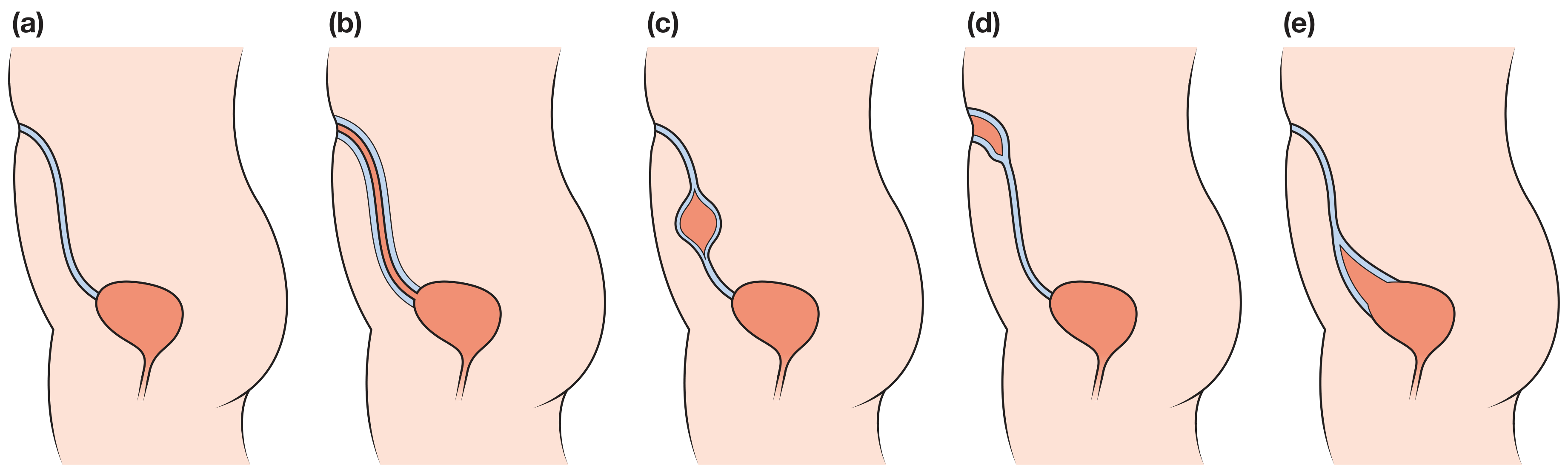
The urachus is a tube that exists in the fetus connecting the bladder to the belly button. It should fully close before birth. When the umbilical (top) end fails to close while the rest does, it forms a urachal sinus - a small blind-ended pouch at the navel that can collect fluid, get infected, and discharge through the umbilicus.
BLADDER ----[closed]---- [open pocket] ---- UMBILICUS (discharge here)
^ Urachal Sinus
Your Symptoms Match Perfectly
| Symptom/Finding | What It Indicates |
|---|---|
| 7x7mm hypoechoic heterogeneous nodule at umbilicus | The infected sinus/remnant |
| Discharge from belly button | Sinus tract draining through umbilicus |
| Pain when pressed | Infection/inflammation of the sinus |
| No intraperitoneal communication | Contained - has not spread into abdomen |
| Normal bladder on scan | Urachal connection to bladder is closed |
| Adult male | More commonly affected than females |
This presentation - umbilical discharge + periumbilical pain + hypoechoic nodule in an adult male - is described in published case reports as the classic presentation of an infected urachal sinus.
⚠️ This Requires Prompt Medical Attention
Why it cannot be ignored:
- The sinus is currently infected - bacteria can spread
- Without treatment, it can progress to a urachal abscess (a larger, more dangerous infection)
- Rarely, untreated urachal remnants can undergo malignant transformation (urachal carcinoma)
- Antibiotics alone are not sufficient - they treat the infection temporarily but the structural problem remains
What Treatment Is Needed
Step 1 - Immediate (Right Now)
- See a surgeon or urologist urgently - do not delay
- Antibiotics will be prescribed to control the current infection (commonly amoxicillin-clavulanic acid or a broad-spectrum antibiotic)
- The doctor may also perform a fistulogram (dye injection into the sinus) or order a CT scan to map the full extent of the tract
Step 2 - Definitive Treatment (Surgery)
- Surgical excision of the urachal remnant is the standard and only curative treatment
- This is done either:
- Laparoscopically (minimally invasive, preferred) - 3 small cuts, faster recovery
- Open surgery - if complicated or infected
- The entire urachal tract from umbilicus toward the bladder is removed to prevent recurrence
- Recovery is typically smooth with no recurrence after complete excision
What to Tell Your Doctor
"I have discharge and pain at my belly button. An ultrasound showed a 7x7mm hypoechoic heterogeneous nodule at the umbilicus with no intraperitoneal communication. All other findings - kidneys, bladder, and prostate - are normal."
Red Flags - Go to Emergency Department Immediately If:
- 🔴 Fever or chills develop
- 🔴 The area becomes very red, swollen, or warm (spreading cellulitis)
- 🔴 Discharge becomes foul-smelling or frankly purulent (pus)
- 🔴 Severe abdominal pain develops
- 🔴 You feel generally unwell or weak
Bottom line: This is almost certainly an infected urachal sinus - a congenital remnant that has become symptomatic. It is a structural problem that needs surgical removal. Please see a surgeon soon, as antibiotics alone will not cure this. The good news is that with proper treatment, the outcome is excellent and full recovery is expected.
Surgical management
Searching sources...
Finding Sources
Searching the Web
urachal sinus surgical management excision laparoscopic adult technique steps
Finding Sources
Finding Sources
Reading File
Reading File
Reading File
Reading File
Reading File
Loading Image
Here is the complete surgical management of an infected urachal sinus, sourced from Campbell-Walsh-Wein Urology and Bailey & Love's Surgery:
Surgical Management of Infected Urachal Sinus
Understanding the Anatomy First
Your condition is (d) in the diagram below - a urachal sinus: a blind-ending tract at the umbilicus that does NOT connect to the bladder:
Phase 1 - Pre-operative Management (Control the Infection First)
Before any surgery, the active infection must be treated:
| Step | Action |
|---|---|
| Antibiotics | Broad-spectrum IV antibiotics (e.g. amoxicillin-clavulanic acid, or piperacillin-tazobactam if severe) |
| Drainage | If an abscess has formed, incision and drainage (I&D) is done first as a temporizing measure |
| Imaging | CT scan of the abdomen/pelvis to map the full extent of the tract and rule out extension toward the bladder |
| Cystoscopy | May be performed to confirm the bladder end is closed (no vesical communication) |
| Wait | Surgery is deferred until the acute infection has settled - operating in infected tissue increases complication risk |
Note: Conservative drainage alone has a 30% recurrence rate. Definitive surgery is mandatory. - Campbell-Walsh-Wein Urology
Phase 2 - Definitive Surgery: Complete Surgical Excision
This is the only curative treatment. The goal is to remove the entire urachal remnant with no tissue left behind (to prevent recurrence and malignant transformation).
Option A - Laparoscopic Excision (Preferred in Adults)
Setup:
- Patient positioned supine, general anaesthesia
- Urinary catheter inserted to distend the bladder (aids identification)
- A small catheter or feeding tube may be placed into the sinus tract through the umbilicus for intraoperative identification
Port placement (standard 3-port technique):
[10mm Camera port] - midline, above umbilicus
[5mm working port] [10mm working port]
left upper quad right upper quad
Surgical steps:
- Peritoneal entry - the space of Retzius (pre-peritoneal space between peritoneum and abdominal wall fascia) is entered
- Identification - the urachal remnant is identified running from the umbilicus toward the bladder dome, flanked by the medial umbilical ligaments
- Umbilical dissection - the urachus is divided and circumscribed at the umbilicus, leaving the skin of the umbilicus intact for cosmesis
- Downward dissection - the remnant is dissected inferiorly toward the bladder extraperitoneally
- Bladder cuff excision - even if the sinus does not reach the bladder, a small cuff of bladder dome tissue is excised with the specimen to ensure complete removal
- Bladder closure - the bladder defect is closed in two watertight layers
- Specimen sent for histopathology - mandatory to exclude urachal carcinoma
- Catheter - left in overnight, removed next morning
Advantages of laparoscopic approach:
- Less pain, smaller scars
- Faster recovery (discharge in 1-2 days)
- Cosmetically superior (umbilicus preserved)
- Equivalent oncological clearance to open surgery
Option B - Open Surgery
Used when laparoscopy is not available, when previous surgeries make adhesions likely, or when the infection is complex.
Incision: Transverse or midline infraumbilical incision
Steps:
- Rectus fascia incised longitudinally, muscles spread apart
- Bladder distended with balloon catheter for identification
- Urachus isolated with vessel loop
- Extraperitoneal dissection from umbilicus down to bladder dome
- Bladder cuff excised with electrocautery
- Bladder closed in two layers
- Catheter left overnight
Option C - Robotic-Assisted Laparoscopy
- Available in advanced centres
- 4-port configuration
- Useful for obese patients or difficult anatomy
- Similar outcomes to standard laparoscopy
What Gets Removed
The specimen includes:
- The urachal sinus/remnant in its entirety
- The medial umbilical ligaments on both sides (which flank the urachus)
- A cuff of bladder dome (even if the bladder is not involved - to ensure complete excision)
- The umbilical skin may be removed in some cases (umbilectomy) if the sinus opens directly into it - though surgeons prefer to preserve the umbilicus for cosmesis when possible
Post-operative Care
| Detail | |
|---|---|
| Hospital stay | 1-2 days (laparoscopic), 3-5 days (open) |
| Catheter | Removed next day (laparoscopic) or Day 2-3 (open) |
| Antibiotics | Continued orally for 5-7 days post-op |
| Activity | Light activity in 1 week; full activity in 3-4 weeks |
| Follow-up | Histopathology review at 2 weeks |
| Recurrence | Near zero after complete excision |
Why Complete Excision Is Non-Negotiable
"Urachal anomalies have a small risk of malignant transformation to adenocarcinoma, with high mortality rates. Complete surgical excision is therefore recommended for both symptomatic and asymptomatic cases."
- Bailey & Love's Short Practice of Surgery, 28th Ed.
"It is important to remove all anomalous tissue. This avoids recurrences or stone formation and prevents the rare event of later transformation into a malignant adenocarcinoma."
- Campbell-Walsh-Wein Urology
Summary Flowchart
Infected Urachal Sinus
|
▼
IV Antibiotics + Drainage (if abscess)
|
▼
CT Abdomen/Pelvis + Cystoscopy
|
▼
Infection settled?
|
YES
|
▼
Laparoscopic Urachal Excision + Bladder Cuff
|
▼
Histopathology of specimen
|
▼
Full recovery - no recurrence expected
The prognosis after complete excision is excellent, and recurrence is extremely rare when the entire tract including the bladder cuff is removed.
This is a shared conversation. Sign in to Orris to start your own chat.