
Causes of chest congestion
Chest congestion refers to the buildup of mucus and fluid in the airways and lungs, making breathing feel heavy or blocked. Common causes include viral upper respiratory infections (the common cold, flu), allergies, asthma, bronchitis, and sinusitis. Less common causes include pneumonia, GERD, heart failure, and environmental irritants like smoke or pollution.
Searching sources...
Reading File
Searching the Web
causes of chest congestion 2025
Finding Sources
Finding Sources
Finding Sources
Finding Sources
Finding Sources
Finding Sources
Here is a structured breakdown of the causes of chest congestion, drawn from authoritative medical textbooks and clinical references:
Causes of Chest Congestion
Chest congestion is the subjective feeling of heaviness, pressure, or fullness in the chest due to excess or thickened mucus in the airways, airway inflammation, or fluid in/around the lungs. It spans a wide range of causes from mild infections to serious cardiac conditions.
1. Infections (Most Common)
Viral respiratory infections are the leading cause. These trigger airway inflammation, mucosal swelling, and excess mucus production.
- Common cold (rhinovirus, adenovirus) - causes nasal and upper chest congestion
- Influenza (flu) - more severe airway inflammation, often with productive cough
- COVID-19 - causes lower respiratory tract inflammation, mucus buildup, and potential progression to pneumonia
- RSV (Respiratory Syncytial Virus) - especially in infants and elderly; triggers acute airway inflammation, edema, bronchospasm, and mucus production, as noted in Tintinalli's Emergency Medicine
- Pneumonia (bacterial, viral, fungal) - consolidation of lung tissue with purulent secretions
- Acute bronchitis - viral infection of the bronchi causing cough and mucus buildup
- Bronchiolitis - viral (predominantly RSV) lower respiratory tract infection in children; causes edema, epithelial necrosis, bronchospasm, and increased mucus in the bronchioles
2. Obstructive Lung Diseases
- Asthma - allergens, pollutants, viruses, and bacteria activate airway inflammatory cells, triggering bronchospasm, mucus production, and airway hyperresponsiveness. Th2-mediated cytokine pathways drive mucus hypersecretion - Fishman's Pulmonary Diseases and Disorders
- COPD (Chronic Obstructive Pulmonary Disease) - airway inflammation, increased mucus production, and air trapping cause chronic congestion and dyspnea. Exacerbations worsen sputum production and purulence - Rosen's Emergency Medicine
- Chronic bronchitis - defined by productive cough for 3+ months per year for 2 consecutive years; excess mucus production is the hallmark
- Bronchiectasis - permanent bronchial dilation from chronic airway infection/inflammation; characterized by cough and copious sputum production - Sabiston Textbook of Surgery
3. Allergic & Environmental Causes
- Allergic rhinitis / hay fever - post-nasal drip leads to irritation and perceived chest congestion
- Allergen exposure - dust mites, mold, pet dander, pollen triggering airway inflammation
- Air pollution and irritants - smoke (cigarette, wildfire), chemical fumes, and particulates activate airway inflammatory cells and stimulate mucus production
- Occupational lung disease - prolonged exposure to dust, asbestos, or chemical vapors
4. Cardiac Causes
- Heart failure (left-sided) - elevated pulmonary venous pressure causes pulmonary edema and pleural effusion, producing a sensation of chest heaviness and breathlessness - Miller's Anesthesia; Goldman-Cecil Medicine
- Pulmonary edema - fluid accumulation in the alveoli (often due to acute cardiac decompensation) causes frothy sputum and severe chest congestion
5. Other Pulmonary Causes
- Pulmonary embolism - can present with pleuritic chest pain and dyspnea mimicking congestion
- Pleural effusion - fluid around the lungs (from infection, malignancy, or heart failure) causes pressure and heaviness
- Lung cancer - obstructs airways, causing mucus buildup distal to the tumor
6. Gastrointestinal Cause
- GERD (Gastroesophageal Reflux Disease) - chronic acid reflux irritates the larynx and lower airways, triggering mucus hypersecretion and a persistent cough that mimics chest congestion
7. Other / Less Common Causes
- Cystic fibrosis - genetic defect in CFTR leads to abnormally thick, sticky mucus that clogs airways
- Primary ciliary dyskinesia - impaired mucociliary clearance leads to chronic mucus retention
- Sinusitis - post-nasal drip from sinus infections can irritate the lower airways
When to Seek Medical Care
Seek prompt evaluation if chest congestion is accompanied by:
- High fever or chills
- Coughing up blood or dark/rust-colored sputum
- Chest pain
- Shortness of breath at rest
- Symptoms lasting more than 2-3 weeks without improvement
Sources: Fishman's Pulmonary Diseases and Disorders, Rosen's Emergency Medicine, Tintinalli's Emergency Medicine, Sabiston Textbook of Surgery, Goldman-Cecil Medicine 2025, Murray & Nadel's Textbook of Respiratory Medicine; Cleveland Clinic, Everyday Health
ExplainRESPIRATORYSYSTEM
The respiratory system is responsible for gas exchange - bringing oxygen into the body and expelling carbon dioxide. It consists of the upper airway (nose, pharynx, larynx), lower airway (trachea, bronchi, bronchioles), and the lungs (alveoli). Breathing is driven by the diaphragm and intercostal muscles, and regulated by the brainstem.
Searching sources...
Reading File
This is a broad anatomical/physiological topic. I'll run parallel searches across the library for anatomy, physiology, and mechanics of breathing.
Finding Sources
Finding Sources
Finding Sources
Reading File
Finding Sources
Finding Sources
Reading File
Finding Sources
Finding Sources
Reading File
Reading File
Now I have rich textbook content. Let me fetch the airway structure image and the gas exchange diagram to include visuals.
Loading Image
Loading Image
Loading Image
Excellent - I have two strong diagrams and rich textbook content. Now let me compile the full, detailed explanation.
The Respiratory System
The respiratory system's core job is gas exchange - delivering oxygen (O₂) from the atmosphere to every cell in the body and expelling carbon dioxide (CO₂) produced by cellular metabolism. It also warms and humidifies inhaled air, filters particles, and plays a role in pH regulation, vocalization, and immune defense.
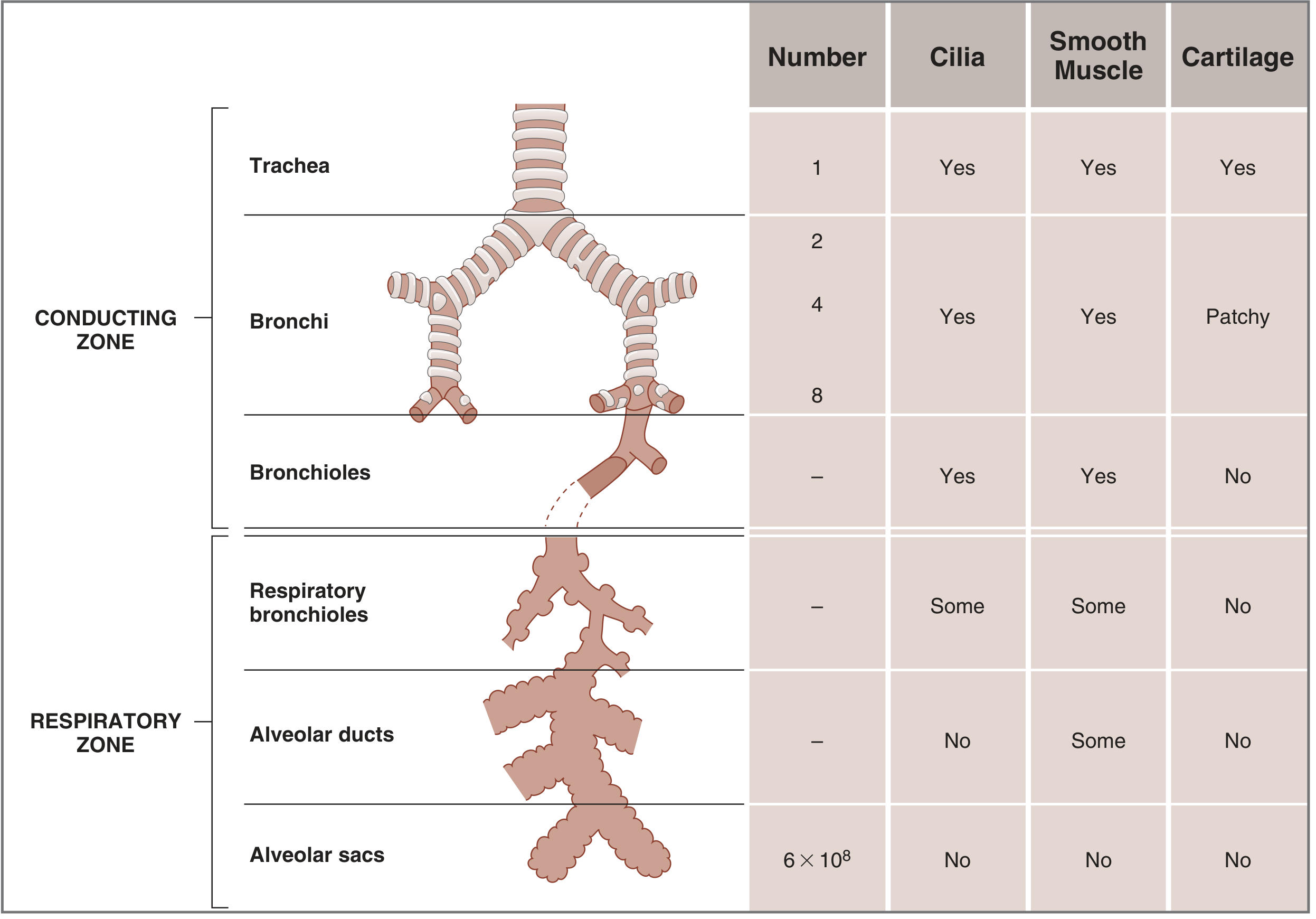
1. Overview: Two Major Zones
The entire airway tree is divided into two functional zones:
| Zone | Structures | Function |
|---|---|---|
| Conducting zone | Nose → Pharynx → Larynx → Trachea → Bronchi → Bronchioles | Air conduction, warming, humidification, filtration |
| Respiratory zone | Respiratory bronchioles → Alveolar ducts → Alveolar sacs → Alveoli | Gas exchange (O₂/CO₂) |
The trachea is "generation 0." By the time the airways branch 23 times, they culminate in ~600 million alveolar sacs. - Costanzo Physiology, 7th Ed.
2. Structural Components
A. Upper Airway
- Nose & Nasal cavity - filters large particles (hairs, turbinates), warms and humidifies inspired air
- Pharynx - common pathway for air and food; divided into nasopharynx, oropharynx, and laryngopharynx
- Larynx - contains the vocal cords; acts as a valve protecting the lower airway during swallowing; site of phonation
B. Trachea
- Begins at the lower border of the cricoid cartilage and extends 10-13 cm to the carina
- Held open by C-shaped cartilaginous rings (anteriorly and laterally); the posterior membranous wall is flexible
- The cricoid is the narrowest part: ~17 mm in men, ~13 mm in women
- At the carina, it bifurcates into right and left mainstem bronchi. The right bronchus is more vertical (foreign bodies tend to lodge here). - Morgan & Mikhail's Clinical Anesthesiology, 7th Ed.
C. Bronchi and Bronchioles (Conducting Zone)
- Airways branch progressively (generations 1-16), getting narrower with each division
- Lined with ciliated and mucus-secreting cells - cilia beat rhythmically upward to sweep inhaled particles out (mucociliary escalator)
- Walls contain smooth muscle with dual autonomic control:
- Sympathetic (β₂ receptors) → bronchodilation (e.g., albuterol works here)
- Parasympathetic (muscarinic receptors) → bronchoconstriction
- Bronchi have cartilage; bronchioles (generation 17+) do not - they rely entirely on smooth muscle tone and elastic recoil to stay open. - Costanzo Physiology, 7th Ed.
D. Respiratory Zone (Alveoli)
- Respiratory bronchioles are transitional - they have some alveoli budding off their walls
- Alveolar ducts are entirely lined with alveoli; alveolar sacs terminate the tree
- Each lung contains ~300 million alveoli; each alveolus is ~200 µm in diameter
- The alveolar wall is extremely thin - maximizing surface area (~70 m², the size of a tennis court) and minimizing diffusion distance for rapid O₂/CO₂ exchange - Costanzo Physiology, 7th Ed.
3. Gas Exchange - How It Works
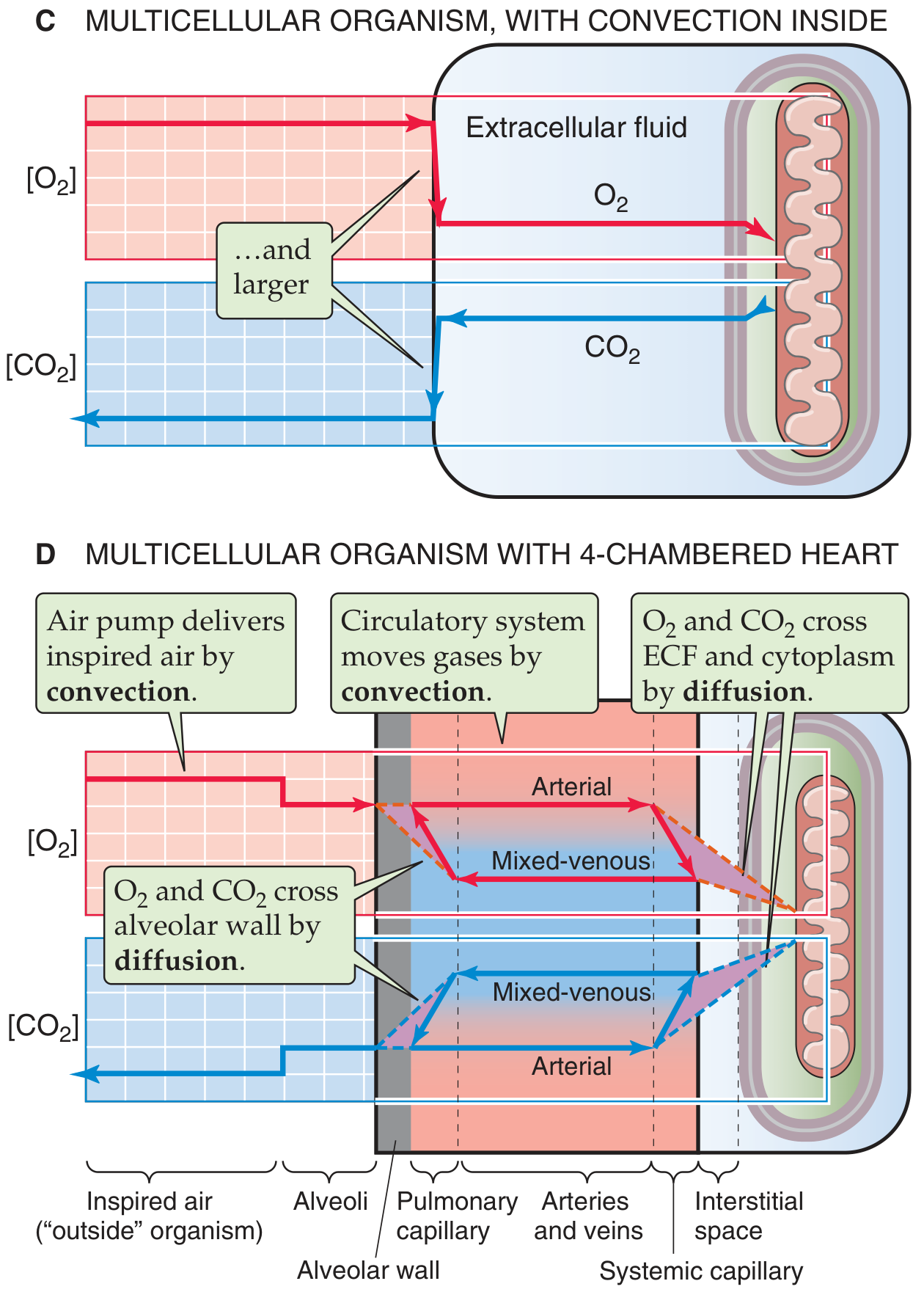
Gas exchange occurs in two steps:
- Convection - the respiratory muscles pump air into the alveoli; the heart pumps blood through the pulmonary capillaries
- Diffusion - O₂ and CO₂ cross the thin alveolar wall down their partial pressure gradients
- O₂ moves from alveoli → pulmonary capillary blood (alveolar PO₂ ~100 mmHg; venous PO₂ ~40 mmHg)
- CO₂ moves from blood → alveoli (venous PCO₂ ~46 mmHg; alveolar PCO₂ ~40 mmHg)
Oxygenated blood then travels via pulmonary veins → left heart → systemic circulation → delivers O₂ to tissues. - Medical Physiology (Boron & Boulpaep)
4. Mechanics of Breathing
Inspiration (active)
- The diaphragm is the primary muscle of inspiration, accounting for ~70-75% of tidal volume. It is innervated by the phrenic nerve (C3-C5).
- Diaphragm contraction pulls the floor of the thorax downward, expanding lung volume and dropping intrapulmonary pressure below atmospheric → air flows in
- The external intercostal muscles lift the ribs outward (bucket-handle motion), further expanding the chest
- During heavy breathing/exercise, accessory muscles are recruited: sternocleidomastoid, scalene, and pectoralis muscles - Murray & Nadel's Textbook of Respiratory Medicine; Morgan & Mikhail's Clinical Anesthesiology
Expiration (passive at rest)
- At rest, expiration is passive - the elastic recoil of the lungs and chest wall drives air out
- During exercise or forced breathing, abdominal muscles (rectus abdominis, obliques, transversus) and internal intercostals actively compress the thorax - Morgan & Mikhail's Clinical Anesthesiology, 7th Ed.
5. Control of Breathing
The ventilatory system is controlled by a complex neural network:
| Controller | Location | Role |
|---|---|---|
| Voluntary breathing | Cerebral cortex (parietal) | Conscious control (speech, breath-holding) |
| Automatic breathing | Pons + Medulla (brainstem) | Rhythm generation, baseline drive to breathe |
| Spinal cord | Cervical & thoracic cord | Relays signals from brain to respiratory muscles |
| Phrenic nerve | C3-C5 | Drives the diaphragm |
Feedback regulation - the brainstem's respiratory centers respond to:
- Central chemoreceptors (medulla) - detect rising CO₂ / falling pH in cerebrospinal fluid → stimulate breathing
- Peripheral chemoreceptors (carotid and aortic bodies) - respond to falling O₂, rising CO₂, and acidosis
- Stretch receptors in the lungs - prevent over-inflation (Hering-Breuer reflex)
The system can precisely maintain CO₂ levels and acid-base balance despite large swings in metabolic demand (e.g., during vigorous exercise). - Murray & Nadel's Textbook of Respiratory Medicine, 2-Volume Set
6. Additional Functions
Beyond gas exchange, the respiratory system serves several other roles:
- pH regulation - adjusting ventilation rate changes CO₂ levels, directly affecting blood pH
- Vocalization - airflow across the vocal cords in the larynx produces sound
- Immune defense - cilia + mucus trap particles; alveolar macrophages phagocytose pathogens
- Filtration - nasal hairs, turbulence in the nose, and the mucociliary escalator remove particles before they reach the alveoli
- Olfaction - nasal mucosa contains olfactory receptors
Summary Diagram (Airway Tree)
| Structure | Generation | Cilia | Cartilage | Gas Exchange |
|---|---|---|---|---|
| Trachea | 0 | Yes | Yes | No |
| Main bronchi | 1 | Yes | Yes | No |
| Lobar/segmental bronchi | 2-4 | Yes | Patchy | No |
| Bronchioles | 5-16 | Yes | No | No |
| Respiratory bronchioles | 17-19 | Some | No | Partial |
| Alveolar ducts | 20-22 | No | No | Yes |
| Alveolar sacs/Alveoli | 23 | No | No | Yes |
Sources: Costanzo Physiology 7th Ed., Murray & Nadel's Textbook of Respiratory Medicine, Morgan & Mikhail's Clinical Anesthesiology 7th Ed., Medical Physiology (Boron & Boulpaep), Miller's Anesthesia 10th Ed.
This is a shared conversation. Sign in to Orris to start your own chat.