I now have strong foundational content from Bailey & Love's Surgery and Schwartz's Principles. Let me now compile comprehensive answers to all the questions in the PDF.Here is a complete, exam-ready answer guide for all the major questions from the BPT/BOT Paper XII - General Surgery past year papers (2015-2025):
BPT/BOT Paper XII - General Surgery: Past Year Questions & Answers (2015-2025)
QUESTION 1: Define and Classify Shock. Discuss Management of Hemorrhagic/Hypovolemic Shock
(Repeated every year - highest priority)
Definition
Shock is a state of inadequate tissue perfusion resulting in decreased delivery of oxygen and metabolic substrates to cells, with failure to remove cellular waste products. It represents a failure of oxidative metabolism.
Classification of Shock (Table)
| Type | Cause | Example |
|---|
| Hypovolemic | Loss of circulating volume | Hemorrhage, burns, dehydration |
| Cardiogenic | Pump failure | MI, cardiac tamponade, arrythmia |
| Septic (Distributive) | Vasodilation, maldistribution | Septicemia, anaphylaxis |
| Neurogenic | Loss of vasomotor tone | Spinal cord injury |
| Obstructive | Obstruction to blood flow | Pulmonary embolism, tension pneumothorax |
(Schwartz's Principles of Surgery, Table 5-1)
Classes of Hemorrhagic Shock (ATLS Classification)
| Class | Blood Loss | % Blood Volume | HR | BP | Consciousness |
|---|
| Class I | <750 mL | <15% | <100 | Normal | Normal/anxious |
| Class II | 750-1500 mL | 15-30% | 100-120 | Normal | Anxious |
| Class III | 1500-2000 mL | 30-40% | 120-140 | Decreased | Confused |
| Class IV | >2000 mL | >40% | >140 | Very low | Lethargic/unconscious |
For a patient with >30% blood loss (Class III-IV):
- This is a life-threatening emergency.
Management of Hemorrhagic Shock - "ABCDE" Approach
A - Airway: Secure airway with cervical spine protection. Intubate if GCS < 8.
B - Breathing: High-flow O2. Treat tension pneumothorax immediately.
C - Circulation/Hemorrhage Control:
- Direct pressure on external wounds
- IV access - two large-bore (16G) peripheral IVs
- Fluid resuscitation:
- Initial bolus: 1-2L warm crystalloid (Ringer's Lactate preferred over NS)
- Damage Control Resuscitation: Blood products in 1:1:1 ratio (PRBC : FFP : Platelets)
- Permissive hypotension - target SBP 80-90 mmHg until surgical hemorrhage control (not in TBI)
- Avoid over-resuscitation with crystalloid (causes ARDS, coagulopathy)
- Transfusion trigger: Hb <7 g/dL (or <10 in cardiac patients/elderly)
- Surgical hemorrhage control - source control is paramount
D - Disability: Neurological assessment (AVPU/GCS)
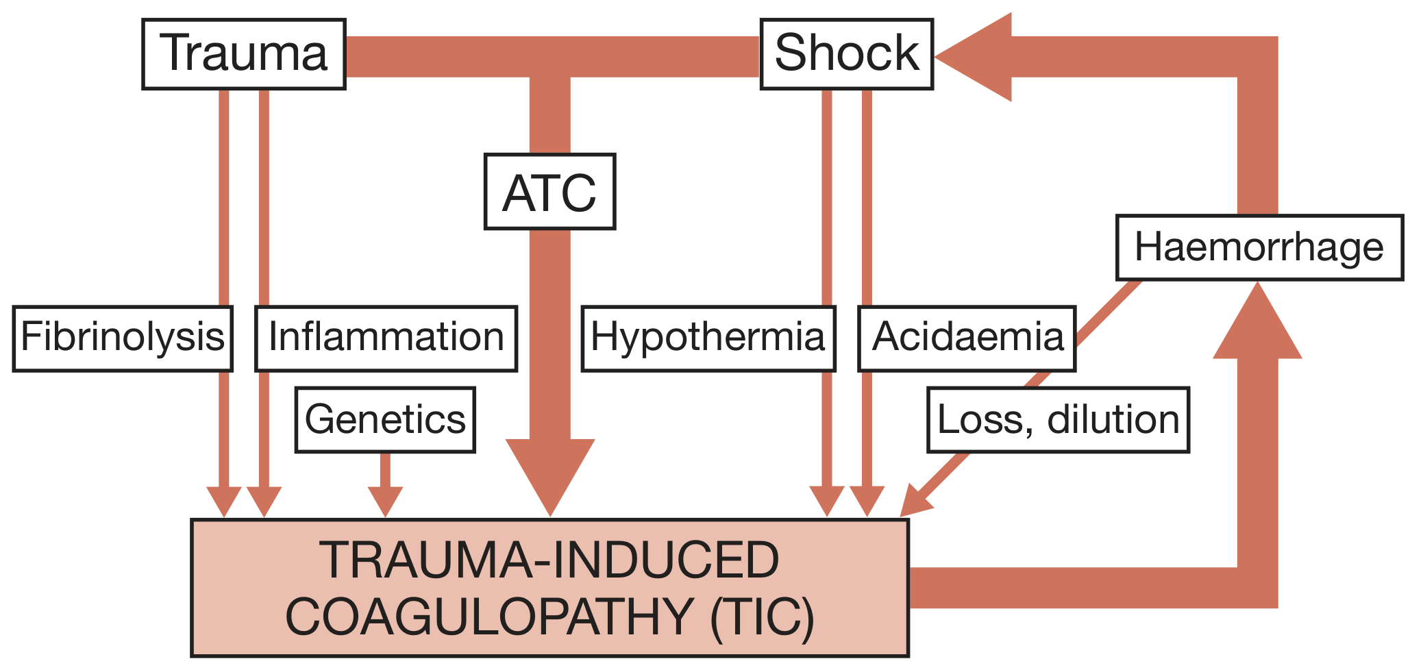
E - Exposure: Full exposure, prevent hypothermia (triad of death: hypothermia + coagulopathy + acidosis)
Monitoring response: HR, BP, urine output (>0.5 mL/kg/hr), lactate clearance, base deficit.
QUESTION 2: Factors Affecting Wound Healing & Management of Chronic Ulcers
Wound Healing Types
- Primary intention: Wound edges reapproximated soon after injury (sutured/stapled)
- Secondary intention: Wound left open, heals from base by granulation
- Tertiary intention (Delayed primary): Wound cleaned, then closed after 4-5 days
Phases of Wound Healing
- Haemostasis (immediate): Platelet plug, clot formation, vasoconstriction
- Inflammatory phase (0-5 days): Neutrophils then macrophages clean debris; growth factor release
- Proliferative phase (5 days-3 weeks): Fibroblasts deposit collagen, angiogenesis, re-epithelialization
- Remodeling phase (3 weeks-2 years): Collagen remodeling; wound reaches ~80% tensile strength
Factors Affecting Wound Healing
Local Factors:
- Wound infection (most common cause of delayed healing)
- Poor blood supply / ischemia
- Foreign body / necrotic tissue
- Wound tension
- Wound size and depth
- Previous irradiation
Systemic Factors:
- Malnutrition (protein, Vit C, Zinc deficiency)
- Diabetes mellitus (impairs neutrophil function, microangiopathy)
- Steroids / immunosuppressants
- Chemotherapy
- Advanced age
- Anemia / hypovolemia
- Malignancy
Management of Chronic Ulcers
Chronic ulcers fail to heal within 3 months. Causes include venous insufficiency, arterial disease, diabetic neuropathy, pressure.
General Principles (TIME Framework):
- T - Tissue debridement (remove necrotic/sloughy tissue - surgical, enzymatic, or larval therapy)
- I - Infection control (swab, treat with topical/systemic antibiotics; silver dressings)
- M - Moisture balance (appropriate dressings - hydrocolloid, foam, alginate)
- E - Edge advancement (stimulate epithelial migration; skin grafting if needed)
Type-specific management:
- Venous ulcers: Four-layer compression bandaging, leg elevation, treat venous reflux
- Arterial ulcers: Revascularization (angioplasty or bypass), avoid compression
- Diabetic ulcers: Glycemic control, offloading, debridement, check for osteomyelitis
- Pressure ulcers: Pressure relief, nutritional support, wound care
Complications of chronic wounds: Marjolin's ulcer - rare but aggressive squamous cell carcinoma arising in chronic wounds; requires biopsy if phenotypic change occurs.
QUESTION 3(a): Complications of Blood Transfusion
Immediate (within 24 hours):
| Complication | Mechanism |
|---|
| Acute hemolytic reaction | ABO incompatibility; most dangerous - rigors, fever, backache, hemoglobinuria, renal failure |
| Febrile non-hemolytic reaction | Antibodies against WBC antigens; fever/chills without hemolysis |
| Allergic reaction | Antibodies to plasma proteins; urticaria to anaphylaxis |
| TRALI (Transfusion Related Acute Lung Injury) | Donor antibodies vs. recipient neutrophils; non-cardiogenic pulmonary oedema within 6 hours |
| Transfusion-associated circulatory overload (TACO) | Fluid overload; pulmonary oedema |
| Septic reaction | Bacterial contamination of blood product |
Delayed:
- Delayed hemolytic reaction (1-2 weeks): Antibodies to minor blood group antigens (Kell, Duffy)
- Post-transfusion purpura: Thrombocytopenia 5-10 days post-transfusion
- Graft-versus-host disease (GVHD): Donor T-lymphocytes attack immunocompromised recipient
- Alloimmunization: Formation of new antibodies
- Iron overload: In chronically transfused patients
Massive Transfusion Complications:
- Hypothermia (transfuse warmed blood)
- Hypocalcemia (citrate chelates calcium)
- Hyperkalemia
- Coagulopathy (dilutional)
- Metabolic acidosis
Infectious complications: HIV, Hepatitis B/C, CMV, syphilis (screened but residual risk)
QUESTION 3(b): Surgical Site Infections (SSI)
Definition
Infection occurring within 30 days of surgery (or 90 days if implant placed) at or near the surgical incision.
Classification:
- Superficial incisional SSI: Involves skin and subcutaneous tissue only
- Deep incisional SSI: Involves deep soft tissue (fascia and muscle)
- Organ/space SSI: Involves any organ or space opened/manipulated during surgery
Common Organisms:
- S. aureus (most common), S. epidermidis, Gram-negative rods (E. coli, Pseudomonas), Enterococcus
Risk Factors:
- Wound classification (contaminated/dirty wounds have highest risk)
- Prolonged operating time
- Diabetes, obesity, malnutrition
- Immunosuppression
- Pre-existing infection
- Emergency surgery
- Poor surgical technique (dead space, haematoma)
Prevention:
- Pre-operative hair removal (clipping, not shaving)
- Antiseptic skin preparation (chlorhexidine + alcohol)
- Prophylactic antibiotics within 60 min of incision, stopped within 24 hours
- Maintain normothermia and normoglycemia intraoperatively
- Aseptic technique, minimize dead space
Treatment:
- Superficial: Open wound, culture, antibiotics if spreading cellulitis
- Deep: Surgical debridement + antibiotics
- Vacuum-assisted wound closure (VAC) for large wounds
QUESTION 3(c): Tension Pneumothorax
Definition
Air accumulates in the pleural space under pressure through a one-way valve mechanism, causing mediastinal shift, compression of the opposite lung, and kinking of great vessels - a life-threatening emergency.
Causes:
- Blunt or penetrating chest trauma
- Iatrogenic (central line insertion, positive pressure ventilation)
- Spontaneous (in ventilated patients especially)
Clinical Features ("5 Ds"):
- Dyspnoea (severe)
- Distended neck veins (JVD due to impaired venous return)
- Decreased breath sounds on affected side
- Deviation of trachea away from affected side
- Dullness to hyperresonance on percussion of affected side
- Hypotension, tachycardia, cyanosis, cardiovascular collapse
Diagnosis:
- Clinical diagnosis - do NOT wait for X-ray in emergency
- CXR (if stable): Absent lung markings, mediastinal shift, depressed diaphragm on affected side
Treatment - Immediate:
- Needle decompression (first): 14G needle into 2nd intercostal space, mid-clavicular line on affected side - converts to open pneumothorax, relieves pressure
- Definitive: Chest drain (tube thoracostomy) in 5th intercostal space, anterior axillary line
QUESTION 3(d): Fluid Replacement in Burns
Initial Assessment - Wallace Rule of Nines:
| Area | %TBSA |
|---|
| Head & neck | 9% |
| Each arm | 9% |
| Anterior trunk | 18% |
| Posterior trunk | 18% |
| Each leg | 18% |
| Perineum | 1% |
(In children - Lund & Browder chart is more accurate)
Resuscitation Formula:
Parkland Formula (most widely used):
4 mL × Weight (kg) × %TBSA burned = Total fluid in first 24 hours
- Use Ringer's Lactate
- First half in first 8 hours from time of burn (not from admission)
- Second half over next 16 hours
Example: 70 kg patient with 40% TBSA burn:
- 4 × 70 × 40 = 11,200 mL in 24 hours
- First 8 hours: 5,600 mL
- Next 16 hours: 5,600 mL
Note: Only burns >20% TBSA (or >10% in children/elderly) need IV resuscitation. Superficial burns (<20% TBSA) managed with oral rehydration.
Monitor adequacy of resuscitation:
- Urine output: 0.5-1 mL/kg/hr in adults, 1 mL/kg/hr in children
- Avoid both under-resuscitation (acute renal failure) and over-resuscitation (oedema, abdominal compartment syndrome)
QUESTION 4(a): Classification and Management of Burns
Classification by Depth:
| Degree | Layer Involved | Appearance | Pain | Healing |
|---|
| Superficial (1st degree) | Epidermis only | Erythema, dry | Painful | 3-5 days |
| Superficial partial thickness (2nd degree) | Epidermis + superficial dermis | Blisters, wet, pink | Very painful | 14-21 days |
| Deep partial thickness | Epidermis + deep dermis | White/red, mottled, less wet | Less painful | >21 days, may need grafting |
| Full thickness (3rd degree) | All layers | Leathery, white/brown/black, dry | Painless (nerve destruction) | Needs grafting |
| 4th degree | Bone/tendon/muscle | Charred | Painless | Major reconstruction |
Burns in Special Areas requiring special attention:
Face, hands, feet, genitalia, perineum, major joints, circumferential burns (escharotomy needed).
Management of Major Burns:
Initial (First Aid):
- Stop burning process (remove clothing/chemicals, cool with 15-20°C running water for 20 min)
- Do NOT use ice (causes vasoconstriction and deeper injury)
Primary Survey (ABCDE):
- Airway - highest priority; inhalation injury present if: singed nasal hair, hoarse voice, carbonaceous sputum, facial burns; early intubation before oedema occludes airway
- Breathing - 100% O2 (for CO poisoning)
- Circulation - IV access (2 large bore), start Parkland formula
- Disability - GCS
- Exposure - calculate %TBSA, keep warm
Burn Wound Care:
- Superficial: Dressings (silver sulfadiazine, Mepitel, Aquacel Ag)
- Deep/Full thickness: Early excision and skin grafting within 3-5 days reduces infection and mortality
- Escharotomy for circumferential full-thickness burns (compartment syndrome)
Systemic Management:
- Nasogastric tube (ileus common) + early enteral nutrition
- Foley catheter for urine output monitoring
- Analgesia (IV morphine/ketamine)
- DVT prophylaxis
- Tetanus prophylaxis
- H2 blockers (prevent Curling's ulcer - stress ulcer of duodenum)
QUESTION 4(b): Advantages & Disadvantages of Laparoscopic and Robotic Surgery
Laparoscopic Surgery:
Advantages:
- Smaller incisions - reduced wound complications
- Less postoperative pain - less analgesic requirement
- Shorter hospital stay and faster return to work
- Reduced blood loss
- Less adhesion formation
- Better cosmesis
- Magnified view improves visualization
Disadvantages:
- Loss of tactile feedback (haptics)
- Two-dimensional view (traditional laparoscopy)
- Long learning curve
- Expensive equipment, specialized training needed
- Risk of trocar injuries (bowel, vessels)
- CO2 pneumoperitoneum risks: hypercarbia, deep vein thrombosis, gas embolism
- Limited range of motion of instruments
Robotic Surgery (e.g., da Vinci system):
Advantages over open and laparoscopy:
- Three-dimensional, high-definition visualization
- Wristed instruments - 7 degrees of freedom (superior to laparoscopy's 4)
- Tremor filtration (improves precision)
- Better ergonomics for surgeon
- Useful in confined spaces (prostate, deep pelvis, mediastinum)
- Reduced blood loss, shorter catheterization time (e.g., prostatectomy)
- Shorter hospital stay
Disadvantages:
- No tactile feedback (even more than laparoscopy)
- Very high cost (equipment + maintenance)
- Large size of robot
- Long setup/docking time
- Requires highly specialized training
- Not suitable for emergencies requiring rapid conversion
QUESTION 5: Signs & Symptoms in Each Trimester of Pregnancy
(Note: This is included in the general surgery paper as obstetric surgical content)
First Trimester (0-12 weeks):
- Amenorrhea (missed period - first sign)
- Nausea/morning sickness (hCG peak)
- Breast tenderness and enlargement
- Urinary frequency (uterus compresses bladder)
- Fatigue, dizziness
- Chadwick's sign: Bluish discolouration of vulva and vagina
- Goodell's sign: Softening of cervix
- Hegar's sign: Softening of lower uterine segment
Second Trimester (13-26 weeks):
- Quickening (first fetal movements felt, ~18-20 weeks in primi, ~16-18 in multi)
- Abdominal enlargement becomes obvious
- Disappearance of nausea
- Increased appetite
- Braxton-Hicks contractions begin (painless, irregular)
- Fundal height rises (umbilicus level at ~20 weeks)
- Pigmentation (linea nigra, chloasma/mask of pregnancy)
- Striae gravidarum
Third Trimester (27-40 weeks):
- Progressive abdominal distension
- Dependent oedema (legs, ankles)
- Dyspnoea (diaphragm pushed up)
- Heartburn (relaxed lower oesophageal sphincter)
- Urinary frequency returns (fetal head descends, lightening)
- Braxton-Hicks become more frequent
- Backache (relaxin + shifted centre of gravity)
- Engagement of fetal head (~36 weeks in primigravida)
QUESTION 6(a): Types of Hearing Aids and Their Indications
Types of Hearing Aids:
| Type | Description | Indication |
|---|
| Behind-the-ear (BTE) | Device sits behind ear, connected by tubing to ear mould | Most common; suitable for all ages and all degrees of hearing loss |
| In-the-ear (ITE) | Custom-fit, fills outer ear | Mild to severe HL; cosmetically preferred |
| In-the-canal (ITC) | Smaller, fits partially in canal | Mild to moderate HL |
| Completely-in-canal (CIC) | Nearly invisible, deep in canal | Mild to moderate HL; best cosmesis; not for severe HL |
| Receiver-in-canal (RIC/RITE) | Speaker sits in canal, processor behind ear | Good sound quality; mild to severe HL |
| Bone-anchored hearing aid (BAHA) | Surgically implanted titanium implant; bypasses outer/middle ear | Conductive HL, mixed HL, single-sided deafness |
| Cochlear Implant | Electronic device surgically implanted; stimulates cochlear nerve | Severe to profound sensorineural HL where hearing aids inadequate |
General Indications:
- Conductive hearing loss (treatable cause should be managed first)
- Sensorineural hearing loss (bilateral ≥25-40 dB loss)
- Mixed hearing loss
- Age-related hearing loss (presbycusis)
- Functional hearing impairment affecting daily communication
QUESTION 6(b): Common Inflammation of the Eye (Conjunctivitis)
Conjunctivitis - Definition & Types:
Inflammation of the conjunctiva. Most common eye condition worldwide.
| Type | Cause | Features | Treatment |
|---|
| Bacterial | S. aureus, H. influenzae, Pneumococcus | Purulent discharge, sticky eyelids in morning, conjunctival hyperaemia | Topical antibiotics (ciprofloxacin, chloramphenicol) |
| Viral | Adenovirus (most common), HSV | Watery discharge, follicles, preauricular lymph node, highly contagious | Self-limiting; lubricating drops; antiviral for HSV |
| Allergic | Atopy, dust, pollen | Bilateral, itching, chemosis, watery discharge | Antihistamine drops, mast cell stabilizers, avoid allergen |
| Gonococcal | N. gonorrhoeae | Hyperacute - copious purulent discharge, risk of corneal perforation | IV/IM ceftriaxone (systemic), irrigation |
| Neonatal (Ophthalmia neonatorum) | Chlamydia, Gonococcus | Neonatal; notifiable disease | Specific antibiotics |
| Chlamydial (Trachoma) | Chlamydia trachomatis | Follicular conjunctivitis, leading infectious cause of blindness | Oral/topical azithromycin |
QUESTION: Degloving Injury & Skin Grafts
Degloving Injury:
Traumatic separation of skin and subcutaneous tissue from underlying fascia, creating a large tissue defect. Caused by road traffic accidents (shearing force, e.g., tyre running over a limb).
Management:
- Wound assessment, debridement of devitalized tissue
- Replace avulsed skin as full-thickness graft if viable
- Vacuum-assisted closure (VAC) as temporary coverage
- Split-skin grafting for large areas
Types of Skin Grafts:
| Feature | Split-Skin Graft (SSG) | Full-Thickness Graft (FTG) |
|---|
| Layers taken | Epidermis + part of dermis | Epidermis + entire dermis |
| Donor site | Heals by re-epithelialization (reusable) | Must be primarily sutured |
| Take (graft survival) | Better (thinner, easier diffusion) | Poorer (requires better vascularity) |
| Contracture | More secondary contracture | Less contracture |
| Cosmetic result | Poorer (shiny, mismatch) | Better colour and texture match |
| Use | Large areas, infected/contaminated wounds, donor site dressing | Small areas, face, hands, eyelids, cosmetically important sites |
| Harvesting tool | Dermatome (Watson, Humby) | Scalpel, freehand |
QUESTION: Classify and Manage Hernia (Inguinal Hernia)
Classification of Hernia:
By nature:
- Reducible: Contents return to abdomen spontaneously or manually
- Irreducible (Incarcerated): Cannot be reduced back
- Obstructed: Bowel in sac is obstructed (no blood supply compromise yet)
- Strangulated: Blood supply cut off - surgical emergency
- Sliding: Part of sac is formed by viscus (e.g., caecum, bladder)
By location: Inguinal, femoral, umbilical, incisional, epigastric, Spigelian, obturator
By direction (inguinal hernia):
| Feature | Direct | Indirect |
|---|
| Passes through | Hesselbach's triangle | Deep inguinal ring |
| Relation to IEA | Medial | Lateral |
| Age | Older adults (acquired) | Young males (congenital) |
| Cause | Weak transversalis fascia | Patent processus vaginalis |
| Descent into scrotum | Rare | Common |
| Strangulation risk | Lower | Higher |
Management of Inguinal Hernia:
Surgical options:
- Tension repair (Bassini, Shouldice): No mesh; primary tissue approximation; higher recurrence
- Mesh repair (Lichtenstein - tension-free): Gold standard for open repair; low recurrence (<1%)
- Laparoscopic repair (TEP - Total Extraperitoneal or TAPP - Transabdominal Preperitoneal): Less postoperative pain, faster return to work; preferred for bilateral/recurrent hernias
Emergency (strangulated hernia): Resuscitation + urgent surgery; bowel resection if non-viable.
QUESTION: Uterine Prolapse - Classification, Clinical Features & Management
Classification (POP-Q / Baden-Walker / Degrees):
| Degree | Description |
|---|
| 1st degree | Cervix descends into vagina but not to introitus |
| 2nd degree | Cervix reaches introitus |
| 3rd degree (Procidentia) | Cervix and uterus outside introitus |
| 4th degree | Complete eversion of uterus |
Types of associated prolapse: Cystocele (bladder), Rectocele (rectum), Enterocele (small bowel)
Clinical Features:
- Feeling of something coming down PV
- Dragging/heaviness in pelvis
- Urinary symptoms: stress incontinence, frequency, difficulty voiding
- Difficulty with defecation
- Backache (lumbar, relieved by lying down)
- Superficial dyspareunia or inability to have intercourse
- Decubitus ulcer on prolapsed cervix (if chronic)
Management:
Conservative (Non-surgical):
- Pelvic floor exercises (Kegel exercises) - mild cases
- Pessary (Ring pessary): Mechanical support; for elderly/medically unfit, postpartum, or as temporary measure
- Oestrogen cream (postmenopausal) - improves tissue tone
- Weight reduction, treat chronic cough/constipation
Surgical:
- Fothergill/Manchester repair: For 2nd degree prolapse with long cervix; amputation of cervix + anterior colporrhaphy
- Vaginal hysterectomy + pelvic floor repair: Definitive; for completed family
- Sacrocolpopexy (abdominal/laparoscopic): Mesh fixation for vault prolapse after hysterectomy
- Le Fort's operation: Obliterative surgery for elderly frail women who don't want/can't have intercourse
QUESTION: Abdominal Incisions and Incisional Hernia
Types of Abdominal Incisions:
| Incision | Direction/Location | Use |
|---|
| Midline (median) | Vertical, through linea alba | Most common; upper/lower/full abdomen; quick, extensile |
| Paramedian | Vertical, 2.5 cm lateral to midline | Historically used; less common today |
| Kocher's (subcostal) | Oblique below right costal margin | Hepatobiliary, cholecystectomy |
| Lanz | Horizontal in RIF (McBurney's point) | Appendicectomy; best cosmesis |
| McBurney | Oblique in RIF | Appendicectomy; classic |
| Pfannenstiel | Transverse suprapubic | Gynaecological, obstetric (LSCS), bladder |
| Roof-top (bilateral subcostal) | Chevron | Liver transplant, stomach, spleen |
| Rutherford-Morrison | Right iliac fossa, oblique | Renal transplant |
| Thoracoabdominal | Chest + abdomen | Oesophagogastric junction tumours |
Criteria for ideal incision:
- Adequate exposure of operative field
- Extensile if needed
- Minimal damage to nerves, muscles, blood vessels
- Secure closure without risk of dehiscence
- Good cosmetic result
- Heals with minimal hernia risk
Incisional Hernia:
Hernia through a previous surgical scar.
Risk factors: Obesity, wound infection, poor nutrition, steroid use, diabetes, chronic cough, constipation, urinary obstruction, poor surgical technique (wrong suture, inadequate closure).
Management:
- Small/asymptomatic: Conservative (truss)
- Surgical: Mesh repair (onlay, sublay/retromuscular, IPOM - laparoscopic intraperitoneal onlay mesh); primary suture repair has 30-50% recurrence vs. <10% with mesh
QUESTION: Types of Anaesthesia and Their Complications
Classification:
1. General Anaesthesia (GA):
- Patient unconscious; loss of consciousness, analgesia, muscle relaxation
- Stages (Guedel's): Analgesia → Excitement/Delirium → Surgical anaesthesia → Respiratory paralysis
- Components (Triad): Hypnosis + Analgesia + Muscle relaxation
- Agents: Inhalational (sevoflurane, isoflurane), IV (propofol, ketamine, thiopentone)
- Complications: Airway obstruction, aspiration (Mendelson syndrome), laryngospasm, malignant hyperthermia (rare but life-threatening with suxamethonium/volatile agents), post-op nausea/vomiting (PONV), awareness under anaesthesia
2. Regional Anaesthesia:
- Spinal (Subarachnoid block): LA injected into subarachnoid space (L3-L4 or below); rapid onset; used for lower limb, perineal, lower abdominal surgery. Complications: spinal headache (PDPH), hypotension, high block
- Epidural: LA in epidural space; slower onset; used for labour analgesia, major surgery, post-op pain. Continuous catheter technique. Complications: dural tap, infection, epidural haematoma
- Brachial plexus block: Upper limb surgery
- Nerve blocks: Digital, femoral, sciatic etc.
3. Local Anaesthesia:
- Infiltration, topical, or nerve block
- Agents: Lignocaine (lidocaine), Bupivacaine
- Complications: Toxicity (CNS: circumoral numbness, convulsions; CVS: arrhythmias, cardiac arrest); maximum dose must not be exceeded
4. Monitored Anaesthesia Care (MAC)/Sedation:
- Patient sedated but maintains airway; for minor procedures
QUESTION: Glasgow Coma Scale (GCS)
| Component | Response | Score |
|---|
| Eye Opening | Spontaneous | 4 |
| To voice | 3 |
| To pain | 2 |
| None | 1 |
| Verbal | Oriented | 5 |
| Confused | 4 |
| Inappropriate words | 3 |
| Sounds | 2 |
| None | 1 |
| Motor | Obeys commands | 6 |
| Localizes pain | 5 |
| Withdraws | 4 |
| Flexion (decorticate) | 3 |
| Extension (decerebrate) | 2 |
| None | 1 |
Total: 3-15
- GCS 13-15: Mild head injury
- GCS 9-12: Moderate
- GCS 3-8: Severe (intubate)
- GCS <8: Intubate and ventilate
QUESTION: Keloid vs. Hypertrophic Scar
| Feature | Hypertrophic Scar | Keloid |
|---|
| Growth beyond wound margins | No | Yes |
| Regression | May regress spontaneously | Does not regress |
| Onset | Within 4-8 weeks of injury | Months after injury |
| Skin types | Any | Darker skin types more common |
| Sites | Any wound | Face, ears, deltoid, sternum |
| Itching/tenderness | Present | More prominent |
| Treatment | Silicone gel, compression, steroids | Excision + radiotherapy/steroids; high recurrence |
QUESTION: Gangrene (Gas Gangrene)
Types of Gangrene:
- Dry gangrene: Ischaemic; no infection; shrivels and demarcates; no odour (arterial disease/diabetes)
- Wet gangrene: Infected ischaemic tissue; rapid spread; putrid odour; systemic sepsis
- Gas gangrene: Clostridial infection producing gas in tissues
- Synergistic gangrene (Fournier's gangrene): Perineal area; mixed organisms
Gas Gangrene:
Causative organism: Clostridium perfringens (most common), C. novyi, C. septicum
Source: Contaminated wounds, soil, devitalised tissue (anaerobic conditions)
Clinical Features:
- Sudden severe pain in wound (out of proportion)
- Brownish/purple discolouration of skin, bronze discolouration
- Blistering with brownish/bloody fluid
- Crepitus (gas in tissue - felt/heard on palpation)
- Foul-smelling (butyric odour)
- Tachycardia, high fever, toxaemia, shock
- Rapidly progressive
Investigations: X-ray shows gas in tissues; Gram stain shows large Gram-positive rods
Treatment:
- Immediate radical surgical debridement - wide excision of all affected tissue; amputation if limb involved
- High-dose IV Penicillin G (+ metronidazole for mixed infection)
- Hyperbaric oxygen therapy (HBO) - adjunct; O2 toxic to anaerobes
- IV fluids, blood transfusion, resuscitation
- Antitoxin (limited evidence)
QUESTION: Primary and Secondary Survey in Trauma (ATLS)
Primary Survey - "ABCDE":
| Step | Assessment | Action |
|---|
| A - Airway | Obstruction? C-spine injury? | Jaw thrust, chin lift; cervical collar; intubation |
| B - Breathing | Pneumothorax, haemothorax, flail chest? | O2, needle decompression, chest drain, ventilation |
| C - Circulation | Shock? Bleeding? | IV access, fluids/blood, tourniquet, surgical control |
| D - Disability | GCS, pupils, lateralizing signs? | CT head if GCS <15 or deteriorating |
| E - Exposure | Hidden injuries? Temperature? | Full exposure, prevent hypothermia |
Secondary Survey:
- Performed after primary survey and initial resuscitation
- Head-to-toe physical examination (head, face, neck, chest, abdomen, pelvis, extremities, back, neurological)
- Detailed history: AMPLE (Allergies, Medications, Past history, Last meal, Events)
- Consider: FAST scan (Focused Assessment with Sonography in Trauma) for haemoperitoneum
- Adjuncts: Chest X-ray, pelvis X-ray, FAST, CT trauma series
QUESTION: Pneumothorax
Types:
- Simple pneumothorax: Air in pleural space; no mediastinal shift
- Open pneumothorax (sucking chest wound): Chest wall defect; air enters through wound
- Tension pneumothorax: One-way valve mechanism; mediastinal shift; life-threatening (see above)
- Haemopneumothorax: Blood + air
Management:
- Simple small (<20%): Conservative (O2, monitoring)
- Simple large (>20%) or symptomatic: Chest drain (tube thoracostomy in 5th ICS, AAL)
- Open: Seal wound with 3-sided occlusive dressing, then chest drain
- Tension: Immediate needle decompression, then chest drain
QUESTION: Flail Chest
Definition:
Fracture of 3 or more consecutive ribs in 2 or more places, creating a free-floating segment that moves paradoxically (in with inspiration, out with expiration).
Pathophysiology:
- Paradoxical motion impairs ventilation
- Underlying pulmonary contusion is the main cause of hypoxia (not just the mechanical flap)
Clinical Features:
- Paradoxical chest wall movement (seen on examination)
- Severe pain, dyspnoea
- Crepitus over fracture sites
- Hypoxia, respiratory failure
Management:
- Analgesia (most important - epidural, intercostal nerve blocks, IV opioids) to enable deep breathing
- Physiotherapy, deep breathing exercises
- Supplemental O2
- Mechanical ventilation with PEEP if respiratory failure develops
- Internal fixation of ribs in severe cases
QUESTION: Complications of Hernia
- Irreducibility (Incarceration)
- Obstruction (bowel obstruction in sac)
- Strangulation (emergency: ischaemic bowel)
- Hydrocele (in children with patent processus vaginalis)
- Inflammation/infection
- Maydl's hernia (W hernia) - strangulation of bowel within abdomen while sac contents appear viable
- Richter's hernia - only part of bowel wall in sac; partial strangulation
QUESTION: SIRS and Sepsis
SIRS (Systemic Inflammatory Response Syndrome):
2 or more of:
- Temperature >38°C or <36°C
- Heart rate >90 bpm
- Respiratory rate >20/min or PaCO2 <32 mmHg
- WBC >12,000 or <4,000 or >10% bands
Sepsis (Sepsis-3 Definition, 2016):
Life-threatening organ dysfunction caused by a dysregulated host response to infection.
- Clinically: SOFA score increase ≥2
- Quick SOFA (qSOFA): ≥2 of: altered mental status, RR ≥22, SBP ≤100
Septic Shock:
Sepsis + vasopressor requirement to maintain MAP ≥65 mmHg + serum lactate >2 mmol/L despite adequate fluid resuscitation.
Management (Surviving Sepsis Bundle - 1-hour bundle):
- Blood cultures before antibiotics
- Broad-spectrum antibiotics within 1 hour
- 30 mL/kg crystalloid for hypotension or lactate ≥4
- Norepinephrine (noradrenaline) for MAP <65
- Measure lactate; remeasure if initial >2
QUESTION: Damage Control Resuscitation (DCR)
A strategy for haemorrhagic shock management:
- Permissive hypotension (SBP 80-90 mmHg) until surgical haemorrhage control
- Haemostatic resuscitation: Blood products in balanced ratios (1:1:1 - PRBC:FFP:Platelets)
- Minimise crystalloid to prevent dilutional coagulopathy, hypothermia, oedema
- Tranexamic acid within 3 hours of injury (antifibrinolytic)
- Damage control surgery: Temporary measures (packing, shunts) to control haemorrhage; definitive repair after resuscitation in ICU
QUESTION: Types of Urinary Incontinence and Management
| Type | Mechanism | Features |
|---|
| Stress incontinence | Weak pelvic floor/sphincter; raised intra-abdominal pressure | Leakage with cough, sneeze, exercise; commonest in women post-partum/menopausal |
| Urge incontinence | Detrusor overactivity (overactive bladder) | Sudden urge, unable to delay micturition |
| Mixed | Combination of stress + urge | Both features |
| Overflow incontinence | Bladder over-distension (BPH, neurogenic) | Continuous dribbling; in men with BPH |
| Functional incontinence | Normal bladder; inability to reach toilet (mobility/cognitive) | Common in elderly |
Management:
Conservative:
- Pelvic floor exercises (Kegel) - effective for stress incontinence
- Bladder retraining - for urge incontinence
- Lifestyle: weight loss, reduce caffeine/alcohol, fluid management
- Incontinence pads
Medical:
- Urge: Anticholinergics (oxybutynin, tolterodine), beta-3 agonist (mirabegron)
- Stress: Duloxetine (SNRI)
- Overflow (BPH): Alpha-blockers (tamsulosin), 5-alpha reductase inhibitors (finasteride)
Surgical:
- Stress: Mid-urethral sling (TVT - tension-free vaginal tape), colposuspension (Burch)
- Urge resistant to medication: Botox injection into detrusor, neuromodulation (sacral nerve stimulation)
- BPH: TURP (Transurethral resection of prostate)
QUESTION: Intracranial Haemorrhages
| Type | Location | Cause | CT appearance |
|---|
| Extradural (Epidural) | Between skull and dura | Middle meningeal artery tear; temporal bone fracture | Biconvex (lens-shaped) hyperdense collection; "lucid interval" |
| Subdural | Between dura and arachnoid | Bridging vein tear; elderly, alcohol | Concave (crescent-shaped) collection; may cross suture lines |
| Subarachnoid | Subarachnoid space | Berry aneurysm rupture; AVM | Hyperdense blood in cisterns/sulci; "worst headache of life" |
| Intracerebral | Brain parenchyma | Hypertension; trauma; AVM | Heterogeneous haematoma within brain |
Extradural haematoma - classic feature: Lucid interval (patient unconscious, then regains consciousness, then deteriorates again as haematoma expands)
Treatment: Surgical evacuation (burr holes/craniotomy) for large/symptomatic haemorrhages
QUESTION: Types of Blood Groups and Haemolytic Disease of the Newborn
ABO Blood Group System:
| Blood Group | Antigen on RBC | Antibody in Serum |
|---|
| A | A | Anti-B |
| B | B | Anti-A |
| AB (Universal recipient) | A + B | None |
| O (Universal donor) | None | Anti-A + Anti-B |
Rh System:
- RhD antigen - most important; 85% population is RhD positive
- Rh incompatibility causes haemolytic transfusion reactions and HDN
Haemolytic Disease of the Newborn (HDN):
- Cause: Rh-negative mother, Rh-positive baby; maternal anti-D antibodies cross placenta and destroy fetal RBCs
- First pregnancy: Usually unaffected (sensitization event)
- Subsequent Rh+ pregnancies: Progressive disease
Clinical features in neonate:
- Anaemia, jaundice (within 24 hours)
- Hepatosplenomegaly
- Hydrops fetalis (severe - ascites, oedema, heart failure - in utero)
- Kernicterus (unconjugated bilirubin deposits in basal ganglia - permanent brain damage)
Prevention: Anti-D immunoglobulin (Rh immunoglobulin) given to Rh-negative mothers at 28 weeks, 34 weeks, and within 72 hours of delivery
Treatment: Phototherapy, exchange transfusion in severe cases; intrauterine transfusion for hydrops
Indications for Blood Transfusion:
- Hb <7 g/dL (symptomatic anaemia)
- Hb <8 in cardiac patients or elderly
- Acute haemorrhagic shock
- Symptomatic anaemia regardless of Hb
- Pre-operative in elective cases
- Haemoglobin < 10 g/dL in patients requiring surgery
QUESTION: Common Causes of Blindness and Prevention
Global Causes (WHO):
- Cataract - most common cause of blindness worldwide; treatable
- Glaucoma - 2nd most common; largely irreversible
- Age-related macular degeneration (AMD)
- Corneal opacity (trachoma, vitamin A deficiency, trauma)
- Diabetic retinopathy
- Trachoma (leading infectious cause)
- Onchocerciasis (river blindness)
- Vitamin A deficiency (xerophthalmia) - leading cause in developing countries in children
Prevention:
- Cataract: Early detection, cataract surgery programs (NPCB in India)
- Glaucoma: Regular tonometry screening, early IOP-lowering treatment
- Diabetic retinopathy: Glycemic control, annual fundus screening, laser photocoagulation
- Trachoma: "SAFE" strategy - Surgery, Antibiotics (azithromycin), Facial cleanliness, Environment improvement
- Vitamin A deficiency: Supplementation programs, dietary education
- Trauma: Eye protection, safety glasses
QUESTION: Refractive Errors of Vision
| Condition | Defect | Cause | Correction |
|---|
| Myopia (Short-sightedness) | Far objects blurred; near objects clear | Long eyeball / strong lens - image forms in front of retina | Concave (diverging) lens |
| Hypermetropia (Long-sightedness) | Near objects blurred; far objects may be clear | Short eyeball / weak lens - image behind retina | Convex (converging) lens |
| Astigmatism | Blurred vision at all distances | Irregular corneal curvature | Cylindrical lens |
| Presbyopia | Near vision lost with age | Loss of lens accommodation (ciliary muscle weakness) >40 yrs | Convex reading glasses (bifocals) |
Surgical correction: LASIK, LASEK, PRK (reshape cornea), phakic IOL, refractive lens exchange
KEY TOPICS FREQUENCY ANALYSIS (Most Repeated):
| Topic | Years Asked |
|---|
| Shock (classification + management) | 2015, 2016, 2018, 2019, 2020 (as SN), 2021, 2022, 2023, 2024, 2025 |
| Burns | 2015, 2016, 2018, 2020, 2024, 2025 |
| Wound healing + chronic ulcers | 2015, 2016, 2019, 2022, 2023, 2024, 2025 |
| Uterine prolapse | 2015, 2016, 2017, 2018, 2019, 2020, 2021, 2022 |
| Hernia | 2017, 2018, 2020, 2021, 2022, 2023 |
| Pneumothorax | 2018, 2019, 2020, 2021, 2023, 2025 |
| Anaesthesia types | 2015, 2016, 2017, 2019, 2022 |
| Blood transfusion complications | 2015, 2016, 2019, 2022, 2023, 2024, 2025 |
| Hearing impairment/aids | 2015, 2016, 2017, 2018, 2020, 2021, 2024, 2025 |
| Skin grafts | 2016, 2018, 2020, 2021, 2023, 2024 |
| Abdominal incisions | 2017, 2021, 2022, 2024 |
Focus on shock, burns, wound healing, uterine prolapse, and hernia - these are the guaranteed questions in every exam.
Sources: Schwartz's Principles of Surgery 11th Ed; Bailey and Love's Short Practice of Surgery 28th Ed; Current Surgical Therapy 14th EdAbove is a complete answer guide for all major questions in your BPT/BOT Paper XII - General Surgery past year papers (2015-2025). Here is a quick summary of what was covered:
What Was Answered (Summary)
The PDF contains 10 years of question papers (2015-2025). Every question has been answered with exam-ready explanations. Topics covered:
Section A - Trauma & Emergency Surgery
- Shock - Definition, classification (5 types), ATLS Classes I-IV, full management of hemorrhagic shock
- Burns - Classification by depth, Wallace Rule of Nines, Parkland formula, airway management, surgical management
- Tension Pneumothorax - Pathophysiology, clinical features, immediate needle decompression
- Flail Chest - Paradoxical movement, management with analgesia + ventilation
- Primary & Secondary Survey (ATLS ABCDE)
- Damage Control Resuscitation - Permissive hypotension, 1:1:1 blood products, tranexamic acid
Section B - Wound & Soft Tissue
- Wound Healing - 4 phases, factors affecting healing (local + systemic)
- Chronic Ulcers - TIME framework, venous/arterial/diabetic/pressure ulcer management
- Surgical Site Infections - Classification, organisms, prevention, treatment
- Skin Grafts - SSG vs FTG comparison table, degloving injury
- Keloid vs Hypertrophic Scar
- Gas Gangrene - Clostridium, features, treatment (debridement + penicillin + HBO)
Section C - Elective Surgery
- Hernia - Classification, direct vs indirect inguinal hernia, Lichtenstein mesh repair
- Incisional Hernia - Risk factors, mesh repair
- Abdominal Incisions - 9 types with uses, criteria for ideal incision
- Uterine Prolapse - Degrees, features, pessary/surgical management (Manchester, vaginal hysterectomy)
- Laparoscopic vs Robotic Surgery - Pros and cons
Section D - ENT & Ophthalmology (included in this paper)
- Hearing Aids - 7 types (BTE, ITE, CIC, BAHA, Cochlear implant) with indications
- Conjunctivitis - Bacterial, viral, allergic, gonococcal, trachoma
- Refractive Errors - Myopia, hypermetropia, astigmatism, presbyopia
- Blindness - Causes and prevention (SAFE strategy for trachoma)
Section E - Miscellaneous
- Blood Transfusion Complications - Immediate + delayed + massive transfusion
- SIRS & Sepsis - Definitions, Sepsis-3, Surviving Sepsis bundle
- Anaesthesia - General, spinal, epidural, local with complications
- GCS - Full scoring table
- Intracranial Haemorrhages - Extradural (biconvex, lucid interval), subdural, SAH, intracerebral
- Urinary Incontinence - 5 types, conservative/medical/surgical management
- Haemolytic Disease of Newborn - Rh incompatibility, anti-D prophylaxis
- Pregnancy Signs by Trimester