
Immunity -types -components of immune system (mps,lymphoid component),Antigen,antibodies, development of humoral immunity, development of cell medicated immunity, hypersensitivity,tissue transplant, immunodeficiency disorders,hb oxygen dissociation curve.Include all topics and from guyton only
Reading File
Finding Sources
Good, Guyton and Hall is ISBN 9780443111013. Now let me search for all the major topics simultaneously.
Finding Sources
Finding Sources
Finding Sources
Finding Sources
Finding Sources
Finding Sources
All in block5.md. Let me read the main immunity chapter and Hb dissociation curve sections now in parallel.
Reading File
Reading File
Excellent content retrieved. Let me continue reading the immunity sections and Hb dissociation curve shifts:
Reading File
Reading File
Reading File
Now let me also search for the MPS (mononuclear phagocyte system) and innate immunity sections, and the transplant/immunodeficiency content:
Finding Sources
Finding Sources
Reading File
Reading File
Now let me fetch the key diagrams for oxygen dissociation curve and the immunity diagram, then read the transplant section:
Loading Image
Reading File
Now let me fetch the shift-of-curve diagram and also read the immunodeficiency section from block10:
Loading Image
Reading File
Now I have all the content I need. Let me also quickly check for immunodeficiency content in the main immunity chapter that I already read, and get the innate immunity section:
Reading File
I now have comprehensive content from all sections. Let me compile the full notes.Here are comprehensive notes from Guyton and Hall Textbook of Medical Physiology covering all requested topics:
IMMUNITY — Guyton & Hall
1. TYPES OF IMMUNITY
A. Innate (Non-specific) Immunity
The body possesses a general capacity to resist invading organisms through:
- Skin and mucosal barriers
- Phagocytosis by neutrophils and macrophages
- Acid secretions in the stomach
- Normal bacterial flora of the gut
- Chemical substances in blood (lysozyme, complement, natural killer cells)
B. Acquired (Adaptive) Immunity
Develops after invasion by a specific foreign agent. Two forms:
| Feature | Humoral Immunity | Cell-Mediated Immunity |
|---|---|---|
| Mediator | Antibodies (circulating globulins) | Activated T lymphocytes |
| Cell of origin | B lymphocytes | T lymphocytes |
| Target | Extracellular pathogens, toxins | Intracellular pathogens, fungi, transplants, tumors |
| Other name | B-cell immunity | T-cell immunity |
C. Active vs. Passive Immunity
- Active immunity: The person's own immune system forms antibodies/T cells in response to an antigen (natural infection or vaccination)
- Passive immunity: Temporary protection from infused antibodies or activated T cells obtained from an immunized donor; antibodies last 2–3 weeks in recipient
2. COMPONENTS OF THE IMMUNE SYSTEM
A. Mononuclear Phagocyte System (MPS) / Reticuloendothelial System
The monocyte-macrophage system (also called the reticuloendothelial system) comprises: monocytes, mobile macrophages, fixed tissue macrophages, and specialized endothelial cells in bone marrow, spleen, and lymph nodes. Almost all originate from monocytic stem cells.
Tissue Macrophage Locations:
| Location | Cell Name | Function |
|---|---|---|
| Skin/subcutaneous tissue | Histiocytes | Divide in situ during local infection |
| Lymph node sinuses | Tissue macrophages | Trap particulate matter from lymph |
| Lung alveoli | Alveolar macrophages | Phagocytize inhaled particles; form giant cell capsules around indigestible material (e.g., TB bacilli, silica, carbon) |
| Liver sinusoids | Kupffer cells | Destroy bacteria entering via portal circulation from gut |
| Spleen red pulp | Splenic macrophages | Phagocytize old and abnormal RBCs and bloodborne debris |
| Bone marrow | Macrophages | Filter blood |
Splenic Function: Blood passes out of capillaries into red pulp cords, slowly squeezes through the trabecular meshwork (lined with macrophages), and re-enters venous sinuses — an exceptional mechanism for phagocytizing old RBCs and debris.
B. Lymphoid Component
Lymphocytes are the cells of acquired immunity. Without them, a newborn dies of overwhelming infection within days.
Locations of Lymphoid Tissue:
- Lymph nodes — most extensive site; intercept antigens in peripheral tissues
- Spleen — intercepts blood-borne antigens
- Submucosal areas of GI tract — intercept antigens entering from the gut
- Tonsils and adenoids — intercept antigens via upper respiratory tract
- Thymus — T-cell maturation/preprocessing
- Bone marrow — B-cell maturation/preprocessing
Two types of lymphocytes:
T Lymphocytes (thymus-processed):
- Formed in bone marrow → migrate to thymus → undergo rapid proliferation and differentiation
- Thymus removes T cells that react against the body's own antigens (self-tolerance)
- Types:
- Helper T cells (CD4+) — secrete lymphokines (IL-2, IL-3, IL-4, IL-5, IFN-γ); activate B cells, cytotoxic T cells, macrophages
- Cytotoxic T cells (CD8+) — directly kill virus-infected cells and cancer cells by releasing perforins and cytotoxic substances
- Suppressor T cells — suppress activity of other T and B cells (prevent autoimmunity)
- Memory T cells — long-lived; rapid response to re-exposure
B Lymphocytes (bone marrow-processed):
- Preprocessed in the fetal liver and bone marrow
- On antigen stimulation → differentiate into plasma cells → secrete antibodies
- Also produce memory B cells
3. ANTIGENS
- Definition: Chemical substances (usually proteins or large polysaccharides) that initiate acquired immunity — they are "antibody generators"
- Molecular weight requirement: Usually ≥ 8000 daltons
- Epitopes: Regularly recurring molecular groups on the antigen surface that determine antigenicity (stereochemical basis)
- Both types of acquired immunity (humoral and cell-mediated) are initiated by antigens
4. ANTIBODIES (Immunoglobulins)
Produced by plasma cells (differentiated B lymphocytes). They are globulin molecules in blood plasma.
Classes of Immunoglobulins
| Class | Features |
|---|---|
| IgG | Most abundant (~75%); crosses placenta; secondary immune response |
| IgM | Pentamer; first antibody produced (primary response); activates complement |
| IgA | Found in secretions (saliva, tears, colostrum, GI fluid); mucosal immunity |
| IgE | Mediates atopic allergies; binds mast cells and basophils; triggers histamine release |
| IgD | Found on B-cell surface; role in B-cell activation |
Mechanism of Antibody Action
- Agglutination — cross-links multiple antigens, causing clumping
- Precipitation — forms insoluble antigen-antibody complexes
- Neutralization — covers toxic sites of antigens
- Lysis — direct rupture of bacterial or other cell membranes
- Opsonization — coating antigen to enhance phagocytosis
Complement System
- A system of ~20 plasma proteins activated by antigen-antibody complexes
- Classical pathway: activated by IgG or IgM bound to antigen
- Alternative pathway: activated by polysaccharides or IgA
- End result: membrane attack complex (MAC) → cell lysis
- Other effects: chemotaxis (C5a), opsonization (C3b), vasodilation/increased permeability (C3a, C5a — anaphylatoxins)
5. DEVELOPMENT OF HUMORAL IMMUNITY
- Antigen exposure: Antigen enters lymphoid tissue and is processed by antigen-presenting cells (APCs), especially macrophages
- B-cell activation: Antigen binds specific B-cell receptors; Helper T cells (CD4+) provide co-stimulation via lymphokines (especially IL-4, IL-5)
- Clonal expansion: Activated B cells proliferate to form a clone
- Plasma cell differentiation: Clone members become plasma cells → secrete large quantities of specific antibodies into lymph → blood
- Memory B cells: Some clone members become long-lived memory cells
- Primary vs. Secondary response:
- Primary: Lag period of days; relatively small antibody production; mainly IgM initially
- Secondary (anamnestic): Rapid (within hours), massive antibody response; mainly IgG; due to memory B cells
Role of Helper T cells in Humoral Immunity:
- Most antigens require helper T-cell co-stimulation for B-cell activation
- Lymphokines released by helper T cells (IL-2, IL-4, IL-5) promote B-cell proliferation and differentiation
- T-independent antigens (some polysaccharides) can stimulate B cells directly without T-cell help
6. DEVELOPMENT OF CELL-MEDIATED IMMUNITY
- Antigen presentation: APCs present antigen fragments on MHC class I (to CD8+ T cells) or MHC class II (to CD4+ helper T cells)
- T-cell activation: Antigen binds T-cell receptor (TCR); helper T cells release IL-2 (key autocrine/paracrine growth factor)
- Clonal expansion: Activated T cells proliferate
- Effector functions:
- Cytotoxic T cells — directly kill target cells by releasing perforins (punch holes in membrane) and granzymes; induce apoptosis
- Helper T cells — release lymphokines: IL-2 (T-cell growth), IFN-γ (macrophage activation), IL-3, GM-CSF (enhance phagocyte production)
- Activated macrophages — increased phagocytic and killing capacity
- Memory T cells: Persist long-term; rapid recall response
Types of activated T cells and their lymphokines:
- Interleukin-2 (IL-2): Causes proliferation of T cells (autocrine)
- Interleukin-3: Stimulates growth of all blood cells
- Interferon-γ (IFN-γ): Activates macrophages, enhances cytotoxicity
- Migration inhibitory factor: Keeps macrophages at site of infection
- Lymphotoxin (TNF-β): Directly kills cells
Tolerance (Self-tolerance):
- T cells that would react against self-antigens are destroyed (clonal deletion) in the thymus
- B cells reactive to self-antigens are deleted in the bone marrow
- Failure → autoimmune disease
7. HYPERSENSITIVITY REACTIONS
Type I — Anaphylactic / Atopic (IgE-mediated)
- Mechanism: Antigen → IgE production → IgE binds to mast cells and basophils → re-exposure causes degranulation → histamine, leukotrienes, prostaglandins, bradykinin release
- Clinical: Hay fever, asthma, urticaria, anaphylaxis
- Note: Certain people have atopic tendency — they produce excess IgE against ordinary substances (allergens)
- Anaphylaxis: Most severe form — systemic vasodilation, increased capillary permeability → circulatory shock; can be rapidly fatal
- Treatment: Epinephrine (antagonizes effects), antihistamines
Type II — Cytotoxic (Antibody-mediated)
- Mechanism: IgG or IgM antibodies against cell surface antigens → complement activation → cell lysis or phagocytosis
- Examples: Transfusion reactions, Rh hemolytic disease, autoimmune hemolytic anemia
Type III — Immune Complex (Serum Sickness type)
- Mechanism: Antigen-antibody complexes deposited in tissues → complement activation → inflammation
- Examples: Post-streptococcal glomerulonephritis, SLE vasculitis
Type IV — Delayed-Type Hypersensitivity (Cell-mediated)
- Mechanism: Activated T cells (not antibodies) — sensitized by prior exposure → on re-exposure, T cells infiltrate tissues within 24–72 hours → release cytokines → macrophage invasion → tissue damage
- Examples: Poison ivy contact dermatitis, tuberculin reaction, organ rejection
- Note: The toxin of poison ivy itself causes little harm; the danger is from the T-cell response it provokes
8. TISSUE TRANSPLANTATION
Graft Types
- Autograft: Donor = Recipient (same individual) — no rejection
- Isograft: Identical twins — no rejection
- Allograft: Same species, different individual — rejection risk
- Xenograft: Different species — strong rejection
Basis of Rejection — HLA System
- The most important antigens causing rejection are Human Leukocyte Antigens (HLAs) — part of the Major Histocompatibility Complex (MHC)
- Each person has 6 HLA antigens on their tissue cell membranes
- There are about 150 different HLAs, creating >1 trillion possible combinations
- Rejection is mediated primarily by cytotoxic T cells (CD8+) recognizing foreign HLA
- Tissue typing: HLAs identified on lymphocytes using antisera + complement — dye uptake test for membrane damage
Prevention of Rejection
Immunosuppressive therapy includes:
| Agent | Mechanism |
|---|---|
| Glucocorticoids | Inhibit cytokine genes, especially IL-2 |
| Azathioprine | Cytotoxic to lymphoid system; blocks T-cell and B-cell formation |
| Cyclosporine / Tacrolimus | Inhibit calcineurin → block IL-2 and other cytokine transcription in T cells; highly specific (spare much of immune system) |
| Antilymphocyte antibodies / Anti-IL-2 receptor antibodies | Immunosuppressive antibody therapy |
Risks of immunosuppression:
- Rampant bacterial and viral infections
- Cancer incidence increased several-fold (immune surveillance impaired)
9. IMMUNODEFICIENCY DISORDERS
Primary (Congenital) Immunodeficiencies
- Complete lack of lymphocytes → newborn dies of overwhelming bacterial infection within days (unless treated)
- Demonstrate that lymphocytes are essential to survival
Key examples:
- DiGeorge syndrome — thymic aplasia → no T cells → impaired cell-mediated immunity
- Bruton's agammaglobulinemia — no B cells → no antibodies (IgM, IgG, IgA, IgE all absent)
- Severe Combined Immunodeficiency (SCID) — both T and B cells absent
Secondary (Acquired) Immunodeficiencies
-
AIDS (HIV infection):
- HIV specifically attacks and destroys helper T cells (CD4+ cells)
- Without helper T cells: B cells cannot produce antibodies, cytotoxic T cells cannot be activated
- Result: Susceptibility to opportunistic infections (Pneumocystis jirovecii pneumonia, CMV, toxoplasmosis) and cancers (Kaposi's sarcoma)
-
Iatrogenic immunodeficiency: From immunosuppressive drugs (see transplantation above)
-
Neonatal antibody deficiency: Gamma globulins fall to <½ of original level by end of first month (maternal antibodies being catabolized faster than infant produces its own); protection from inherited antibodies lasts ~6 months
10. HEMOGLOBIN-OXYGEN DISSOCIATION CURVE
The Curve
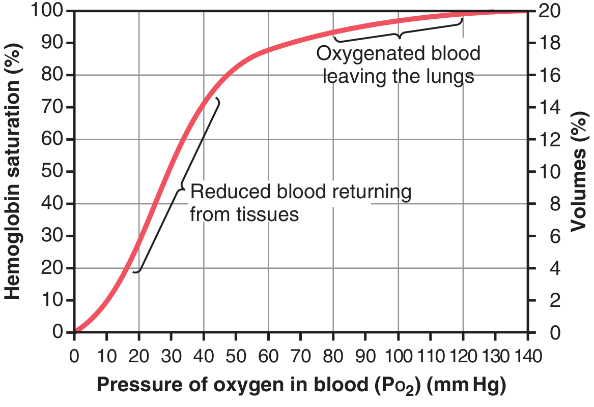
- Shape: Sigmoid (S-shaped) — due to cooperative binding (each O₂ bound increases affinity for next)
- X-axis: PO₂ (mm Hg); Y-axis: % Hemoglobin saturation (left) and Volume% O₂ (right)
Key Reference Points
| Blood | PO₂ | Saturation |
|---|---|---|
| Pulmonary/systemic arterial blood | ~95 mm Hg | 97% |
| Mixed venous blood | ~40 mm Hg | 75% |
| Exercise/heavy activity | ~15 mm Hg (tissue) | ~19% |
O₂ delivery calculation:
- 15 g Hb/100 mL blood × 1.34 mL O₂/g Hb = 20.1 mL O₂/100 mL blood (at 100% saturation = 20 vol%)
- Normal delivery: 19.4 mL (arterial) − 14.4 mL (venous) = ~5 mL O₂ per 100 mL blood per pass
- During exercise: up to 15 mL O₂/100 mL blood can be extracted
Shifts of the Dissociation Curve
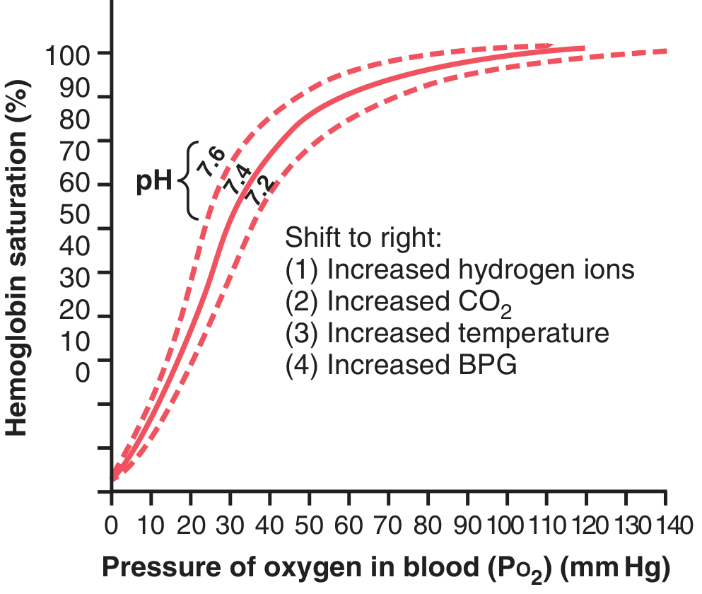
Right Shift (↓ affinity for O₂ → more O₂ released to tissues):
- ↑ H⁺ (↓ pH) — Bohr effect
- ↑ CO₂ (PCO₂)
- ↑ Temperature
- ↑ 2,3-Bisphosphoglycerate (BPG/DPG)
Left Shift (↑ affinity for O₂ → more O₂ retained by Hb):
- ↓ H⁺ (↑ pH)
- ↓ CO₂
- ↓ Temperature
- ↓ BPG
- Fetal Hb (HbF) — has higher O₂ affinity than adult HbA
- CO poisoning — CO shifts curve left AND reduces Hb available for O₂ binding
Bohr Effect
- When blood passes through tissues: CO₂ diffuses in → ↑ PCO₂ → ↑ H₂CO₃ → ↑ H⁺ → right shift → more O₂ released to tissues
- In the lungs: CO₂ diffuses out → ↓ PCO₂ → ↓ H⁺ → left shift → more O₂ loaded onto Hb from alveoli
BPG Effect
- Normal BPG always keeps curve slightly right-shifted
- In prolonged hypoxia → BPG increases markedly → curve shifts further right → O₂ released at up to 10 mm Hg higher tissue PO₂ than without BPG
- Important adaptation to hypoxia (altitude, poor tissue perfusion)
During Exercise
- ↑ CO₂ + ↑ H⁺ + ↑ temperature in exercising muscle → major right shift in muscle capillaries
- Allows O₂ release at PO₂ as high as 40 mm Hg even when 70% O₂ already removed
- In lungs: opposite shift → more O₂ loaded
SUMMARY TABLE — Immunity at a Glance
| Topic | Key Point |
|---|---|
| Innate | Non-specific; first line; skin, phagocytes, complement |
| Acquired | Specific; B cells (humoral) + T cells (cell-mediated) |
| Antigen | MW ≥ 8000; protein/polysaccharide; has epitopes |
| Antibody | 5 classes (IgG, IgM, IgA, IgE, IgD); made by plasma cells |
| Humoral | B cells → plasma cells → antibodies; memory B cells |
| Cell-mediated | Cytotoxic T cells kill by perforins; helper T cells release lymphokines |
| Tolerance | Self-reactive T cells deleted in thymus; B cells in bone marrow |
| Hypersensitivity | Type I (IgE/mast cell); II (cytotoxic Ab); III (immune complex); IV (T cell/delayed) |
| Transplant | HLA typing; rejected by cytotoxic T cells; prevented by cyclosporine/tacrolimus |
| Immunodeficiency | AIDS = helper T cell destruction by HIV; congenital = SCID, DiGeorge, Bruton |
| Hb-O₂ curve | Sigmoid; right shift = ↑CO₂, ↓pH, ↑temp, ↑BPG; left shift = opposite |
*All content sourced from: Guyton and Hall Textbook of Medical Physiology, 14th Edition (Guyton & Hall, Elsevier)*These are comprehensive notes covering all your requested topics directly from Guyton and Hall Textbook of Medical Physiology. Here's a quick orientation:
What's covered:
- Types of Immunity — Innate vs. Acquired (Humoral + Cell-mediated), Active vs. Passive
- Components of Immune System
- MPS (Monocyte-Macrophage/Reticuloendothelial System) — Histiocytes, Kupffer cells, alveolar macrophages, splenic macrophages, lymph node macrophages
- Lymphoid component — T cells (helper, cytotoxic, suppressor, memory) + B cells; locations: lymph nodes, spleen, thymus, bone marrow, gut, tonsils
- Antigens — MW ≥8000, proteins/polysaccharides, epitopes
- Antibodies — 5 classes (IgG/M/A/E/D), mechanisms of action, complement system
- Humoral Immunity Development — B cell → plasma cell pathway, primary vs. secondary response, role of helper T cells
- Cell-Mediated Immunity Development — MHC presentation, T-cell activation, perforins, lymphokines (IL-2, IFN-γ, etc.), self-tolerance
- Hypersensitivity — Type I (IgE/anaphylaxis), II (cytotoxic), III (immune complex), IV (delayed/T-cell)
- Tissue Transplantation — HLA system, graft rejection mechanism, immunosuppressants (cyclosporine, tacrolimus, azathioprine, glucocorticoids)
- Immunodeficiency — AIDS (HIV kills helper T cells), congenital (SCID, DiGeorge, Bruton's), neonatal antibody gap
- Hb-O₂ Dissociation Curve — Sigmoid shape, key values (95/40 mmHg → 97%/75% saturation), right vs. left shift factors (Bohr effect, BPG, temperature, CO₂), clinical applications
This is a shared conversation. Sign in to Orris to start your own chat.