
Types of lung carcinoma
lung carcinoma types histology squamous cell adenocarcinoma small cell
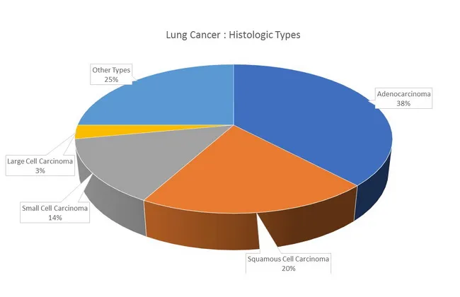
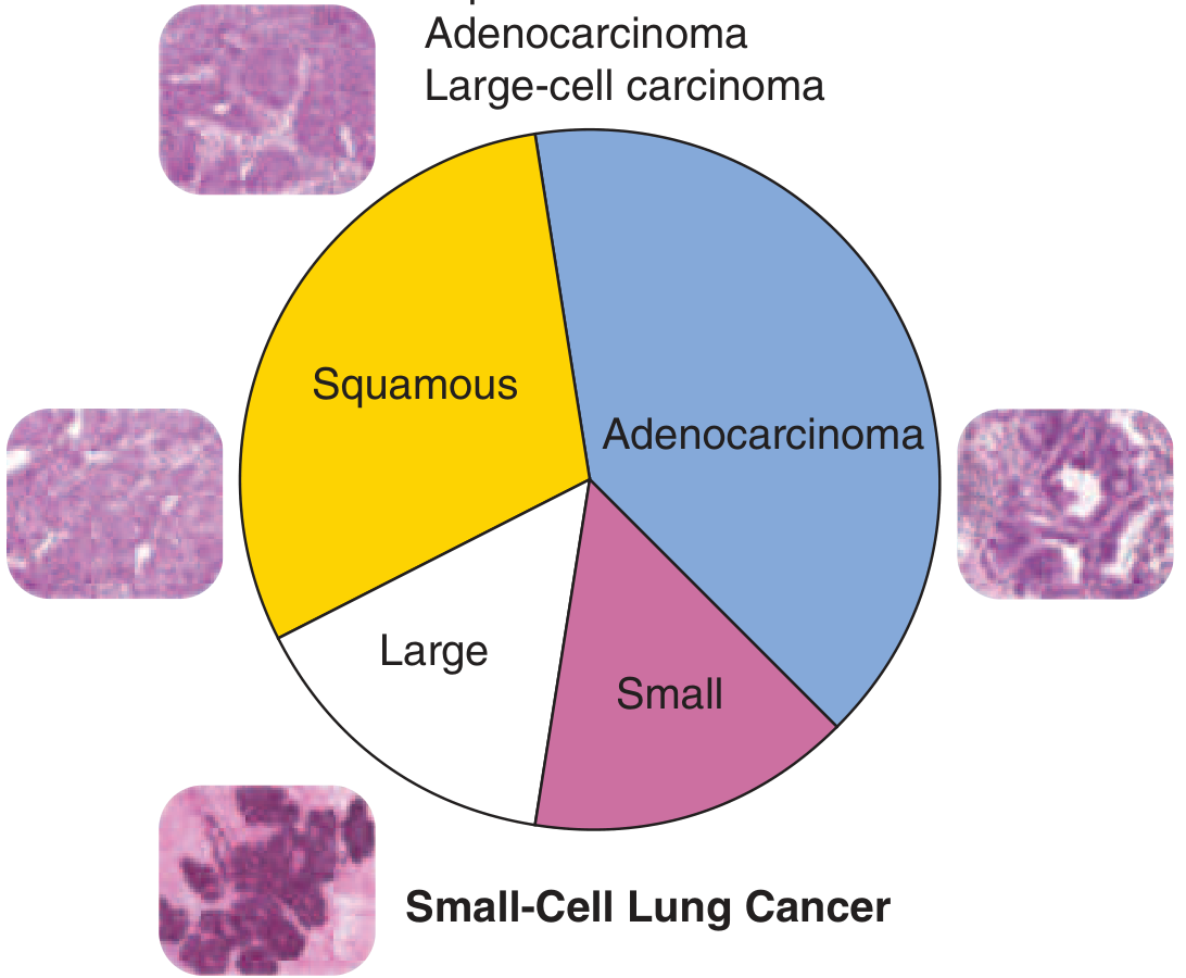
Lung cancer histologic types distribution infographic presenting major histologies and their relative frequencies. The chart is a non-imaging visualization with labeled segments. It shows adenocarcinoma as the most common histology (~38%), followed by squamous cell carcinoma (~20%), small cell carcinoma (~14%), large cell carcinoma (~3%), and other types (~25%). This representation highlights epidemiology: adenocarcinoma is more frequent in women and never-smokers and tends to arise in peripheral airways, whereas squamous and small cell carcinomas originate in proximal airways and show strong smoking associations. Mixed histologies occur in about 10% of cases (e.g., adenocarcinoma with squamous cell). In never-smokers, adenocarcinoma frequently harbors EGFR mutations, while KRAS mutations are more linked to smoking history; TP53 mutations are more common in smoking-related cancers. Clinical relevance includes informing diagnostic workups, guiding biopsy targeting, and shaping research into histology-specific biology and molecular profiles. This image serves educational, epidemiologic, and research contexts in pathology, oncology, pulmonology, and medical education, and supports discussions about tumor heterogeneity, histology-driven therapy implications, and population-based cancer statistics.
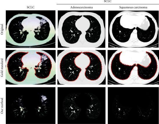
This diagnostic imaging set presents axial thoracic CT scans illustrating three types of lung cancer: Small Cell Lung Cancer (SCLC), Adenocarcinoma, and Squamous Cell Carcinoma. The content is organized into three comparative rows: 'Original' scans, 'Gold Standard' manual segmentations, and automated segmentation results ('Our method'). The SCLC image displays an irregular, large area of increased density in the upper right lung field, consistent with a consolidated mass. The adenocarcinoma scan reveals a more localized, peripheral rounded nodule in the left lung parenchyma. The squamous cell carcinoma scan shows a central lesion with significant soft tissue density near the mediastinum. The 'Gold Standard' row utilizes a red dotted contour to delineate the lung parenchyma boundaries, including the identified pathological regions. This comparison highlights the capability of deep learning algorithms in Lung Parenchyma Segmentation (LPS), specifically the extraction of texture features and nodule morphology for differential diagnosis between SCLC and Non-Small Cell Lung Cancer (NSCLC) subtypes in clinical oncology.
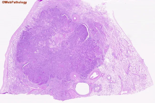
Digital whole-slide histopathology image of a peripherally located lung squamous cell carcinoma captured on a hematoxylin and eosin stained section. The specimen represents peripheral parenchyma with a bronchogenic carcinoma infiltrating the lung periphery. On low- and high-power inspection, nests and irregular clusters of polygonal tumor cells extend from the capsule into adjacent stroma. Tumor cells exhibit eosinophilic cytoplasm, distinct cell borders, and prominent nuclei with coarse chromatin; intercellular bridges may be visible, and occasional keratinization features can be present in well-differentiated foci. The surrounding lung parenchyma shows desmoplastic stromal response and inflammatory infiltrate. The lesion demonstrates peripheral localization, an infiltrative growth pattern, and disruption of normal alveolar architecture. The image supports a histopathologic diagnosis of squamous cell carcinoma due to squamous differentiation, keratinization in places, and intercellular keratin bridges; such morphology is typical in smokers with bronchogenic carcinoma. Clinically, this histology correlates with a smoking history and indicates a solid, non-small cell lung cancer variant with squamous lineage. This image would be valuable for educational purposes, pathology review, differential diagnosis with adenocarcinoma or large cell carcinoma, and in teaching about tumor-stroma interactions in peripheral lung cancers. Correlation with clinical history enhances interpretive accuracy and guides multidisciplinary care in treatment planning.
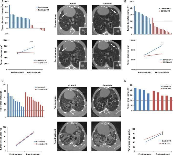
This composite educational graphic illustrates the differential response of lung adenocarcinoma (ADC) and squamous cell carcinoma (SCC) to anti-VEGFR therapies (Sunitinib and DC101) using preclinical mouse models. The data is presented in four panels (A-D). Panels A and B focus on ADC, featuring waterfall plots and line graphs of tumor diameter change. Representative transverse respiratory-gated micro-CT images of the thoracic cavity show ADC lesions (white arrows and high-magnification insets) at pre-treatment and post-treatment stages. In the control group, micro-CT shows visible lesion enlargement, while the Sunitinib-treated group exhibits stabilization of lesion size, corroborated by the flat line plot and waterfall bars below the 20% progression threshold. Panels C and D focus on SCC. Waterfall plots and line graphs depict tumor area change. Micro-CT images demonstrate diffuse tumor masses (outlined by dotted lines). In both control and Sunitinib-treated SCC groups, post-treatment images show significant expansion of the tumor-occupied area, indicating primary resistance to anti-VEGFR2 treatment. This highlights the clinical concept of histology-dependent therapy response in non-small cell lung cancer subtypes.
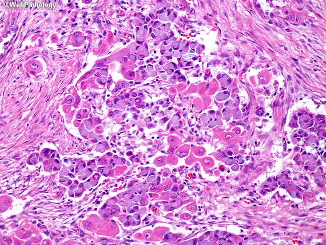
This histopathology slide depicts lung tumor tissue examined by light microscopy following hematoxylin and eosin staining. The specimen is derived from pulmonary parenchyma in a region containing tumor cells. The architecture shows a hybrid neoplasm composed of mucin-producing cells arranged in gland-like clusters and adenomatoid glands intermingled with nests of squamous cells, characterized by dense eosinophilic cytoplasm and rounded to polygonal nuclei. Mucinous cells exhibit bluish cytoplasm and intracytoplasmic mucin; squamous cells display keratinization features and abundant eosinophilic cytoplasm; together they confirm a combined histology with two distinct subtypes. The background stroma contains desmoplastic reaction and inflammatory infiltrate in places. This pattern is typical of adenosquamous carcinoma of the lung, observed in roughly 10% of lung cancers, where prognosis and therapeutic decisions hinge on the more aggressive component. Clinically, such tumors portend a poorer prognosis than pure adenocarcinoma or pure squamous cell carcinoma and may influence choices between surgical resection and systemic therapy. Immunophenotypic profiling (TTF-1 and Napsin A positivity for glandular components; p40/p63 positivity for squamous components) can assist subtyping. Recognize that combined histology carries implications for staging, prognosis, and potential targeted regimens; differential considerations include pure adenocarcinoma with mucin production, pure squamous cell carcinoma, or small cell variants.
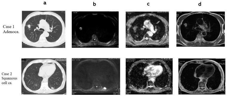
Multi-modal chest imaging comparison of two primary lung cancer types: Adenocarcinoma (Case 1) and Squamous Cell Carcinoma (Case 2). The 4x2 grid displays axial sections across four modalities: (a) Computed Tomography (CT), (b) Diffusion-Weighted Imaging (DWI), (c) Apparent Diffusion Coefficient (ADC) maps, and (d) T2-Weighted Imaging (T2WI). In Case 1, the adenocarcinoma presents as a focal nodule in the right lung field with hyperintensity on DWI and corresponding hypointensity on the ADC map (1.39 x 10^-3 mm^2/s), indicating restricted diffusion. In Case 2, the squamous cell carcinoma is located in the left posterior lung periphery, demonstrating stronger signal suppression on the ADC map (1.04 x 10^-3 mm^2/s) compared to Case 1. T2WI reveals heterogeneous internal architecture for the adenocarcinoma and a more homogeneous appearance for the squamous cell carcinoma. This comparative imaging set illustrates the diagnostic utility of combining structural (CT) and functional (DWI/ADC) MRI sequences to characterize pulmonary nodules and differentiate histological subtypes based on diffusion restriction and T2 contrast ratios.
small cell lung carcinoma histology oat cell neuroendocrine
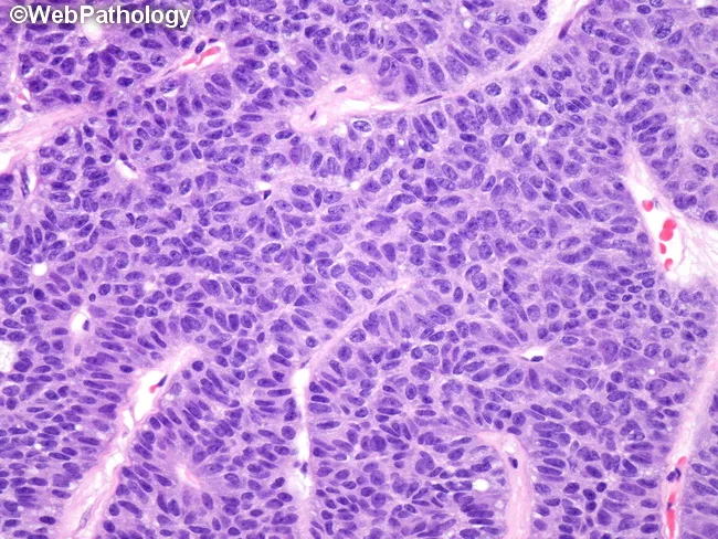
This histopathology image depicts a pulmonary neuroendocrine tumor consistent with carcinoid. The specimen is a lung tissue biopsy prepared as formalin-fixed paraffin-embedded sections, stained with hematoxylin and eosin. Under high magnification, there is a uniform population of small to medium-sized cells arranged in nests and trabeculae (organoid nesting) with rosette-like features and delicate vascular cores. The tumor cells show round to oval nuclei with finely dispersed salt-and-pepper chromatin, pale nucleoli, and scant cytoplasm; mitotic activity is low in typical carcinoids, with limited necrosis. The architectural pattern demonstrates bland cytology with minimal pleomorphism and a relatively low Ki-67 index (if tested), supporting a low-grade neuroendocrine neoplasm. Clinically, such lesions arise in the lung and require differentiation from atypical carcinoids and high-grade neuroendocrine carcinomas (small cell carcinoma or large cell neuroendocrine carcinoma) due to differences in prognosis and treatment approach. Imaging features on histology correlate with indolent behavior, potential for surgical resection, and regional lymph node assessment. Immunohistochemical profiling (not shown) typically reveals neuroendocrine markers such as chromogranin A, synaptophysin, and CD56, with MEN1 involvement noted in a subset of cases. The image illustrates classic histomorphology that pathologists use to classify pulmonary carcinoids and guide clinical management.
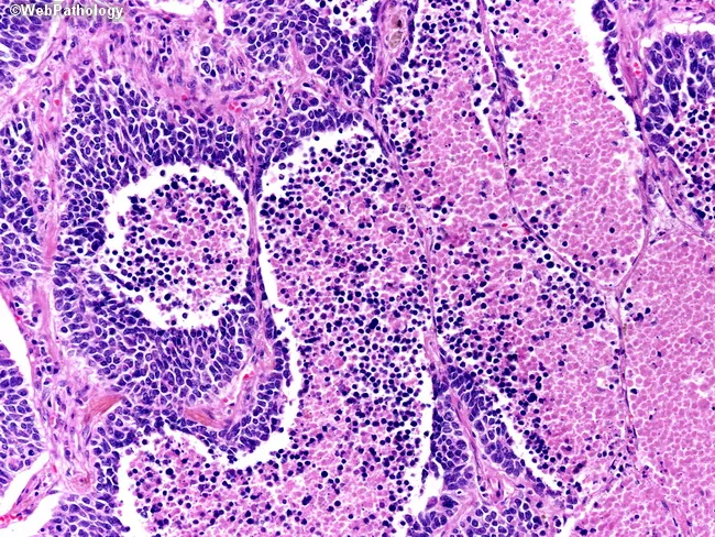
This slide depicts a high-fidelity hematoxylin and eosin stained section of lung tissue analyzed by light microscopy at approximately 400x magnification. The lesion resides within the pulmonary parenchyma with features characteristic of small cell lung carcinoma (SCLC), a high-grade neuroendocrine tumor. Tumor cells form cohesive nests and sheets separated by delicate fibrovascular stroma. Cells are small to medium in size with scant cytoplasm, round to oval nuclei, and finely granular ('salt-and-pepper') chromatin; nuclear molding and frequent mitotic figures are evident. Geographic necrosis is present, producing irregular foci of eosinophilic debris and ghost-like tumor islands, a hallmark of aggressive neoplasia. Background lung tissue shows residual non-neoplastic alveolar parenchyma with inflammatory infiltrates. The overall pattern is consistent with a central, rapidly proliferating tumor commonly associated with early metastasis. Immunophenotypic confirmation (not shown) would typically reveal neuroendocrine markers such as synaptophysin, chromogranin A, and CD56. Clinically, this histology aligns with a diagnosis of primary small cell lung carcinoma and has implications for oncologic management, favoring systemic platinum-based chemotherapy with adjunctive radiotherapy. The image is valuable for educational purposes, differential diagnosis training, and correlation with radiologic findings in lung cancer workups.
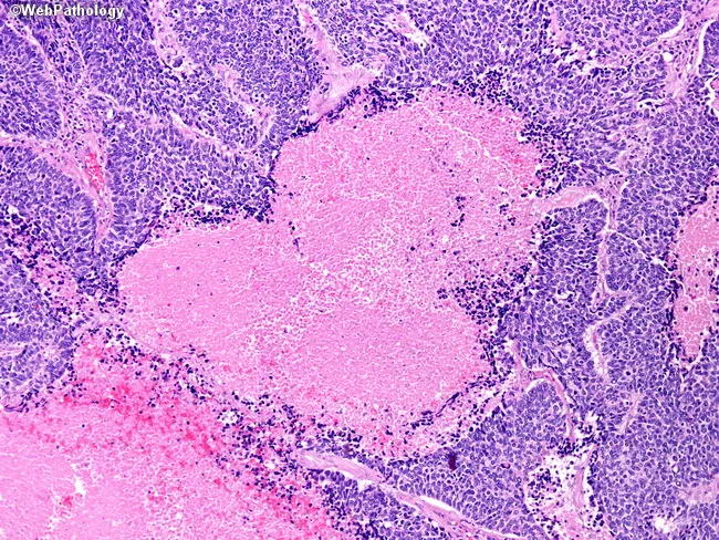
This histopathology image depicts a hematoxylin and eosin-stained lung tissue section showing features characteristic of pulmonary small cell carcinoma with geographic necrosis. The neoplasm is composed of densely packed small round to oval blue cells with scant cytoplasm, ill-defined cell borders, and finely granular, stippled chromatin. Nuclear molding and high mitotic activity are evident in the viable tumor islands, consistent with a high-grade neuroendocrine carcinoma. Within the tumor mass, large pale pink zones of geographic necrosis interrupt cellularity and are surrounded by a rim of residual malignant cells and inflammatory infiltrate. The surrounding non-neoplastic lung parenchyma shows preserved air spaces and alveolar septa without clear organization, highlighting the destructive nature of this tumor. The morphological pattern—sheets and nests of small cells with scant cytoplasm and necrotic foci—supports the diagnosis of small cell lung carcinoma. Clinically, this tumor is aggressive, often centrally located, and associated with paraneoplastic syndromes; histology guides systemic chemotherapy and radiotherapy planning. This image serves as an educational reference for recognizing small cell carcinoma histology, necrotic geography, and the characteristic high-grade appearance in pulmonary tissue. Recognizing this pattern aids differential diagnosis and informs oncologic management. Immunohistochemical confirmation may be pursued if needed.
Types of Lung Carcinoma
WHO Classification (2021)
| Category | Subtypes |
|---|---|
| Adenocarcinoma | Acinar, papillary, micropapillary, solid, lepidic, mucinous |
| Squamous Cell Carcinoma | Keratinizing, non-keratinizing, basaloid |
| Large Cell Carcinoma | Undifferentiated (diagnosis of exclusion) |
| Neuroendocrine Carcinomas | Small cell carcinoma, Large cell neuroendocrine carcinoma |
| Carcinoid Tumors | Typical carcinoid, Atypical carcinoid |
| Mixed / Other | Adenosquamous carcinoma, Sarcomatoid carcinoma, Giant/Spindle cell |
Clinical Grouping: NSCLC vs. SCLC
- Non-Small Cell Lung Cancer (NSCLC) - ~85% of all lung cancers: adenocarcinoma + squamous cell + large cell
- Small Cell Lung Cancer (SCLC) - ~15%: treated entirely differently from the outset
1. Adenocarcinoma (~38% - Most Common Overall)
- Shows glandular differentiation or mucin production
- Growth patterns: acinar, papillary, lepidic, solid, or micropapillary
- Lepidic pattern - tumor cells grow along intact alveolar septa (like a "scaffold"), formerly called bronchioloalveolar carcinoma
- Precursor lesion: atypical adenomatous hyperplasia → adenocarcinoma in situ (AIS, ≤3 cm) → minimally invasive adenocarcinoma → invasive adenocarcinoma
- Tends to metastasize widely at an early stage despite slower growth
- EGFR mutations - ~20% of adenocarcinomas, more common in non-smoking women of Asian origin; sensitive to EGFR inhibitors (erlotinib, osimertinib)
- KRAS mutations - ~30%, mutually exclusive with EGFR
- ALK fusions - 4-6%, targetable with crizotinib/alectinib
- ROS1, HER2, MET - less common but targetable
2. Squamous Cell Carcinoma (~20-25%)
- More common in men; closely correlated with heavy smoking
- Preceded by squamous metaplasia → dysplasia → carcinoma in situ → invasive cancer (well-characterized progression)
- Histology: keratin pearls and intercellular bridges (well-differentiated) to poorly differentiated forms
- May undergo central necrosis with cavitation (important radiologic clue)
- Spreads to hilar lymph nodes first; extrathoracic dissemination is later than other types
- Can be detected early in sputum cytology
3. Small Cell Carcinoma (SCLC) (~14%)
- Also called "oat cell carcinoma" - cells are small, round to fusiform, scant cytoplasm
- Finely granular "salt-and-pepper" chromatin, absent/inconspicuous nucleoli
- Very high mitotic index; extensive necrosis common
- Azzopardi effect - "crush artifact" in small biopsies, DNA staining blue from fragile tumor cells
- Virtually always has loss of function mutations in BOTH TP53 and RB
- Has almost always metastasized to hilar and mediastinal lymph nodes by diagnosis - surgery is NOT curative
- ACTH → Cushing syndrome
- ADH → SIADH
- Gastrin-releasing peptide
- Lambert-Eaton myasthenic syndrome (anti-VGCC antibodies)

4. Large Cell Carcinoma (~10%)
- Diagnosis of exclusion - undifferentiated epithelial tumor lacking features of neuroendocrine carcinoma, glandular, or squamous differentiation
- Large nuclei, prominent nucleoli, moderate cytoplasm
- Peripheral location, tends to be a bulky mass
- No targetable mutations identified consistently
- Relatively poor prognosis
5. Neuroendocrine Tumors - Carcinoid Tumors
| Type | Grade | Mitoses | Necrosis | Behavior |
|---|---|---|---|---|
| Typical Carcinoid | Low | <2/10 HPF | Absent | Indolent; good prognosis |
| Atypical Carcinoid | Intermediate | 2-10/10 HPF | May be present | Intermediate prognosis |
| Large Cell Neuroendocrine Carcinoma | High | >10/10 HPF | Prominent | Aggressive, like SCLC |
| Small Cell Carcinoma | High | Very high | Extensive | Most aggressive |
6. Other Tumor Types
- Adenosquamous carcinoma - combined glandular and squamous components; poorer prognosis than either pure type alone
- Sarcomatoid carcinoma - includes spindle cell and giant cell variants; rare and aggressive
- Mucoepidermoid carcinoma, Adenoid cystic carcinoma - arise from bronchial glands (salivary gland-type tumors)
SCLC vs. NSCLC - Key Comparison
| Feature | SCLC | NSCLC |
|---|---|---|
| Microscopy | Small cells, scant cytoplasm, fine chromatin, diffuse sheets | Abundant cytoplasm, coarse chromatin, prominent nucleoli, glandular/squamous architecture |
| Neuroendocrine markers (CD56, synaptophysin, chromogranin) | Present | Absent |
| RB mutations | ~90% | ~20% |
| TP53 mutations | ~90% | ~50% |
| EGFR mutations | Absent | ~20% (adenocarcinoma) |
| KRAS mutations | Rare | ~30% (adenocarcinoma) |
| ALK fusions | Absent | 4-6% (adenocarcinoma) |
| Response to chemotherapy | Often complete but always recurs | Incomplete response |
| Response to checkpoint inhibitors | Poor | Good (PD-L1 driven) |
| Surgery | Not curative | Curative for early stages |
Smoking and Histologic Association
- Squamous cell and small cell - most strongly linked to smoking (proximal airway origin)
- Adenocarcinoma - still smoking-related but is the dominant type in never-smokers; associated with EGFR mutations (especially in Asian non-smoking women)
- All histologic types can occur in smokers