
adenl.ate cyclase mechanism in enzymatic receptor in short
Reading File
Quick answer: Adenylate cyclase (AC) is a membrane-bound enzyme activated via G-protein coupled receptors. When a ligand binds, it activates a Gs protein, which stimulates AC to convert ATP to cAMP, the second messenger that activates protein kinase A (PKA).
Searching sources...
Finding Sources
Searching Images
adenylate cyclase G protein cAMP second messenger signaling pathway diagram
Reading File
Reading File
Loading Image
Adenylate Cyclase Mechanism in Enzymatic (G-Protein Coupled) Receptors
Adenylate cyclase (AC) is the key effector enzyme in the cAMP second messenger pathway, operating downstream of G-protein coupled receptors (GPCRs). Here is the step-by-step mechanism:
Step-by-Step Mechanism
1. Ligand Binding
A hormone or neurotransmitter (e.g., glucagon, adrenaline, norepinephrine via β-receptors) binds to its specific GPCR on the cell surface.
2. G-Protein Activation (Gs)
- At rest, the stimulatory G-protein (Gs) exists as a trimeric complex (α, β, γ subunits) with GDP bound to the α-subunit.
- Ligand-bound receptor interacts with the Gs complex → the α-subunit releases GDP and binds GTP.
- The Gα-GTP subunit dissociates from the βγ complex.
3. Adenylate Cyclase Activation
- The free Gα-GTP subunit binds to and activates membrane-bound adenylate cyclase.
- AC catalyzes the conversion: ATP → cAMP (3',5'-cyclic AMP) + pyrophosphate.
4. Signal Amplification via PKA
- cAMP binds to the regulatory (R) subunits of Protein Kinase A (PKA), which is normally inactive as a tetramer (2R + 2C subunits).
- Binding of cAMP to R subunits causes a conformational change → catalytic (C) subunits are released and become active.
- Active PKA phosphorylates target proteins on serine/threonine residues, activating some enzymes and inhibiting others.
5. Signal Termination
- The intrinsic GTPase activity of the α-subunit hydrolyzes GTP → GDP, switching off AC.
- cAMP is degraded to 5'-AMP by phosphodiesterase (PDE), terminating the signal.
Inhibitory Pathway (Gi)
Some receptors (e.g., α2-adrenergic, opioid) couple to Gi proteins, which inhibit adenylate cyclase, reducing cAMP production - the opposite effect.
Downstream Effects of PKA Activation
PKA phosphorylates many targets:
- Metabolic enzymes (e.g., activates glycogen phosphorylase, inhibits glycogen synthase)
- Ion channels (rapid effects on excitability)
- CREB (cAMP Response Element Binding protein) - enters the nucleus and regulates gene transcription via CRE elements (longer-lasting effects)
Diagram
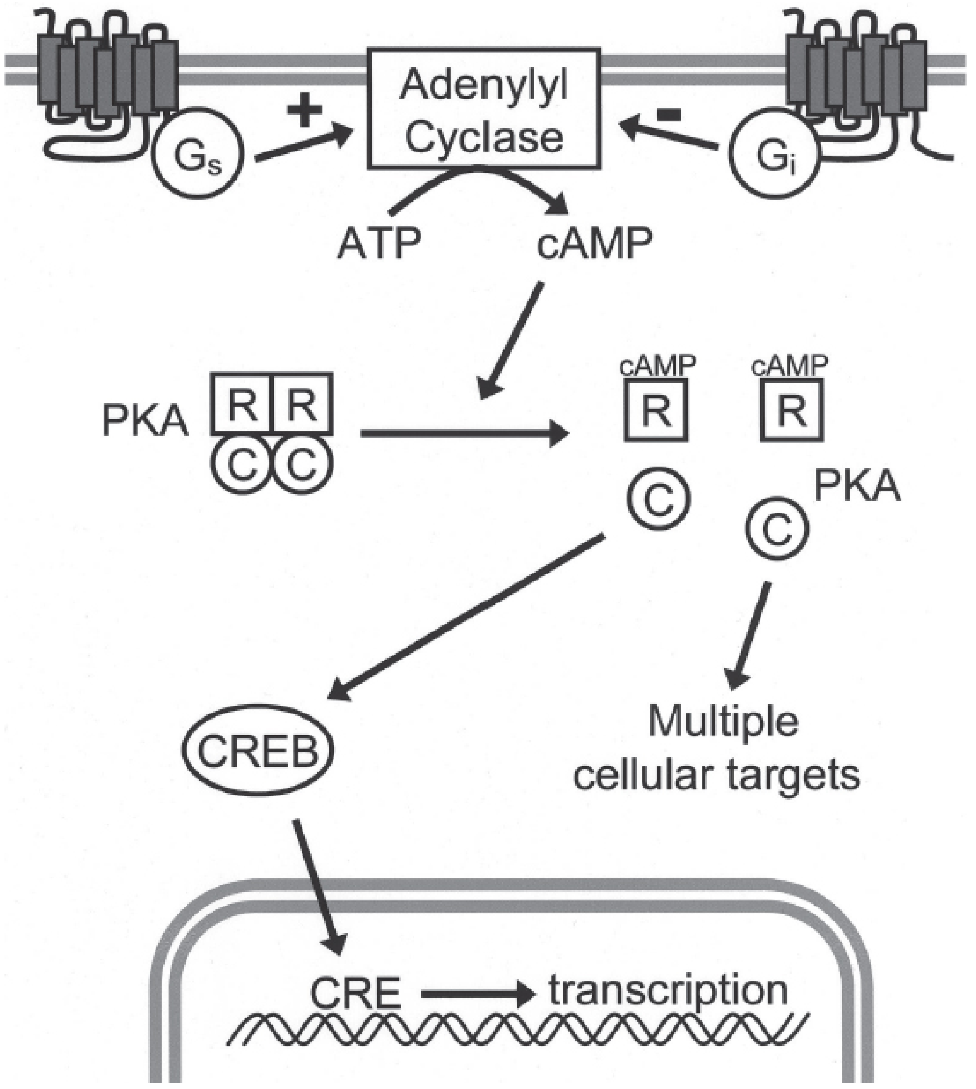
FIGURE: Basic organization of the cAMP signaling pathway - Kaplan & Sadock's Comprehensive Textbook of Psychiatry
Summary Table
| Step | Component | Event |
|---|---|---|
| 1 | GPCR | Ligand binds receptor |
| 2 | Gs protein (α-subunit) | GDP → GTP; α-subunit dissociates |
| 3 | Adenylate cyclase | ATP → cAMP |
| 4 | PKA | R subunits release C subunits → protein phosphorylation |
| 5 | PDE | cAMP → 5'-AMP (signal off) |
Key examples of hormones using this pathway: Glucagon, Epinephrine (β-receptors), ACTH, TSH, PTH, FSH, LH, ADH (V2 receptor).
Sources: Basic Medical Biochemistry - A Clinical Approach 6e, p. ~1793; Kaplan & Sadock's Comprehensive Textbook of Psychiatry, p. 564-565
adenylate cyclase function in heart
Quick answer: In the heart, adenylate cyclase is activated mainly by β1-adrenergic receptors (via Gs proteins) in response to catecholamines (norepinephrine, epinephrine). The resulting cAMP/PKA signaling increases heart rate (chronotropy), contractility (inotropy), and conduction velocity (dromotropy).
Searching sources...
Finding Sources
Finding Sources
Reading File
Reading File
Finding Sources
Finding Sources
Reading File
Reading File
Loading Image
Loading Image
Adenylate Cyclase Function in the Heart
Adenylate cyclase (AC) sits at the center of cardiac adrenergic signaling. Its activation controls heart rate, contractility, and relaxation through cAMP/PKA-mediated phosphorylation of calcium-handling proteins.
Trigger: β1-Adrenergic Stimulation
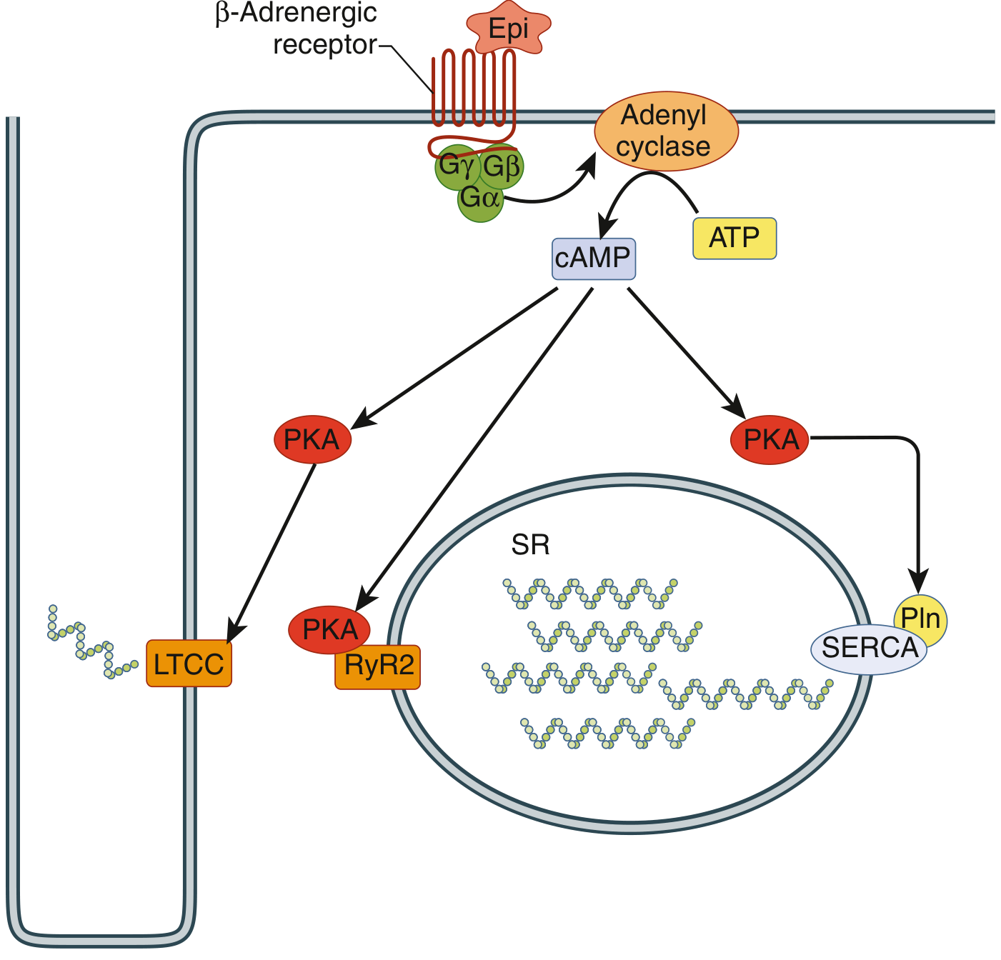
Catecholamines (norepinephrine from sympathetic nerves; epinephrine from the adrenal medulla) bind β1-adrenergic receptors (predominant in the heart) on the sarcolemma.
- β1-receptor → Gs protein (αs subunit) → activates adenylate cyclase
- AC converts ATP → cAMP
- cAMP activates Protein Kinase A (PKA)
PKA Phosphorylates 4 Key Cardiac Targets
| Target | Location | Effect |
|---|---|---|
| L-type Ca²⁺ channels (LTCC) | Sarcolemma | Increased Ca²⁺ influx → stronger contraction |
| Ryanodine receptor (RyR2) | SR membrane | Enhanced Ca²⁺ release from SR |
| Phospholamban (PLN) | SR membrane | Relieves SERCA2A inhibition → faster Ca²⁺ reuptake → faster relaxation + increased SR Ca²⁺ load |
| Troponin I (TnI) | Myofilament | Reduces Ca²⁺ sensitivity → aids relaxation |
Net Cardiac Effects
- Positive Chronotropy (increased heart rate) - cAMP speeds pacemaker If current in SA node; phosphorylation of SR proteins accelerates spontaneous Ca²⁺ cycling in pacemaker cells
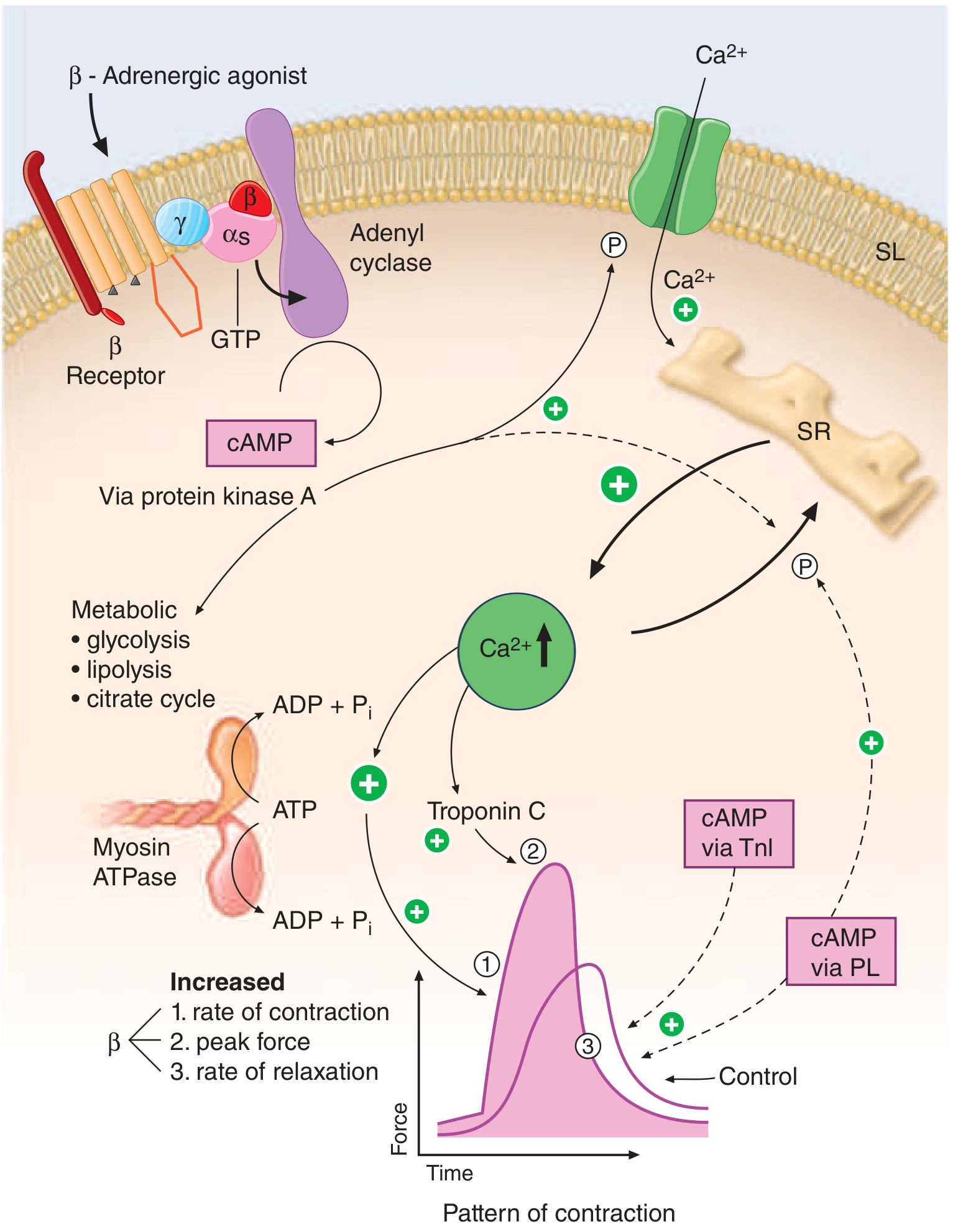
- Positive Inotropy (increased contractility) - more Ca²⁺ enters and is released, greater force of contraction
- Positive Lusitropy (faster relaxation) - phospholamban phosphorylation activates SERCA2A, pulling Ca²⁺ back into the SR faster; TnI phosphorylation reduces myofilament Ca²⁺ sensitivity
- Positive Dromotropy (faster conduction) - enhanced AV node conduction
- Metabolic boost - PKA also stimulates glycolysis, lipolysis, and the citrate cycle to meet increased energy demand
Inhibitory Counterpart (Gi)
- Parasympathetic (ACh via M2 receptors on cardiac pacemaker and myocytes) → Gi → inhibits AC → ↓ cAMP → bradycardia and reduced contractility
- α2-adrenergic receptors also couple to Gi and inhibit AC
Diagram 1 - Cardiomyocyte β-adrenergic → Adenylate Cyclase → Ca²⁺ handling
Cardiomyocyte signaling - Goldman-Cecil Medicine
Diagram 2 - Full β-adrenergic → cAMP → PKA pathway showing inotropic and lusitropic effects
Inotropic and lusitropic effects of β-adrenergic stimulation - Harrison's Principles of Internal Medicine 22E
Clinical Relevance
| Drug/Condition | Mechanism | Effect on AC |
|---|---|---|
| β-blockers (metoprolol, carvedilol) | Block β1-receptor | Reduce AC activation → lower HR and contractility |
| PDE3 inhibitors (milrinone) | Prevent cAMP degradation | Prolong AC's product → increase contractility (used in acute heart failure) |
| Dobutamine | β1-agonist | Directly stimulates AC activation → positive inotropy |
| Heart failure | Downregulation of β1-receptors and AC | Blunted cAMP response → reduced reserve |
Sources: Goldman-Cecil Medicine, p. 412-413; Harrison's Principles of Internal Medicine 22E, p. 1892-1893; Medical Physiology (Boron), Table 23-1
can adenylate cyclase cause tachycardia
Quick answer: Yes - adenylate cyclase activation directly causes tachycardia. When β1-adrenergic receptors are stimulated (e.g., by adrenaline), adenylate cyclase raises cAMP in SA node pacemaker cells, accelerating the rate of spontaneous depolarization and increasing heart rate.
Searching sources...
Finding Sources
Finding Sources
Reading File
Reading File
Yes - Adenylate Cyclase Directly Causes Tachycardia
Here is the full explanation of how and why:
The Mechanism: AC → cAMP → Faster SA Node Firing
Step-by-step in pacemaker cells (SA node):
- Catecholamines (norepinephrine / epinephrine) bind β1-adrenergic receptors on SA node pacemaker cells
- Gs protein → activates adenylate cyclase → ↑ cAMP
- cAMP acts on two key ion channels in the SA node:
| Channel | Effect of ↑cAMP | Result |
|---|---|---|
| HCN channels (If - "funny current") | cAMP directly binds HCN and increases If (Na⁺/K⁺ inward current) | Speeds up phase 4 diastolic depolarization |
| L-type & T-type Ca²⁺ channels | PKA phosphorylation → ↑ ICa | Further accelerates depolarization slope; shifts threshold to more negative voltage |
- Net result: Phase 4 (diastolic depolarization) becomes steeper → SA node fires more frequently → tachycardia
Two Clocks Accelerated by cAMP
The SA node uses two interdependent pacemaking mechanisms, both driven by cAMP:
- Membrane clock (Voltage clock): cAMP directly binds the HCN channel (If), increasing the diastolic inward current that depolarizes the cell toward threshold
- Calcium clock: cAMP/PKA phosphorylates RyR2 and phospholamban → enhanced spontaneous SR Ca²⁺ cycling → triggers more Na⁺/Ca²⁺ exchanger (NCX) inward current → further depolarization
Both clocks are "wound up" by adenylate cyclase activation.
Opposite: How AC Inhibition Causes Bradycardia
Parasympathetic stimulation (ACh via M2 receptors) activates Gi protein → inhibits adenylate cyclase → ↓ cAMP → If decreases, ICa decreases → slower phase 4 depolarization → bradycardia. M2 also directly opens GIRK K⁺ channels, hyperpolarizing the cell further.
Summary Table: AC Activity and Heart Rate
| AC Activity | Trigger | cAMP Level | Heart Rate |
|---|---|---|---|
| Activated (Gs) | Sympathetic, β1-agonists, adrenaline, stress | ↑↑ | Tachycardia |
| Inhibited (Gi) | Parasympathetic (ACh), β-blockers, adenosine | ↓↓ | Bradycardia |
Clinical Conditions Linked to AC-Driven Tachycardia
| Condition / Drug | How AC is Over-Activated |
|---|---|
| Pheochromocytoma | Excess catecholamines → constant β1 stimulation → AC overdrive |
| Thyrotoxicosis | Thyroid hormone upregulates β-receptors and AC sensitivity |
| Dobutamine / Salbutamol (β-agonists) | Directly activate β1/β2 → AC → ↑ cAMP → tachycardia (side effect) |
| PDE inhibitors (milrinone) | Prevent cAMP breakdown → same effect as prolonged AC activation |
| Anesthesia awareness / pain / anxiety | Sympathetic surge → NE/Epi release → AC activation |
Drug That Blocks the cAMP Effect on If (Without Blocking AC)
Ivabradine specifically blocks HCN channels (the direct target of cAMP in the SA node) - it slows heart rate without blocking AC or affecting contractility. This confirms that the cAMP → If pathway is the key tachycardia mechanism.
Sources: Medical Physiology (Boron & Boulpaep), p. 792; Fuster and Hurst's The Heart 15th Ed., p. SA node automaticity section
This is a shared conversation. Sign in to Orris to start your own chat.