
Cervical incompitence explain detail for neet pg
Reading File
Now let me search the medical library for cervical incompetence content in parallel:
Finding Sources
Finding Sources
Good. Let me search specifically in OB/GYN textbooks for dedicated sections:
Finding Sources
Finding Sources
Now let me read the dedicated cervical cerclage sections from the best sources:
Reading File
Reading File
Excellent content. Let me read more from both files for the full picture:
Reading File
Reading File
Excellent content. Now let me read the procedure details and get more clinical content from both sources:
Reading File
Finding Sources
Let me read the Creasy & Resnik section on cervical factors (the most authoritative MFM textbook):
Reading File
Reading File
I now have comprehensive content. Let me fetch the ultrasound image shown in Creasy & Resnik to include in the answer:
Loading Image
I now have all the material needed to write a comprehensive NEET PG answer. Let me compile it:
Cervical Incompetence (Cervical Insufficiency) - NEET PG
Definition
Cervical incompetence (now preferred term: cervical insufficiency) is defined as the inability of the uterine cervix to retain a pregnancy in the second trimester in the absence of uterine contractions. Classically, this means:
- Painless cervical dilation
- Leading to recurrent second-trimester pregnancy loss
- In the absence of other identifiable causes (labor, abruption, infection, ROM)
Diagnosis is one of exclusion - it identifies women with anatomic/structural cervical weakness, not those where preterm parturition has already begun for other reasons. - Creasy & Resnik's Maternal-Fetal Medicine
Incidence
- Exact incidence is uncertain due to lack of clear diagnostic criteria
- Estimated 1 in 100 pregnancies (approximately 1%)
- Responsible for approximately 15-20% of all mid-trimester losses
Etiology / Risk Factors
| Category | Examples |
|---|---|
| Congenital | Mullerian duct anomalies, connective tissue disorders (Ehlers-Danlos), DES (diethylstilbestrol) exposure in utero |
| Acquired / Traumatic | Prior cervical surgery (cone biopsy, LEEP/LLETZ), forceful cervical dilation (D&C), obstetric lacerations, prolonged second stage |
| Structural | Cervical length abnormalities (shortened cervix), deficiencies in cervical collagen and elastin |
| Functional | Multiple gestations, low pre-pregnancy weight |
- Most common cause: Prior surgical trauma from cone biopsy, loop electrosurgical excision procedure (LEEP), or forceful mechanical dilation
- In utero DES exposure is a classic NEET PG association
Pathophysiology
The cervix is normally competent because of:
- High collagen and elastin content in stroma
- Adequate length (normal: 3-4 cm in mid-pregnancy)
- Internal os acting as a functional sphincter
In cervical insufficiency:
- Structural weakness of the cervical stroma
- Painless dilation occurs under the weight of the growing pregnancy
- Funneling of membranes into the cervical canal occurs ("beaking" or "funneling" sign on USG)
- Progressive dilation without contractions leads to second-trimester expulsion
Clinical Features (Classic Presentation)
- History of recurrent, painless mid-trimester losses (14-28 weeks)
- Losses tend to occur at progressively earlier gestation with each pregnancy
- Minimal prodrome - patient may notice increased pelvic pressure or watery vaginal discharge
- Sudden expulsion of a live fetus with intact or minimal contractions
- On examination: dilated os with bulging membranes ("bag of waters" visible through os)
Key distinguishing feature from preterm labor: No painful uterine contractions
Diagnosis
1. Clinical/Historical Diagnosis
Classic diagnostic criteria (all 4 must be present traditionally):
- Painless cervical dilation
- Recurrent (2 or more) losses
- Second-trimester gestation
- Absence of other causes
2. Transvaginal Ultrasound (TVU) - Gold Standard Investigation
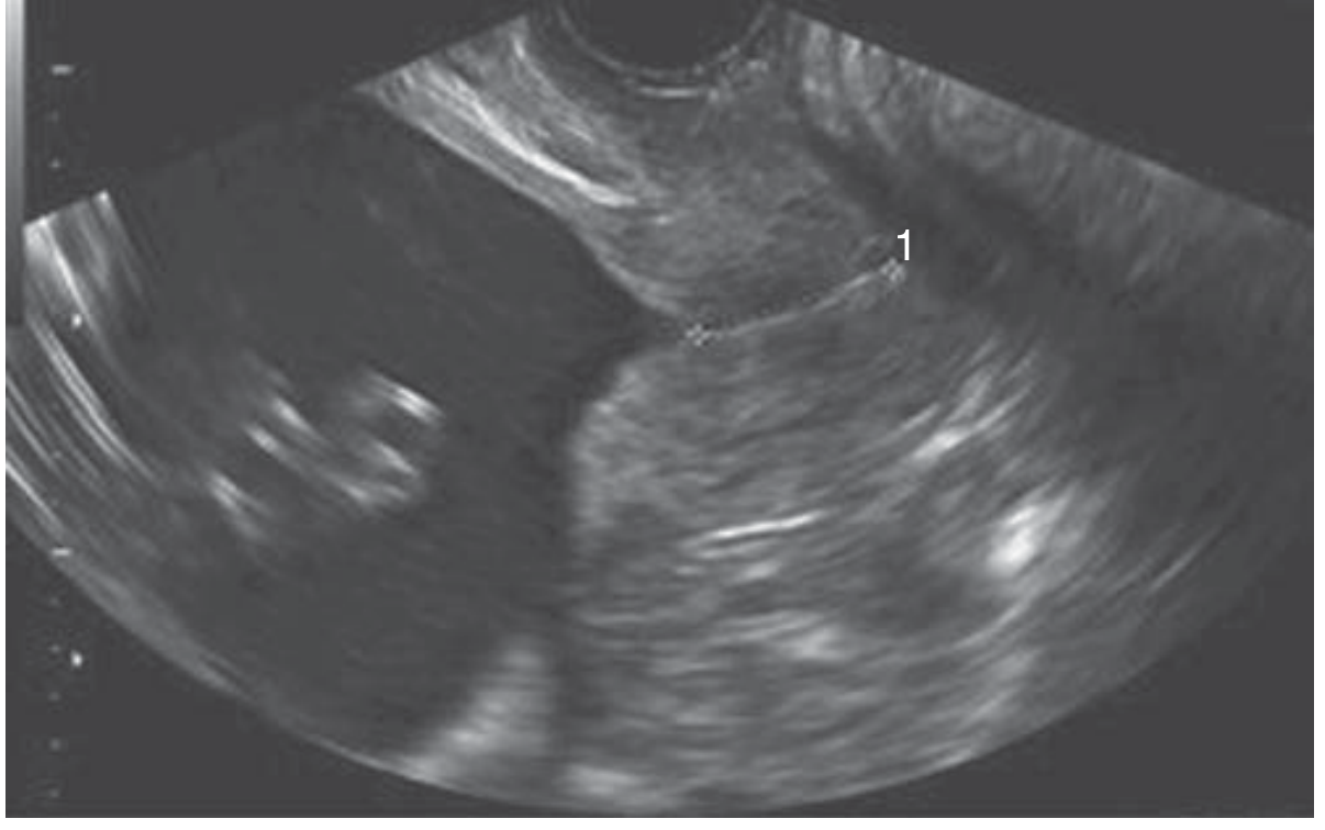
TVU showing Y-shaped funneling at the internal os, with cervical length of 21.3 mm (~5th percentile) - Creasy & Resnik's Maternal-Fetal Medicine
USG Findings of cervical incompetence:
- Short cervical length: <25 mm at 16-24 weeks (action threshold)
- Funneling: V-shaped, U-shaped, or Y-shaped funneling at internal os
- Beaking sign: Wedge-shaped protrusion of membranes into internal os
- Sludge: Amniotic fluid debris (sign of intra-amniotic infection/inflammation)
Cervical length cut-offs (NEET PG high-yield):
| Finding | Significance |
|---|---|
| <25 mm at 22-24 wks (10th percentile) | Abnormal - standard action threshold |
| <20 mm (5th percentile) | Significantly short |
| Funneling >25% of total cervical length | Associated with preterm birth |
- In women with prior preterm birth + CL <25 mm: ~60% risk of preterm birth
- In general population with CL ≤25 mm: ~30-40% risk of preterm birth
3. Non-Pregnant Diagnosis (less reliable)
- Hegar dilator test: Passage of Hegar No. 8 dilator without resistance through internal os in non-pregnant state (positive test, but poor specificity)
- Balloon traction test: Not commonly used today
- HSG: May show incompetent os
Treatment
A. Cervical Cerclage (Main Treatment)
Definition: Placement of a non-absorbable suture circumferentially around the cervix to reinforce it and prevent dilation.
ACOG recommends: Elective cerclage at 14-16 weeks gestation (after documenting fetal viability by USG).
Types of Cerclage:
| Type | Description | Approach |
|---|---|---|
| McDonald cerclage (most common) | Purse-string suture at cervicovaginal junction; simplest technique | Transvaginal |
| Shirodkar cerclage | Higher placement with bladder/rectum reflection; suture buried under mucosa | Transvaginal |
| Transabdominal cerclage (TAC) | Used when transvaginal has failed, absent or severely shortened cervix; laparoscopic or open | Transabdominal |
McDonald vs Shirodkar:
- McDonald: Simpler, placed at cervicovaginal junction, can be removed easily at 37 weeks
- Shirodkar: Higher placement (closer to internal os), suture buried under mucosa, often requires general anesthesia for removal
Three Indications for Cerclage Placement:
| Type | Indication | Timing |
|---|---|---|
| History-indicated (HIC) | History of painless cervical dilation leading to 2nd trimester loss; prior cerclage with preterm birth | 11-15 weeks |
| Ultrasound-indicated (UIC) | Short CL <25 mm on TVU in singleton with prior preterm birth | Before 24 weeks |
| Exam/Physical-indicated | Painless cervical dilation found on exam <24 weeks without labor/PROM/abruption | <24 weeks, urgently |
Perioperative Considerations:
- Amniocentesis: Consider for UIC and exam-indicated (rule out subclinical infection)
- Indomethacin: Consider for exam-indicated cerclage (tocolysis)
- Antibiotics: Perioperative for exam-indicated
Cerclage Removal:
- Removed at 37 weeks gestation
- Or immediately at onset of preterm labor, PROM, or clinical chorioamnionitis
- Removal at onset of labor is important to prevent cervical lacerations
Success Rate:
- 75-90% success in preventing subsequent losses when used after recurrent pregnancy loss
-
- Pfenninger & Fowler's Procedures for Primary Care
Contraindications to Cerclage:
Absolute:
- Nonviable pregnancy
- Ruptured membranes
- Active preterm labor
- Undiagnosed vaginal bleeding
- Acute cervical/intrauterine infection (chorioamnionitis)
Relative:
- Known/suspected fetal anomaly
- Prolapsed membranes through external os (high risk of iatrogenic PROM)
B. Progesterone Therapy (For Short Cervix)
- 17-alpha hydroxyprogesterone caproate (17-OHPC): Weekly IM injections for women with prior preterm birth
- Vaginal micronized progesterone: For asymptomatic short cervix found on USG screening (even without prior PTB history)
- Progesterone and cerclage are complementary in high-risk patients
C. Cervical Pessary
- A mechanical support device placed around cervix
- Proposed as an alternative to cerclage in short cervix
- Cervical incompetence is a proposed indication
D. General Measures
- Pelvic rest (avoid intercourse, heavy exertion)
- Avoid Valsalva maneuvers
- Serial TVU monitoring of cervical length every 1-2 weeks
Complications of Cerclage
- PROM (most common)
- Chorioamnionitis / infection
- Cervical laceration or tear (at removal or with labor)
- Displacement or migration of suture
- Bladder injury
- Preterm labor
- Suture erosion
NEET PG High-Yield Points
| Point | Fact |
|---|---|
| Timing of loss | 14-28 weeks (mid-trimester), painless |
| Most common cause | Prior cervical surgery (LEEP, cone biopsy) |
| Classic association | DES exposure in utero |
| Best diagnostic tool | Transvaginal USG (CL <25 mm at 22-24 wks) |
| Hegar test positive | Can pass No. 8 dilator without resistance |
| Preferred cerclage | McDonald (simplest, most common) |
| ACOG timing for elective cerclage | 14-16 weeks |
| Cerclage removal | 37 weeks gestation |
| Cerclage success rate | 75-90% |
| Absolute contraindication | Ruptured membranes, active labor, infection |
| USG sign | Funneling at internal os (V/U/Y shape) |
| "Beaking sign" | Amniotic membranes funneling into internal os |
| Progesterone use | 17-OHPC (prior PTB) or vaginal progesterone (short CL on USG) |
Key Differentiators for NEET PG
Cervical Incompetence vs Preterm Labor:
| Feature | Cervical Incompetence | Preterm Labor |
|---|---|---|
| Pain | Absent (painless) | Present (contractions) |
| Timing | 14-28 weeks | 28-37 weeks |
| Pattern | Recurrent | May be first episode |
| Cervix | Passively dilated | Ripening with contractions |
| Treatment | Cerclage | Tocolytics, steroids |
Sources:
- Creasy & Resnik's Maternal-Fetal Medicine: Principles and Practice - Cervical Cerclage chapter
- Pfenninger & Fowler's Procedures for Primary Care - Cervical Cerclage chapter
- Schwartz's Principles of Surgery, 11th Ed. - Cervical Insufficiency section
This is a shared conversation. Sign in to Orris to start your own chat.