images of skin legion of sarcodosies

Searching Images

sarcoidosis skin lesions cutaneous

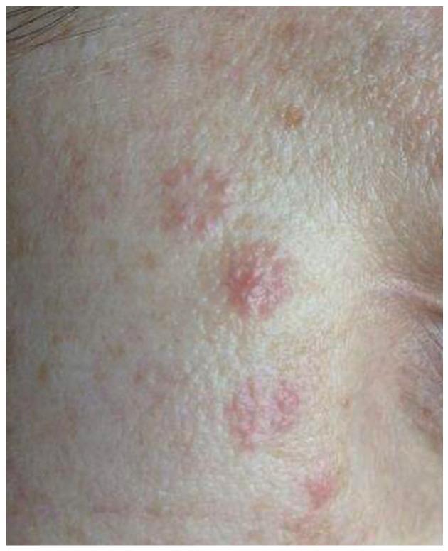
This clinical photograph displays skin lesions on the right cheek of a 38-year-old female patient, illustrating the clinical appearance of cutaneous sarcoidosis during treatment. The visual focus is on a vertical, linear distribution of erythematous, circular to oval-shaped plaques. These lesions appear as discrete, reddish-pink spots with a slightly raised or infiltrated texture. The surrounding skin exhibits a fair tone with mild solar lentigines (freckles) and a relatively smooth texture. Compared to the acute phase of the disease, these lesions show reduced infiltration and erythema, signifying a positive therapeutic response to systemic corticosteroids (prednisone). This image is an educational example of how cutaneous sarcoidosis can present as localized, discoid-like plaques on the face and demonstrates the visible healing process and flattening of granulomatous skin involvement under pharmacological management.
This clinical photograph displays skin lesions on the right cheek of a 38-year-old female patient, illustrating the clinical appearance of cutaneous sarcoidosis during treatment. The visual focus is on a vertical, linear distribution of erythematous, circular to oval-shaped plaques. These lesions appear as discrete, reddish-pink spots with a slightly raised or infiltrated texture. The surrounding skin exhibits a fair tone with mild solar lentigines (freckles) and a relatively smooth texture. Compared to the acute phase of the disease, these lesions show reduced infiltration and erythema, signifying a positive therapeutic response to systemic corticosteroids (prednisone). This image is an educational example of how cutaneous sarcoidosis can present as localized, discoid-like plaques on the face and demonstrates the visible healing process and flattening of granulomatous skin involvement under pharmacological management.

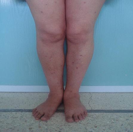
Clinical photograph of the lower extremities demonstrating multiple cutaneous lesions characteristic of systemic sarcoidosis. The image shows a symmetric distribution of red-to-violaceous papules and small nodules on the extensor surfaces of the shins and thighs. The lesions are well-circumscribed, vary in size, and some exhibit focal fine scaling on their surface. Clinically, these granulomatous skin lesions may display a 'lupoid' infiltrate appearance under diascopy (vitropression). The surrounding skin appears unremarkable without evidence of significant edema or ulceration. This visual presentation is an important dermatological manifestation of sarcoidosis, often correlating with multisystem involvement, including pulmonary and hepatic granulomatous disease as indicated by the patient's associated symptoms of dyspnea and elevated serum angiotensin-converting enzyme (ACE) levels.
Clinical photograph of the lower extremities demonstrating multiple cutaneous lesions characteristic of systemic sarcoidosis. The image shows a symmetric distribution of red-to-violaceous papules and small nodules on the extensor surfaces of the shins and thighs. The lesions are well-circumscribed, vary in size, and some exhibit focal fine scaling on their surface. Clinically, these granulomatous skin lesions may display a 'lupoid' infiltrate appearance under diascopy (vitropression). The surrounding skin appears unremarkable without evidence of significant edema or ulceration. This visual presentation is an important dermatological manifestation of sarcoidosis, often correlating with multisystem involvement, including pulmonary and hepatic granulomatous disease as indicated by the patient's associated symptoms of dyspnea and elevated serum angiotensin-converting enzyme (ACE) levels.

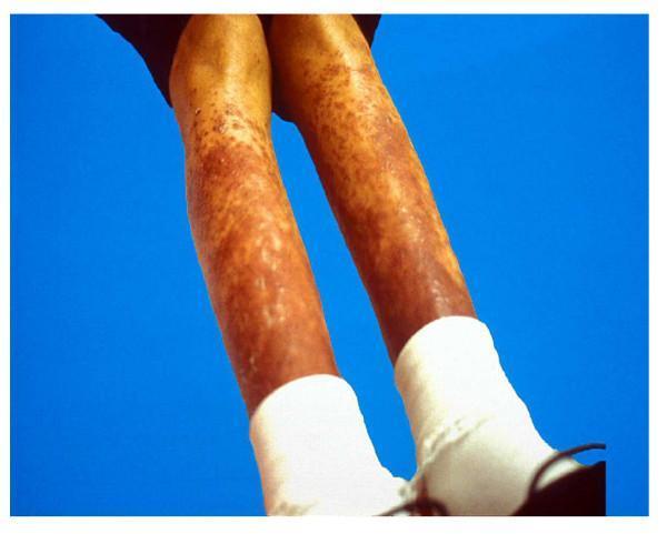
This clinical photograph shows the bilateral lower extremities of a pediatric patient, exhibiting a diffuse cutaneous manifestation of childhood sarcoidosis. The skin displays extensive, erythematous to violaceous (purplish) lesions characterized by a combination of elevated nodules and scaly plaques. These lesions are distributed symmetrically along the shins and calves, ranging from small individual papules to larger, confluent nodular areas. The texture appears irregular, with visible scaling or ichthyosiform changes in several patches. In the context of sarcoidosis, these findings represent cutaneous granulomatous involvement, which occurs in approximately 24-40% of older children. The image serves as an educational example of the diverse dermatological presentations of systemic sarcoidosis, highlighting the importance of skin examination in the diagnostic workup of the disease.
This clinical photograph shows the bilateral lower extremities of a pediatric patient, exhibiting a diffuse cutaneous manifestation of childhood sarcoidosis. The skin displays extensive, erythematous to violaceous (purplish) lesions characterized by a combination of elevated nodules and scaly plaques. These lesions are distributed symmetrically along the shins and calves, ranging from small individual papules to larger, confluent nodular areas. The texture appears irregular, with visible scaling or ichthyosiform changes in several patches. In the context of sarcoidosis, these findings represent cutaneous granulomatous involvement, which occurs in approximately 24-40% of older children. The image serves as an educational example of the diverse dermatological presentations of systemic sarcoidosis, highlighting the importance of skin examination in the diagnostic workup of the disease.

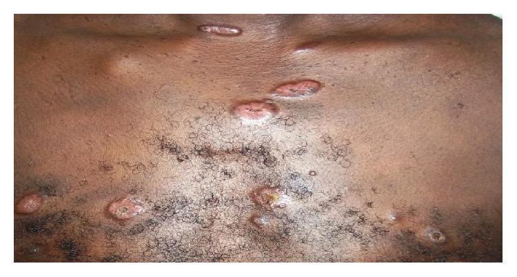
This clinical photograph shows multiple skin lesions on the chest of a dark-skinned patient, consistent with cutaneous sarcoidosis. The primary lesions are well-circumscribed, raised papules and small nodules ranging in color from reddish-pink to brownish-copper. Notable morphological features include central umbilication and crusting in several lesions, suggesting a degree of ulceration or regression. The distribution is scattered across the upper chest, including the infraclavicular and sternal regions. Individual lesions vary in shape from circular to elongated, with surface textures appearing rough or irregular compared to the surrounding healthy skin. These findings are characteristic of the elementary skin lesions found in systemic sarcoidosis, often representing granulomatous infiltration of the dermis. The image serves as an educational example of how systemic inflammatory diseases present dermatologically in patients with higher melanin levels.
This clinical photograph shows multiple skin lesions on the chest of a dark-skinned patient, consistent with cutaneous sarcoidosis. The primary lesions are well-circumscribed, raised papules and small nodules ranging in color from reddish-pink to brownish-copper. Notable morphological features include central umbilication and crusting in several lesions, suggesting a degree of ulceration or regression. The distribution is scattered across the upper chest, including the infraclavicular and sternal regions. Individual lesions vary in shape from circular to elongated, with surface textures appearing rough or irregular compared to the surrounding healthy skin. These findings are characteristic of the elementary skin lesions found in systemic sarcoidosis, often representing granulomatous infiltration of the dermis. The image serves as an educational example of how systemic inflammatory diseases present dermatologically in patients with higher melanin levels.

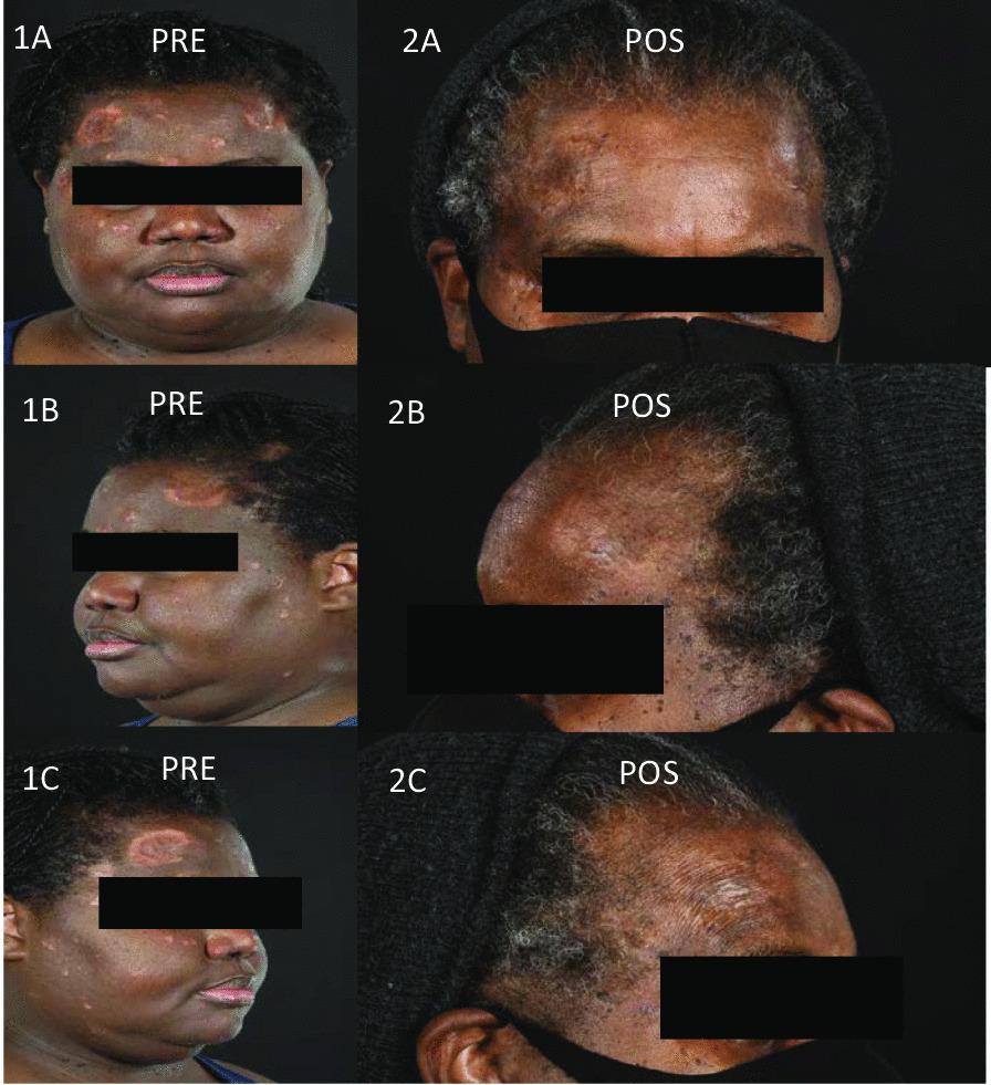
A series of clinical photographs (comparison chart) documenting a case of cutaneous sarcoidosis before and after treatment with infliximab. The pre-treatment images (1A-1C) show a patient with dark skin tones presenting with multifocal, well-demarcated annular lesions, erythematous papules, and indurated plaques. These lesions are primarily distributed across the forehead, temples, and scalp margin, exhibiting central atrophy, scarring, and a mottled pattern of hyperpigmentation and hypopigmentation. The post-treatment images (2A-2C) demonstrate significant clinical improvement. There is a visible resolution of the active inflammatory plaques and papules, leading to a flattened skin surface. However, residual post-inflammatory hypopigmentation and areas of dermal atrophy remain at the sites of the previous active sarcoidosis. The images illustrate the effectiveness of biologic therapy (anti-TNF) in managing recalcitrant cutaneous manifestations of sarcoidosis and the potential for long-term pigmentary and structural skin changes following disease resolution.
A series of clinical photographs (comparison chart) documenting a case of cutaneous sarcoidosis before and after treatment with infliximab. The pre-treatment images (1A-1C) show a patient with dark skin tones presenting with multifocal, well-demarcated annular lesions, erythematous papules, and indurated plaques. These lesions are primarily distributed across the forehead, temples, and scalp margin, exhibiting central atrophy, scarring, and a mottled pattern of hyperpigmentation and hypopigmentation. The post-treatment images (2A-2C) demonstrate significant clinical improvement. There is a visible resolution of the active inflammatory plaques and papules, leading to a flattened skin surface. However, residual post-inflammatory hypopigmentation and areas of dermal atrophy remain at the sites of the previous active sarcoidosis. The images illustrate the effectiveness of biologic therapy (anti-TNF) in managing recalcitrant cutaneous manifestations of sarcoidosis and the potential for long-term pigmentary and structural skin changes following disease resolution.

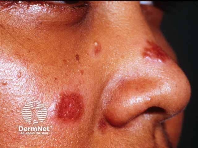
High-resolution clinical photograph of facial skin showing cutaneous sarcoidosis presenting as erythematous-violaceous plaques. The dominant lesion is a well-demarcated, indurated plaque on the cheek with a smaller plaque near the nasal sidewall/bridge. Surfaces appear relatively smooth with minimal scaling and firm, slightly raised borders. Coloration ranges from deep red to purple with subtle gloss; margins are distinctly defined, and the surrounding skin shows mild edema without obvious ulceration. The distribution is central on the midface, involving malar region and nasal area, with unilateral predominance. This image captures typical morphological features of cutaneous sarcoidosis, where granulomatous inflammation produces compact, noncaseating granulomas in the dermis. The imaging modality is clinical photography using a close-up macro lens to maximize color fidelity, border delineation, and lesion texture. Clinically, facial sarcoidosis raises suspicion for systemic involvement; thus, findings should prompt further evaluation for extrafacial sarcoidosis (pulmonary, ocular, lymphatic). Differential diagnoses include granulomatous rosacea, lupus vulgaris, necrobiosis lipoidica diabeticorum, and other granulomatous dermatoses. Knowledge of plaque morphology aids dermatology and pathology training, supports case-based discussion, and enhances databases for AI-assisted recognition of sarcoid skin lesions. This image is suitable for educational, diagnostic, and research applications in dermatology.
High-resolution clinical photograph of facial skin showing cutaneous sarcoidosis presenting as erythematous-violaceous plaques. The dominant lesion is a well-demarcated, indurated plaque on the cheek with a smaller plaque near the nasal sidewall/bridge. Surfaces appear relatively smooth with minimal scaling and firm, slightly raised borders. Coloration ranges from deep red to purple with subtle gloss; margins are distinctly defined, and the surrounding skin shows mild edema without obvious ulceration. The distribution is central on the midface, involving malar region and nasal area, with unilateral predominance. This image captures typical morphological features of cutaneous sarcoidosis, where granulomatous inflammation produces compact, noncaseating granulomas in the dermis. The imaging modality is clinical photography using a close-up macro lens to maximize color fidelity, border delineation, and lesion texture. Clinically, facial sarcoidosis raises suspicion for systemic involvement; thus, findings should prompt further evaluation for extrafacial sarcoidosis (pulmonary, ocular, lymphatic). Differential diagnoses include granulomatous rosacea, lupus vulgaris, necrobiosis lipoidica diabeticorum, and other granulomatous dermatoses. Knowledge of plaque morphology aids dermatology and pathology training, supports case-based discussion, and enhances databases for AI-assisted recognition of sarcoid skin lesions. This image is suitable for educational, diagnostic, and research applications in dermatology.

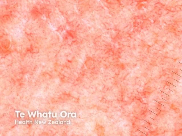
Dermoscopic image of a cutaneous sarcoidosis lesion obtained with polarized dermoscopy. In vivo, magnified view of the affected skin shows a pink to salmon background with diffuse, yellow-orange structureless areas representing granulomatous infiltration of the dermis. Superimposed vascular structures are present, including fine linear and dotted vessels that encircle the yellow areas, reflecting subtle inflammatory vasculopathy. The surface is relatively homogenous with minimal scale, and the lesion may appear slightly translucent due to dermal edema. These dermoscopic features correlate with noncaseating granulomas and granulomatous dermal infiltrates seen histologically, and they help distinguish sarcoidosis from other granulomatous conditions such as lupus vulgaris, granuloma annulare, or infectious etiologies. The pattern is often solitary or localized, without the ulceration or crusting sometimes observed in alternative dermatoses. Recognition of the yellow-orange structureless zones, together with vascular patterns, supports a clinical suspicion of sarcoidosis in patients with compatible systemic signs, such as bilateral hilar lymphadenopathy or skin plaques. Clinically, dermoscopy assists in triaging suspicious lesions for biopsy and guiding systemic evaluation, including chest imaging and laboratory tests (ACE, calcium metabolism). This image emphasizes practical dermoscopic cues for early diagnosis and monitoring response to therapy in cutaneous sarcoidosis.
Dermoscopic image of a cutaneous sarcoidosis lesion obtained with polarized dermoscopy. In vivo, magnified view of the affected skin shows a pink to salmon background with diffuse, yellow-orange structureless areas representing granulomatous infiltration of the dermis. Superimposed vascular structures are present, including fine linear and dotted vessels that encircle the yellow areas, reflecting subtle inflammatory vasculopathy. The surface is relatively homogenous with minimal scale, and the lesion may appear slightly translucent due to dermal edema. These dermoscopic features correlate with noncaseating granulomas and granulomatous dermal infiltrates seen histologically, and they help distinguish sarcoidosis from other granulomatous conditions such as lupus vulgaris, granuloma annulare, or infectious etiologies. The pattern is often solitary or localized, without the ulceration or crusting sometimes observed in alternative dermatoses. Recognition of the yellow-orange structureless zones, together with vascular patterns, supports a clinical suspicion of sarcoidosis in patients with compatible systemic signs, such as bilateral hilar lymphadenopathy or skin plaques. Clinically, dermoscopy assists in triaging suspicious lesions for biopsy and guiding systemic evaluation, including chest imaging and laboratory tests (ACE, calcium metabolism). This image emphasizes practical dermoscopic cues for early diagnosis and monitoring response to therapy in cutaneous sarcoidosis.

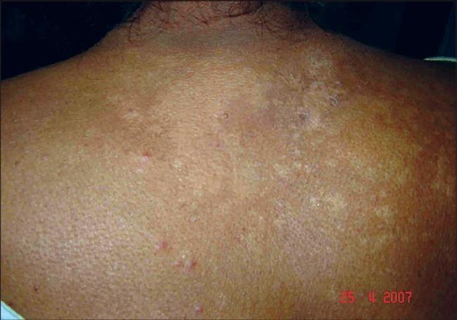
Clinical photograph of a patient's upper back and shoulders showing post-treatment changes of cutaneous sarcoidosis. The skin exhibits a mottled appearance with widespread, irregular areas of hypopigmentation and residual hyperpigmentation. There are visible clusters of light-brown to tan skin tones interspersed with lighter, coalescing patches. A few sparse, small, erythematous papules remain visible, particularly in the lower aspects of the frame. The overall skin texture shows subtle irregularities and resolving surface changes, indicating the healing phase of previously pruritic, coalescent plaques and papules. The image illustrates the clinical outcome following systemic corticosteroid and antimalarial therapy for photosensitive cutaneous sarcoidosis, characterized by significant clearing of active lesions and residual pigmentary alterations.

Clinical photograph of a patient's upper back and shoulders showing post-treatment changes of cutaneous sarcoidosis. The skin exhibits a mottled appearance with widespread, irregular areas of hypopigmentation and residual hyperpigmentation. There are visible clusters of light-brown to tan skin tones interspersed with lighter, coalescing patches. A few sparse, small, erythematous papules remain visible, particularly in the lower aspects of the frame. The overall skin texture shows subtle irregularities and resolving surface changes, indicating the healing phase of previously pruritic, coalescent plaques and papules. The image illustrates the clinical outcome following systemic corticosteroid and antimalarial therapy for photosensitive cutaneous sarcoidosis, characterized by significant clearing of active lesions and residual pigmentary alterations.

Reading File
Here are clinical images of cutaneous sarcoidosis skin lesions:

1. Facial Plaques (Malar/Cheek Region)

Erythematous-violaceous plaques on the cheek - classic cutaneous sarcoidosis
Erythematous-violaceous indurated plaques on the malar/nasal region. The deep red-to-purple color with firm raised borders is characteristic. This distribution (midface) is classic for lupus pernio, the most specific cutaneous form of sarcoidosis.

2. Papules & Nodules on the Chest (Dark Skin)

Papules and nodules on the chest in cutaneous sarcoidosis
Well-circumscribed reddish-pink to brownish-copper papules and nodules on the upper chest with central crusting. Cutaneous sarcoidosis often presents differently in patients with higher melanin levels - lesions may appear more hyperpigmented.

3. Lower Extremity Nodules/Papules

Violaceous papules and nodules on the lower legs in sarcoidosis
Symmetric red-to-violaceous papules and small nodules on the extensor surfaces of shins and thighs. These granulomatous skin lesions often correlate with systemic involvement (pulmonary, hepatic) and elevated serum ACE levels.

4. Pediatric Cutaneous Sarcoidosis

Extensive erythematous-violaceous nodules and plaques on a child's lower legs
Bilateral symmetric violaceous nodules and scaly plaques on the shins of a pediatric patient. Cutaneous involvement occurs in ~24-40% of children with systemic sarcoidosis.

5. Annular/Facial Lesions (Before & After Infliximab)

Before and after treatment comparison of facial sarcoidosis plaques
Annular plaques with central atrophy and hyperpigmentation on the forehead and scalp margin (pre-treatment), with near-complete resolution after anti-TNF (infliximab) therapy. Residual post-inflammatory pigmentary changes persist.

6. Dermoscopy of a Sarcoid Lesion

Dermoscopy of cutaneous sarcoidosis showing yellow-orange structureless zones
Dermoscopic view showing the hallmark salmon/pink background with yellow-orange structureless areas (corresponding to dermal granulomas) and fine linear/dotted vessels. This pattern helps distinguish sarcoidosis from other granulomatous conditions.

7. Facial Plaques Responding to Steroids

Sarcoidosis facial plaques improving after corticosteroid treatment
Linear erythematous plaques on the cheek showing reduced infiltration and erythema following prednisone therapy - useful for monitoring therapeutic response.

Key Morphological Patterns at a Glance

TypeLocationAppearance
Lupus pernioNose, cheeks, earsViolaceous indurated plaques
Papules/nodulesFace, trunk, extremitiesRed-brown, firm
PlaquesAny siteIndurated, scaly or smooth
Annular lesionsFace, trunkRing-shaped with central clearing
Scar sarcoidosisOld scarsInfiltration/induration of scars
Erythema nodosumShinsTender, transient red nodules
Biopsy showing non-caseating granulomas in the dermis is the diagnostic gold standard.
This is a shared conversation. Sign in to Orris to start your own chat.